*Correspondence: M. Kalegari. Departamento de Farmácia, Universidade Federal do Paraná. Av. Prefeito Lothário Meissner, 632 - Jardim Botânico - 80210-170 - Curitiba - PR, Brasil. E-mail: [email protected]
A
rti
Pharmaceutical Sciences vol. 47, n. 3, jul./sep., 2011
Phytochemical constituents and preliminary toxicity evaluation of
leaves from
Rourea induta
Planch. (Connaraceae)
Milena Kalegari
1*, Marilis Dallarmi Miguel
1, Josiane de Fátima Gaspari Dias
1, Ana Luísa Lacava
Lordello
2, Cristina Peitz de Lima
1, Cristina Mayumi Sasaki Miyazaki
1, Sandra Maria Warumby
Zanin
1, Maria Christina dos Santos Verdam
1, Obdulio Gomes Miguel
11Department of Pharmacy, Federal University of Paraná, 2Department of Chemistry, Federal University of Paraná
Most active plants are toxic at high doses and it is therefore important to investigate the preliminary toxicity of plant extracts. The Rourea induta species is a potential drug with no phytochemical or biological studies registered in the literature. Thus, a phytochemical study and a toxicity analysis of the ethanolic extract obtained from the leaves of Rourea induta Planch., Connaraceae, was run. A long chain hydrocarbon, n-tetracosane, and four lavonoids were identiied: quercetin, and three glycosylated derivates, quercetin-3-O-α-arabinofuranoside, quercetin-3-O-β-xyloside and quercetin-3-O-β-galactoside. This is the irst time these have been isolated in thisspecies. The structures were elucidated by 13C NMR, 1H NMR, UV and IR spectroscopy. The toxicity evaluation of extracts was performed by the brine shrimp method and determination of hemolytic activity. The samples demonstrated no toxic potential by the analyzed methods.
Uniterms: Rourea induta/phytochemistry. Connaraceae/phytochemistry. Flavonoids. N-tetracosane. Plant extracts/toxicity.
A maioria das plantas ativas é tóxica em doses elevadas, portanto, é importante a investigação da toxicidade preliminar dos extratos das plantas. A espécie Rourea induta é uma droga potencial que não apresenta estudo itoquímico ou biológico descrito na literatura. Assim, um estudo itoquímico e análises toxicológicas foram realizados com o extrato etanólico obtido das folhas de Rourea induta Planch., Connaraceae. Foram obtidos um hidrocarboneto de cadeia longa, n-tetracosano, e quatro lavonóides, quercetina e três derivados glicosilados, quercetina-3-O-α-arabinofuranosideo, quercetina-3-O-β-xilosideo e quercetina-3-O-β-galactosideo. Esta é a primeira vez que estes compostos são isolados nesta espécie. As estruturas foram elucidadas por espectroscopia de RMN-13C, RMN-1H, ultravioleta e infravermelho. A avaliação da toxicidade dos extratos foi analisada pelo método da Artemia salina e atividade hemolítica. Nenhuma das amostras testadas apresentou um potencial tóxico pelos métodos analisados.
Unitermos: Rourea induta/itoquímica. Connaraceae/itoquímica. Flavonóides. N-tetracosano. Extrato de plantas/toxicidade.
INTRODUCTION
The Connaraceae family comprises approximately 16 genera among 300 to 350 species distributed throughout tropical areas of the world; that consists of the angiosperms characterized by ligneous, trees or shrub species. In Ame-rica, ive genera can be found: Bernardinia, Cnestidium,
Connarus, Pseudoconnarus and Rourea (Forero, 2007).
Rourea is a pantropical genus and comprises 42 species and 19 varieties; the Amazon is its dispersion center (Forero, 1976; Fonseca, Proença, 2002). The Rou-rea induta Planch. species is characterized by small trees and shrubs that grow to a maximum height of 4 meters. It bears greenish and densely ramiied young shrubs. Its cork is widely known for the treatment of Rheumatism (Fonseca, Proença, 2002).
Some important biological activities have been described for species of the Connaraceae family, whereas the Rourea induta species has no registered studies in the literature. Phytochemical and biological activity studies are a starting point contributing toward the valuable detec-tion of the species and its isolated compounds.
Most active plants are toxic at high doses and it is thus important to investigate the preliminary toxicity of plant extracts. To achieve a toxicological evaluation, the brine shrimp method and hemolytic activity determina-tion techniques have been employed for different groups (Oliveira et al., 2009). Against this background the aims of this study were to explore and isolate chemical consti-tuents from the ethanol extract of Roureainduta Planch., Connaraceae, and to perform a toxicity evaluation of the extract and fractions.
MATERIAL AND METHODS
Plant material
The leaf samples from R. induta Planch., Connara-ceae were collected in Rondonópolis city, Mato Grosso State, Brazil, in November 2007. The material was iden-tiied by Gert Hatschbach, botanist of Curitiba Botanical Museum, Brazil, and the exsiccate was registered under voucher number 261574.
Extraction and isolation
The total amount of ethanol extract was obtained from 2 kg of plant material in 95% ethanol, with the use of Soxhlet equipment, and then iltered, concentrated in a ro-tary evaporator to 300mL, and fractioned by liquid-liquid partition with solvents of different polarities. From this mixture, hexane, ethyl acetate and chloroform fractions were obtained. The hexane fraction was used to dissolve the iltered residue, and this was fractioned with hexane in a silica column on the Soxhlet equipment. The fractions hexane 1 and hexane 2 were obtained as a result, yielding 5.7834g (0.289%) for hexane 1, 4.9818g (0.249%) for hexane 2, 39.92g (1.99%) for chloroform and 54.3579g (2.717%) for ethyl acetate, at the end of this procedures.
Hexane fraction 1 underwent column chromato-graphy in silica gel stationery phase 60 Merck 0.063 – 0.200 mm, and hexane/ethyl acetate mobile phase at 5% gradient. White crystals (compound 1)(1.0314 g) were obtained among sub-fractions 1-3.
The chloroform fraction was also submitted to si-lica column chromatography with a hexane/ethyl acetate eluent system at 5% gradient. It attained ethyl acetate to 100% followed by an ethyl acetate/methanol system at 5% gradient. The samples that had some crystallization were submitted to thin-layer chromatography in mobile phase with ethyl acetate, formic acid, acetic acid, water (100:11:11:27) and reactive NEU (reactive 2-aminoe-thylbutyrate). A pure compound (compound 2) (0.1171 g) was identiied between sub-fractions 11-16. Sub-fractions 30-36 and 37-44 were submitted to Sephadex LH-20 with 70% methanol as the eluent, and two compounds were obtained denominated 3 (0.0095 g) and 4 (0.022 g).
During the concentration of ethyl acetate fraction, yellow crystals precipitated, which were puriied by va-cuum iltering and identiied as compound 5 (1.5494 g).
Compound identiication
The 13C NMR and 1H NMR spectrums were obtai-ned on a Brucker®spectrophotometer model AC200 at 300 MHz. Ultraviolet spectroscopy was carried out on a UV-1601 Shimadzu® spectrophotometer from 200 to 450 nm following the Mabry, Markham & Thomas (1970) methodology. IR spectrums were carried out on pastilles of anhydrous potassium bromide (KBr) compressed on Bomem-Hartmann & Braum MB-series equipment and analyzed on a Biored® FTS 3500 GX, 400 to 4000 cm-1. The melting point was measured on Kofler-Reichert Austria equipment.
N-tetracosane (1): White crystals. mp 65 ºC; IR (KBr) νmax 2918. 2848 (CH2) cm-1; 13C NMR (CDCl3; 75 MHz) δ: 32.1 (CH2). 29.9 (CH2). 29.6 (CH2). 22.9 (CH2). 14.3 (CH3. C-1; C-24); 1H NMR (CDCl3; 300 MHz) 0.8 (3H. t); 1.26 (32H. s); EIMS m/z 338 (5). C24H50 (M+). 44 (100) [C3H7+]. 57 (95) [C4H9+]. 71 (67) [C5H11+]. 85 (45) [C6H13+]. 97 (30). 113 (23). 124 (14). 141 (13). 168 (10). 194 (15). 207 (18). 267 (15). 295 (7).
IR (KBr) νmax 3406, 3294 (OH), 1664 (C=O), 1610-1400 (C=C) cm-1; 13C NMR (CD
3OD; 75 MHz) δ 177.2 (C, C-4), 165.3 (C, C-7), 162.3 (C, C-5), 158.1 (C, C-9), 148.5 (C, C-3’), 147.8 (C, C-2), 146 (C, C-4’), 137.1 (C, C-3), 124 (C, C-1’), 121.6 (CH, C-6’), 116.1 (CH, C-5’), 115.9 (CH, C-2’), 104.1 (C, C-10), 99.1 (CH, C-6), 94.4 (CH, C-8); 1H NMR (CD3OD; 300 MHz) 7.72 (1H, d, J=2.1Hz, H-2’), 7.6 (1H, dd; J=8.7; 2.1 Hz, H-6’), 6.8 (1H, d, J=8.4 Hz, H-5’), 6.36 (1H, d, J=2.1 Hz, H-8), 6.17 (1H, d, J=2.1 Hz, H-6).
Quercetin-3-O-α-arabinofuranoside (3): Yellow amorphous powder, mp 205 ºC; UV λmax (log ε) (MeOH) 355 (6.46), 257 (6.57), 267(sh) (6.52) nm; (NaOAc) 370 (6.41), 271 (6.58) nm; (NaOAc/H3BO3) 375 (6.52), 310 (6.10), 261 (6.66) nm; (AlCl3) 436 (6.58), 275 (6.66) nm; (AlCl3/HCl) 400 (6.42), 270 (6.60) nm; IR (KBr) νmax 3427; 3311(OH), 1652 (C=O),1606, 1360 (C=C) cm-1; 13C NMR (CD
3OD; 75 MHz) δ 179.9 (C, C-4), 166.0 (C, C-7),163.1 (C, C-5), 159.4 (C, C-9), 158.5 (C, C-2), 149.9 (C, C-3’), 146.4 (C, C-4’), 134.9 (C, C-3), 123.1 (C, C-1’), 122.9 (CH, C-6’), 116.8 (CH, C-2’), 116.4 (CH, C-5’), 109.5 (CH, C-1’’), 105.6 (C, C-10), 99.9 (CH, C-6), 94.7 (CH, C-8), 87.9 (CH, C-4’’), 83.3 (CH, C-2’’), 78.6 (CH, C-3’’), 62.5 (CH2, C-5’’); 1H NMR (CD3OD; 300 MHz) 7.5 (1H, d, J=3.3 Hz, H-2’), 7.4 (1H, ls, H-6’), 6.9 (1H,
d, J=8.4 Hz ,H-5’), 6.38 (1H, ls ,H-8), 6.2 (1-H, , ls, H-6), 5.58 (1H, d, J=1.0 Hz, H-1’’), 4.3 (1H, d, J=4.3 Hz, H-2’’), 3.9 (1H, dd, J=4.2; 2.4 Hz, H-3’’), 3.8 (1H, dd, J=4.2; 8.5 Hz, H-4’’), 3.5 (2H, m, H-5’’a; H-5’’b).
Quercetin-3-O-β-xyloside (4): Yellow amorphous powder. UV λmax (log ε) (MeOH) 355 (6.65), 257 (6.74) nm; (NaOAc) 371.5 (6.58), 272.5 (6.76) nm; (NaOAc/ H3BO3) 375.5 (6.70), 261 (6.83) nm; (AlCl3) 430 (6.68), 273 (6.82) nm; (AlCl3/HCl) 400 (6.60), 270 (6.78)nm; IR (KBr) νmax 3419, 3327 (OH), 1651 (C=O), 1606-1360 (C=C) cm-1; 13C NMR (CD
3OD; 75 MHz) δ 179.4 (C, C-4), 166.0 (C, C-7), 163.1 (C, C-5), 158.5 (C, C-2), 158.5 (C, C-9), 149.8 (C, C-3’), 146.1 (C, C-4’), 135.4 (C, C-3), 123.3 (CH, C-6’),123.0 (C, C-1’), 117.2 (CH, C-5’), 115.9 (CH, C-2’), 105.6 (C, C-10), 104.5 (CH, C-1’’), 99.8 (CH, C-6), 94.7 (CH, C-8), 77.5 (CH, C-3’’), 75.3 (CH, C-2’’), 71.0 (CH, C-4’’),67.2 (CH2, C-5’’); 1H NMR (CD3OD; 300 MHz) 7.6 (1H, H-2’), 7.57 (1H, d, J=2.1 Hz, H-6’), 6.84 (1H, s,
H-5’), 6.39 (1H, d, J=1.8 Hz, H-8), 6.16 (1H, d, J=2.1 Hz, H-6), 5.17 (1H, d, J=7.2 Hz, H-1’’), 3.78 (1H, dd, J=11.4; 5.0 Hz, H-5’’b), 3.46-3.54 (2H, m, H-3’’ e H-4’’), 3.39 (1H,
m, H-2’’), 3.1 (1H, dd, J=11.5; 9.5Hz, H-5’’a).
Quercetin-3-O-β-galactoside (5): Yellow amor-phous powder, mp 230 oC; UV λmax (MeOH) 257.9 (6.37),
269(sh) (6.36), 297(sh) (6.06), 359.5 (6.27) nm; ( NaOAc) 271.5 (6.30), 372.5 (6.21) nm; (NaOAc/H3BO3) 379.5 (6.32), 262 (6.46) nm; (AlCl3) 272 (6.41), 424.5 (6.22) nm; (AlCl3/HCl) 269 (6.40), 299.5 (sh) (6.00), 400 (6.22) nm; IR (KBr) νmax 3429, 3244 (OH), 1656 (C=O), 1606-1508 (C=C) cm-1; 13C NMR (75 MHz, DMSO-d
6) δ 177.5 (C, C-4), 164.2 (C, C-7), 161.3 (C, C-5), 156.2 (C, C-2), 156.3 (C, C-9), 148.5 (C, C-4’), 144.9 (C, C-3’), 133.5 (C, C-3), 122.1 (CH, C-6’), 121.1 (C, C-1’), 115.9 (CH, C-2’), 115.2 (CH, C-5’), 103.9 (C, C-10), 101.8 (CH, C-1’’), 98.7 (CH, C-6), 93.5 (CH, C-8), 75.9 (CH, C-5’’), 73.2 (CH, C3’’), 71.2 (CH, C-2’’), 67.9 (CH, C-4’’), 60.2 (CH2, C-6’’); 1H NMR (300 MHz, DMSO-d6) 12.62 (1H,
s, 5-OH), 10.87 (1H, s, 7-OH), 9.74 (1H, s, 4’-OH), 9.17 (1H, s, 3’-OH), 7.65 (1H, dd, J= 8.4 e 2.1 Hz, H-6’), 7.52 (1H, d, J= 2.1 Hz, H-2’), 6.82 (1H, d, J=8.4 Hz, H-5’), 6.40 (1H,d, J=2.1 Hz, H-8), 6.20 (1H,d, J=2.1 Hz, H-6), 5.38 (1H, d, J=8.4 Hz, H-1’’), 5.14 (1H, s, 2’’-OH), 4.87 (1H, s, 3’’-OH), 4.44 (2H, s, 4’’, 6’’ – OH).
Brine shrimp bioassay
The brine shrimp (Artemia salina) method was irst described by Meyer et al. (1982). The eggs of brine shrimp hatched within 48 hours in artiicial sea water, producing a large number of larvae. The samples, ethanolic extract, and all the fractions, were tested at concentrations of 1000, 100 and 10 μg/mL. Ten shrimps were transferred to the samples and after 24 hours survivors were counted and percentage of deaths at each dose recorded. Quinidine sulfate was used as a positive control and the solvent used to prepare the samples as the negative control. The test was run in triplicate and the data was analyzed by the Probitos method to determine LD50 with a 95% conidence interval.
Hemolytic activity
absence of hemolysis based on the supernatant tonality after centrifugation. The presence of a red blood cell pre-cipitate indicated a negative result.
The second method tested was agar diffusion using plates with sheep red blood cell agar (Newprov®). The samples were prepared at 1000 μg/mL and impregnated in sterile discs (Whatmann no1, diameter 7 mm). After sol-vent evaporation, the discs were distributed in the plates, which were incubated at 36 oC for 24 hours. Hemolytic halos were then measured and results expressed as mean halos found in duplicate. Discs with the solvents used for sample dilution were used as a negative control and Triton X-100 at 1000μg/mL as a positive control (Eing, 2008).
RESULTS AND DISCUSSION
The hexane fraction of ethanol extract from the leaves of R. induta was subjected to a chromatographic step over silica gel to yield compound 1 (n-tetracosane), while the chloroform fraction was subject to multiple chromatographic steps over silica gel to yield compounds 2, 3 and 4 (quercetin, quercetin-3-O-α-arabinofuranoside and quercetin-3-O-β-xyloside, respectively). Compound 5 (quercetin-3-O-β-galactoside) was obtained during ethyl acetate fraction concentration.
The 1H NMR signals for compound 1, in the range 0.8 – 1.7ppm, indicated a region characteristic of ali-phatic alicyclic compounds with three hydrogen at δ0.8, whereas the signal integration at δ1.26 indicated about 32 hydrogen, corresponding to CH2. The 13C NMR spectrum characterized ive signals in the range 14- 32ppm. The mass spectrum showed a characteristic fragmentation of aliphatic hydrocarbons, with 14 mass units between peaks. The fragmentation standard is determined by C-C fragmentation because hydride (H-) elimination is difi-cult. The spectrum showed higher intensity peaks (44 and 57) that represented the fragments with three and four carbons, and a low intensity molecular ion (m/z) of 338, characteristic of long chain. The melting point was 65 oC a typical value for a hydrocarbon. These results were in line with those of Siddiqui et al. (2004) and denoted that compound 1 was n-tetracosane (C24H50).
For compounds 2-5, the UV spectrum showed typi-cal results for lavonoids, with bands between 256-259 nm relative to ring A and 348-360 relative to ring B (Mabry, Markham, Thomas, 1970), which suffers a hypsochromic effect in comparison to quercetin due to C-3 substitution for compounds 3, 4 and 5 (Santos, Schripsema, Kuster, 2005). For all samples, the addition of NaOAc produced a bathochromic effect of band II, which indicated a free hydroxyl in position 7. H3BO3 led to a bathochromic shift
of band I in the presence of an orthodihydroxyl group in ring B. For 3, 4 and 5, the spectrum with HCl revealed a bathochromic shift for bands I and II. The addition of AlCl3 caused a bathochromic effect of band I between 35-55nm compared with methanol spectrum, typically shifted for 5-hydroxylavons with position 3 replaced. For compound 2 this effect was 57nm, a typical result for 3,5-hydroxyla-vons (Mabry, Markham, Thomas, 1970).
The infrared spectrum revealed two absorption bands in 3430 and 3300 cm-1 that typically result from the OH link axial deformation. Moreover, the existence of an aromatic ring may be observed, evidenced by a set of ban-ds that range between 1600 and 1400 cm-1, which denotes the axial deformation of the aromatic C=C. Bands close to 1650 cm-1 suggest the presence of carbonyl (Silverstein, Bassler, Morril, 1994).
The 13C NMR signals for these compounds were in good agreement with those of quercetin, the difference between them was located at the glycoside linked to the C-3 of the aglycone. This glycoside was identified by the signals at sugar region (60-80ppm) in the spectrum by comparing against those described in the literature. For compound 2, no signals were identiied at this region (Andersen, Markham, 2006). The signals for compound 3 indicated arabinofuranoside (Vvdenskaya et al., 2004), for compound 4 xyloside (Yan et al., 2002) and for the isolated 5 galactoside (Jiang et al., 1990). The 1H NMR signals for these compounds were found in the aromatic region (6-8ppm) and sugar region (3-4.2 ppm). The con-iguration of the sugars were determined by the coupling constant of the anomeric hydrogen, where compound 3 showed the anomeric hydrogen at δ5.58 with J=1.00 Hz, indicating that arabinofuranoside is α (Zhang et al., 2005), compound 4 was δ5.17 with J=7.2 Hz, showing a β coni-guration for xyloside (Yan et al., 2002), and compound 5 was δ5.39 with J=7.8 Hz, a β coniguration for galactoside (Santos, Schripsema, Kuster, 2005).
After the above analysis the results were compared against literature data (Tables I, II and III) and compound
TABLE I - Compound 1 NMR (ppm) experimental data compared
against literature data
Experimental
13C Siddiqui 13et alC . (2004)
32.1 32.1
29.9 29.8
29.6 29.5
22.9 22.8
2 was identiied as quercetin (Andersen, Markham, 2006), 3 as quercetin-3-O-α-arabinofuranoside (Vvdenskaya et al., 2004; Lhuillier, 2007), 4 as quercetin-3-O-β-xyloside (Yan et al., 2002), and 5 as quercetin-3-O-β-galactoside (Jiang et al., 1990), also known as hyperin (Figure 1).
Quercetin and quercetin-3-O-α-arabinofuranoside were previously isolated from the Connaraceae family
spe-cies Byrsocarpus coccineus (Ahamadu et al., 2007), while hyperin was previously isolated in the Rourea microphylla
species (Jiang et al., 1990). However, this is the irst time these have been isolated in the R. induta species. There are no previous reports on the isolation of quercetin-3-O -β-xyloside in the Connaraceae family.
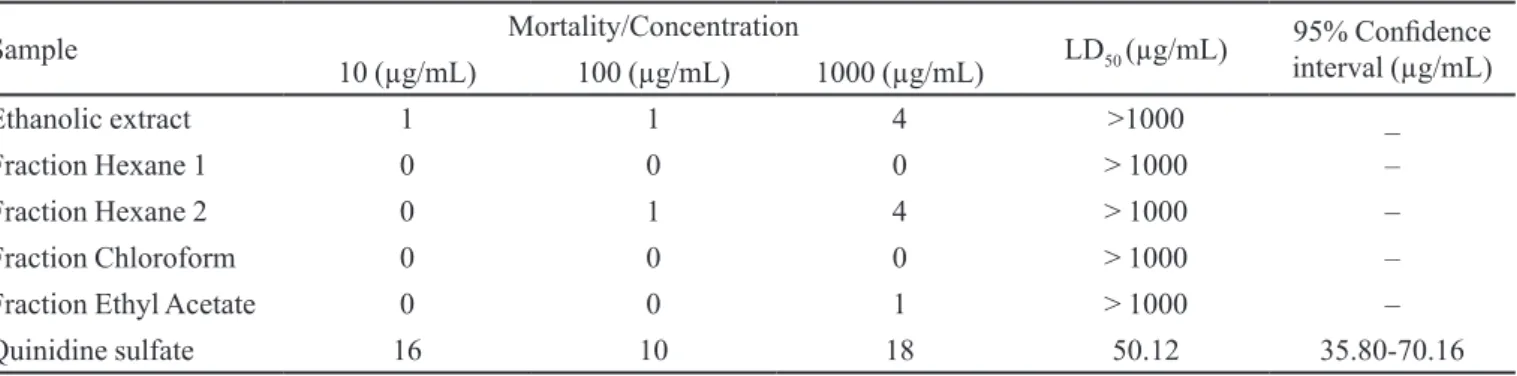
The toxicity evaluation against Artemia salina
TABLE II - Compounds 2 and 3 NMR (ppm) experimental data compared against literature data
Position Quercetin Quercetin-3-O-α-arabinofuranoside
Experimental Andersen,
Markham (2006) Experimental Lhuillier (2007)
13C 1H 13C 13C 1H 13C 1H
2 147.8 - 148.00 158.5 - 158.2
-3 137.1 - 137.21 134.9 - 133.7
-4 177.2 - 177.33 179.9 - 178.8
-5 162.3 - 162.50 163.1 - 161.8
-6 99.1 6.17 d
(J=2.1 Hz) 99.25 99.9 6.2 ls 98.9 (J=2.0 Hz)6.21d
7 165.3 165.34 166.0 - 164.8
-8 94.4 6.36 d
(J=2.1 Hz) 94.40 94.7 6.38 ls 93.5 (J=2.0Hz)6.4 d
9 158.1 - 158.22 159.4 - 157.4
-10 104.1 - 104.52 105.6 - 104.4
-1’ 124.0 - 124.15 123.1 - 121.9
-2’ 115.9 7.72 d
(J=2.1 Hz) 115.99 116.8 (J=3.3 Hz)7.5 d 115.7 (J=2.1 Hz)7.54d
3’ 148.5 - 148.75 149.9 - 148.7
-4’ 146.0 - 146.21 146.4 - 145.1
-5’ 116.1 6.8 d
(J=8.4 Hz) 116.22 116.4 (J=8.4 Hz)6.9 d 115.3 (J=8.3 Hz)6.92d
6’ 121.6 7.6 dd
(J=8.7; 2.1 Hz) 121.67 122.9 7.4 ls 121.8 (J=8.3 e 2.1 Hz)7.5dd
Arabinofuranoside
1’’ - - - 109.5 5.5 s 108.5 5.48d
(J=1.0Hz)
2’’ - - - 83.3 4.3 d
(J=2.4Hz) 82.1 (J=3.0; 1.0Hz)4.35dd
3’’ - - - 78.6 3.9 dd
(J=4.2; 2.4Hz) 77.5 (J=5.0; 3.0Hz)3.93dd
4’’ - - - 87.9 3.8 dd
(J=4.2; 8.5Hz) 86.8 3.89 m
5’’ - - - 62.5 3.5 m (2H) 61.4 3.52dd
(Table IV) demonstrated that all the samples showed a median lethal concentration greater than 1000 μg/mL, the maximum concentration for a sample to be considered active (Meyer et al., 1982).
This bioassay is used to evaluate the acute toxicity and is considered essential as a preliminary assay in the study of compounds with potential biological activities (Amaral, Silva, 2008). Analyzing the results, it can be concluded that neither the ethanolic extract, nor the fractions from R. induta
exhibited toxic potential against the brine shrimp.
For the hemolytic tests, the tested sample showed no hemolytic ability according to the two methods analyzed. In the test using the red blood cell suspension, none of the supernatants showed red coloration and all had a red blood cell precipitate at the tested doses and therefore caused no hemolysis in red blood cells. This result indicates that nei-ther the fractions nor the ethanolic extract are toxic against red blood cells. These results were conirmed by the agar
TABLE III - Compounds 4 and 5 NMR (ppm) experimental data compared against literature data
Position
Quercetin-3-O-xyloside Quercetin-3-O-β-galactoside
Experimental Yanet al. (2002) Experimental Jianget al. (1990)
13C 1H 13C 1H 13C 1H 13C 1H
2 158.5 - 152.0 - 156.2 - 156.28
-3 135.4 - 130.0 - 133.5 - 133.61
-4 179.4 - 179.5 - 177.5 - 177.44
-5 163.1 - 162.0 - 161.3 - 161.15
-6 99.8 (J=2.1 Hz)6.16 d 100.1 6.20 98.7 (J=6.2 1.8 Hz)d 98.62 6.20 s
7 166.0 - 166.3 - 164.2 - 164.02
-8 94.7 (J=1.8 Hz)6.39 d 94.9 6.39 93.5 (J=6.4 2.1 Hz)d 93.45 6.41 s
9 158.5 - 158.6 - 156.3 - 156.28
-10 105.6 - 100.1 - 103.9 - 103.94
-1’ 123.0 - 123.4 - 121.1 - 121.16
-2’ 115.9 7.6 116.2 7.6 115.9 (J=2.4 Hz)7.5 d 115.17 7.53 s
3’ 149.8 - 143.0 - 144.9 - 144.67
-4’ 146.1 - 146.0 - 148.5 - 148.32
-5’ 117.2 6.84 s 117.4 6.85 115.2 (J=8.7 Hz)6.8 d 116.10 (J6.82 =8 Hz)d
6’ 123.3 (J=2.1 Hz)7.57 d 123.2 7.57 122.1 (J=8.4; 2.1 Hz)7.65 dd 121.62 (J7.67 =8 Hz)d
Xylose Galactoside
1’’ 104.5 (J=7.2 Hz)5.17 d 104.7 (J5.18 =6 Hz)d 101.8 (J=7.8 Hz)5.39 d 102.12
2’’ 75.3 3.39 m 75.4 3.4 d 71.2 (J=7.8; 3.9 Hz)3.55 dt 71.26
3’’ 77.5 3.46-3.54 77.7 3.34 s 73.2 3.33 73.27
4’’ 71.0 3.46-3.54 71.2 3.45-3.54 m 67.9 3.64 ls 67.93
5’’ 67.2
3.1 dd (J=11.5; 9.5 Hz)
3.78 dd (J=11.4; 5.0 Hz)
67.4 3.10 3.76 dddd
75.9 3.30 s 75.73
6’’ - - - - 60.2 3.25 e 3.45 (2H) 60.14
diffusion method, on which none of the samples had a hemolytic halo around the impregnated disc, conirming the absence of hemolytic activity. Similarly, the negative controls exhibited no hemolytic halo, whereas the Triton X-100 had a halo of 2 cm.
Red blood cell lyses can cause an elevation in plas-ma hemoglobin leading to harmful effects in the kidneys (nephrotoxicity), and in the cardiovascular system (vaso-motor effect) (Carvalho et al., 2007). Therefore, negative hemolytic activity is a good indicator because it conirms no toxicity to erythrocyte membrane.
The acute toxicity results showed that the Rourea induta extracts had no toxic potential, thus favoring the species for further study such as pharmacological analyses, to determine therapeutic properties of this species.
ACKNOWLEDGEMENTS
We would like to thank Gert Hatschbach, botanist of Museu Botânico de Curitiba, for identifying the species, and the Universidade Estadual de Maringá for the NMR spectrums.
REFERENCES
AHAMADU, A. A.; HASSAN, H. S.; ABUBAKAR, M. U.; AKPULU, N. Flavonoid Glycosides from Byrsocarpus coccineus Leaves. Schum. and Thonn (Connaraceae). Afr. J. Trad.C.A.M., v.4, n.3, p.257-260, 2007.
AMARAL, E.A.; SILVA, R.M.G. Avaliação da toxicidade aguda de angico (Anadenanthera falcata), pau-santo (Kylmeyera coreaceae), aroeira (Myracrodruon urundeuva) e cipó-de-são-jõao (Pyrostegia venusta), por meio do bioensaio com Artemia salina. Perquirere, Rev. Eletr. da Pesquisa. Avaiable at: <http://www.unipam.edu.br/perquirere/ile/ ile/2008_cb/artigo_eni.pdf>. Accessed on: April, 2010.
ANDERSEN, O.M.; MARKHAM, K.R. Flavonoids, chemistry, biochemistry and applications.New York: CRC Press; 2006. 1197 p.
CARVALHO, E.B.; BORGES, E.L.; CARLOS, L.M.B.; SILVA, M.A.M; MAGALHÃES, S.M.M; GOMES, F.V.B.A.F.; CARVALHO, M.J.C.; QUIXADÁ, A.T.S.; PITOMBEIRA, M.H.S. Efeito da bomba de infusão de soluções sobre o grau de hemólise em concentrado de hemácias. Rev. Bras. Hematol. Hemoter., v.29, n.2, p.149-152, 2007.
CARVALHO, M.S.; MORAIS-COSTA, F.; CARDOSO-FILHO, O.; FERREIRA, B.C.; CARVALHO, S.; SILVA, W.A.; OLIVEIRA, D.A. Estudo preliminar da itotoxicidade de Myracrodrum urundeuva fr. Allemao (Anacardiaceae). In: CONGRESSO DE ECOLOGIA DO BRASIL, 9., São Lourenço, 2009. Anais. São Lourenço: SEB, 2009. p.1-2.
EFING, L.M.A.C. Compostos bioativos do material resinoso, subproduto do processamento da erva-mate (Ilex paraguariensis A.St.-Hil.). Curitiba, 2008. 108 f. [Thesis of PhD degree in Food Technology. Federal University of Paraná].
TABLE IV – Artemia salina lethality and LD50
Sample Mortality/Concentration LD50 (µg/mL) 95% Conidence interval (µg/mL)
10 (µg/mL) 100 (µg/mL) 1000 (µg/mL)
Ethanolic extract 1 1 4 >1000 _
Fraction Hexane 1 0 0 0 > 1000 –
Fraction Hexane 2 0 1 4 > 1000 –
Fraction Chloroform 0 0 0 > 1000 –
Fraction Ethyl Acetate 0 0 1 > 1000 –
Quinidine sulfate 16 10 18 50.12 35.80-70.16
FONSECA, L.C.M.; PROENÇA, C.E.B. Connaraceae. In: CAVALCANTI, T.B.; RAMOS, A.E. Flora do Distrito Federal. 2.ed. Brasília: Embrapa Recursos Genéticos e Biotecnologia, 2002. p.41-47.
FORERO, E. Uma nueva especie mexicana de Rourea (Connaraceae). Caldasia, v.29, n.1, p.19-21, 2007.
FORERO, E. A revision of american species of Rourea Subg. Rourea (Connaraceae). Mem. New York Bot. Gard., v.26, n.1, p.1-119, 1976.
JIANG, J-Q.; FANG, S-D.; XU, C-S.; LUO, J-T. Studies on the chemical constituents of Rourea microphylla (Hook. Et Arn) Planch. Acta Bot. Sin., v.32, n.5, p.376-379, 1990.
LHUILLIER, M.A. Contribution a l’etude phytochique de quatre plantes malgaches: Agauri salicifolia HOOK.F ex Oliver, Agauria polyphylla Baker (Ericaceae), Tambourissa trichophyla Baker (Monimiaceae) et Embelia concinna Baker (Myrsinaceae). Toulouse, 2007. 214 f. [Thesis of PhD degree. L’institut National Polytechnique de Toulouse. Sciences des Agroressources, École doctorale: Transferts, Dynamique des Fluides, Energie et Procédés].
LORENZI, H.; SOUZA, V.C. Botânica sistemática: guia ilustrado para identiicação das famílias de angiospermas da lora brasileira, baseado em APG II. Nova Odessa: Instituto Plantarum, 2005. 640 p.
LENZA, E.; FERREIRA, J. N.; CONSOLARO, H.; AQUINO, F.G. Biologia reprodutiva de Rourea induta Planch. (Connaraceae), uma espécie heterostílica de cerrado do Brasil Central. Rev. Bras. Bot.,v.31, n.3, p.389-398, 2008.
MABRY, T. J. MARKHAM, K.R.; THOMAS, M.B. The
systematic identiication of lavonoids. New York: Springer-Verlag, 1970. 354 p.
MEYER, B.N.; FERRIGNI, N.R.; PUTNAM, J.E.; JACOBSEN, L.B.; NICHOLS, D.E; MCLAUGHLIN, J.L. Brine shrimp: a convenient general bioassay for active plant constituents. Planta Med., v.45, n.1, p.31-34, 1982.
OLIVEIRA, V.M.A.; CARNEIRO, A.L.B.; CAUPER, G.S.B.; POHLIT, A.M. In vitro screening of amazonian plants for hemolytic activity and inhibition of platelet aggregation in human blood. Acta Amaz., v.39, n.4, p.973-980, 2009.
RECOR. Reserva ecológica do IBGE. O cerrado brasileiro. Available at:<http://www.recor.org.br/cerrado/cerrado. html>. Accessed on: 07 fev. 2009.
SANTOS, P.M.L. SCHRIPSEMA, J.; KUSTER, R.M. Flavonóides O-glicosilados de Croton campestris St. Hill. (Euphorbiaceae). Rev. Bras. Farmacogn., v.15, n.4, p.321-325, 2005.
SIDDIQUI, B. S.; RASHEED, M.; ILYAS, F.; GULZAR, T.; TARIQ, R. M.; NAQVI, S. N.H. Analysis of Insecticidal Azadirachta indica A. Juss. Fractions. Z. Naturforsch., v.59c, n.1/2, p.104-112, 2004.
SILVERSTEIN, R.M.; BASSLER, F.X.; MORRIL, T.C. Identificação espectrométrica de compostos orgânicos. 5.ed. Rio de Janeiro: Guanabara Koogan, 1994. 387 p.
VVDENSKAYA, I. O.; ROSEN, R. T.; GUIDO, J. E.; RUSSEL, D. J.; MILLS, K. A.; VORSA, N. Characterization of Flavonols in Cranberry (Vacciniummacrocarpon) Powder. J. Agric. Food Chem., v.52, n.2, p.188-195, 2004.
WORLD HEALTH ORGANIZATION. Quality control methods for medicinal plant materials. Geneve, 1998. 127 p.
YAN, X.; MURPHY, B. T.; HAMMOND, G. B.; VINSON, J. A.; NETO, C. C. Antioxidant activities and antitumor screening of extracts from cranberry fruit (Vaccinium macrocarpon). J. Agric. Food Chem., v.50, n.21, p.5844-5849, 2002.
ZHANG, X.; THUONG, P.T.; WENYI, J.; SU, N.D.; DAIEUN, S.; KIHAWAN, B.; SIK, K.S. Antioxidant activity of anthraquinones and lavonoids from lower of Reynoutria sachalinensis. Arch. Pharmacal Res.,v.28, n.1, p.22-27, 2005.
Received for publication on 7th April 2010