*Correspondence: L. M. Lima. LASSBio - Laboratório de Avaliação e Síntese de Substâncias Bioativas, Centro de Ciências da Saúde, Faculdade de Farmácia, Universidade Federal do Rio de Janeiro – UFRJ, Ilha do Fundão. Caixa Postal 68024 - 21944-970 - Rio de Janeiro - RJ, Brazil. E-mail: [email protected]
R
ev
Pharmaceutical Sciences vol. 47, n. 3, jul./sep., 2011
Therapeutic approaches for tumor necrosis factor inhibition
Maria Letícia de Castro Barbosa
1,2, Milla Machado Fumian
1,3, Ana Luísa Palhares de Miranda
1,3,
Eliezer J. Barreiro
1,2,3, Lídia Moreira Lima
1,2,3,*
1LASSBio, Laboratory of Evaluation and Synthesis of Bioactive Substances, Faculty of Pharmacy, Federal University of Rio de Janeiro, 2Chemistry Institute, Federal University of Rio de Janeiro,
3Institute of Biomedical Sciences, Federal University of Rio de Janeiro
Tumor necrosis factor (TNF) consists of an inlammatory cytokine essential for homeostasis and organism defense. Despite its physiological relevance, both increased biosynthesis and release of TNF lead to the exacerbation of inlammatory and oxidative responses, which are related to the pathogenesis of a host of diseases of an inlammatory, autoimmune and/or infectious nature. In this context, effective therapeutic approaches for the modulation of TNF have been the focus of research efforts. Approximately one million individuals worldwide have been treated with biotechnological inhibitors of this cytokine, the so-called anti-TNF biopharmaceuticals. However, given the high risk of infection and the limitations related to cost and administration routes, new therapeutic approaches aimed at biological targets that directly or indirectly modulate the production and/or activation of TNF appear promising alternatives for the discovery of new anti-inlammatory and immunomodulatory orally active drugs and are therefore discussed in this paper.
Uniterms: Tumor necrosis factor. TACE. p38 MAPK. NF-κB. cAMP. Phosphodiesterase. Adenosine receptor.
O fator de necrose tumoral (do inglês, tumor necrosis factor - TNF) consiste em uma citocina inlamatória essencial para a homeostase e defesa do organismo. A despeito de sua relevância isiológica, o aumento da biossíntese e liberação do TNF conduzem à exacerbação das respostas inlamatória e oxidativa, as quais estão relacionadas à patogênese de várias doenças de natureza inlamatória, auto-imune e/ou infecciosa. A busca por abordagens terapêuticas eicientes na modulação do TNF tem sido alvo de diversos esforços de pesquisa. Aproximadamente um milhão de pessoas ao redor do mundo já foi tratado com inibidores biotecnológicos desta citocina, os chamados biofármacos anti-TNF. Entretanto, em face ao elevado risco de infecções e as limitações relacionadas ao custo e a via de administração, novas abordagens terapêuticas com foco em alvos que modulem, de forma direta ou indireta, a produção e/ou ativação do TNF surgem como alternativas promissoras para a descoberta de novos fármacos antiinlamatórios e imunomoduladores ativos por via oral e serão discutidas neste trabalho.
Unitermos: Fator de necrose tumoral. TACE. MAPK p38. NF-κB. AMPc. Fosfodiesterase. Receptor de adenosina.
INTRODUCTION
The functioning of the immune system is finely modulated by the activity of cytokines, pro- and anti-inlammatory mediators (Palladino et al., 2003). Imbalan-ces in this system can lead to the development of chronic
inlammatory conditions, autoimmune diseases and in-creased susceptibility and/or non-resolution of infections (Hanada, Yoshimura, 2002). In immune-mediated inlam-matory disorders (IMIDs), the immune system becomes imbalanced thereby preventing healing of inlammation, which consequently evolves and promotes tissue damage (Lin et al., 2008).
of immune response triggering, by using for instance, the modulation of cytokine signaling pathways as a strategy (Hanada,Yoshimura, 2002).
The use of modulators of the production and bio-logical activity of tumor necrosis factor alpha (TNF-α) in particular has been widely described and discussed in the literature (Lin et al., 2008), since the increased tissue and/or plasma concentration of this cytokine is associ-ated with the establishment and development of several diseases with diverse causes. These diseases can stem from: (a) an autoimmune origin – e.g. Crohn’s disease, diabetes, psoriasis, rheumatoid arthritis and systemic lupus erythematosus; (b) an infectious origin – e.g. hepatitis C, acquired immunodeficiency syndrome (AIDS), septic shock, erythema nodosum leprosum; and (c) a tumor –
e.g. multiple myeloma and ovarian cancer (Marriot et al., 1997; Sampaio et al., 2002; Palladino et al., 2003; Steinwurz, 2003; Sirohi, Powles, 2004; Paul et al., 2006; DasGupta et al., 2009).
Tumor necrosis factor
Tumor necrosis factor (TNF) consists of a pleiotropic cytokine, regulating various cellular and biological events, such as immune and inlammatory responses, cell differen-tiation and proliferation, apoptosis and energy metabolism (Cawthorne, Sethi, 2008). TNF was originally identiied as a substance present in the serum of animals treated with bacterial lipopolysaccharide (LPS), resulting in necrosis of tumors in vivo (Palladino et al., 2003; Paul et al., 2006).
Two types of tumor necrosis factor are currently known: TNF-α and TNF-� (or lymphotoxin), which pres-α and TNF-� (or lymphotoxin), which pres- and TNF-� (or lymphotoxin), which pres-� (or lymphotoxin), which pres- (or lymphotoxin), which pres-ent a striking similarity in their three-dimensional struc-tures, despite the low sequence homology (ca. 33%) found in the primary structure of their proteins (Gray et al., 1984; Pennica et al., 1984; Banner et al., 1993). The activities of the cytokines TNF-α and TNF-� are also very similar, both in vitro and in vivo, although they are antigenically distinct (Sack, 2002).
TNF-α is the most extensively studied cytokine and its main sources invivo are activated monocytes, ibroblasts and endothelial cells. Macrophages, T cells, B lymphocytes, granulocytes, smooth muscle cells, eo-sinophils, chondrocytes, osteoblasts, mast cells, glial cells and keratinocytes also produce TNF-α after stimulation (Mukhopadhyay et al., 2006).
LPS is the most potent stimulus triggering the bio-synthesis of TNF-α by mononuclear phagocytes enabling large quantities of this cytokine to be released in infections by gram-negative bacteria. Other mediators induce its production in different cell types, including interleukin-1
(IL-1), platelet derived growth factor (PDGF), oncostatin M, interferon-� (IFN-�), and the TNF-α itself, which can stimulate or inhibit its own synthesis, depending on the cell type (Mukhopadhyay et al., 2006).
The production of TNF-α begins with the adhesion of a ligand (which usually consists of a microbial compo-nent) to a pattern recognition receptor (PRR) - the toll-like receptor (TLR), for example. This type of receptor is then able to recognize pathogen-associated molecular patterns (PAMPs), starting a cascade of signal transduction leading to the activation of transcription factors, such as the NF-κB (nuclear factor kappa B) (Zuany-Amorin etal., 2002; Lima et al., 2006).
The NF-κB is present in the cytoplasm associ-κB is present in the cytoplasm associ-B is present in the cytoplasm associ-ated with the inhibitory protein IκBα. Activation of the NF-κB signaling pathway leads to phosphorylation and polyubiquitination of the protein IκBα and its subsequent degradation by the 26S proteasome. The degradation of this inhibitory protein releases the NF-κB dimer for translocation to the nucleus, inducing the transcription of several genes associated with the inlammatory response, including those encoding the cytokine TNF-α (Keifer et al., 2001; Suryaprasad, Prindiville, 2003).
The human TNF-α is synthesized as a polypeptide precursor of 233 amino acid residues attached to the cell membrane and is released through regiospeciic proteoly-sis between the amino acids Ala76 and Val77, producing the soluble form of the cytokine (Gearing et al., 1994; Lima et al., 2006; Mukhopadhyay et al., 2006; DasGupta
et al., 2009). The enzyme responsible for the proteolytic cleavage of TNF-α anchored to the cell membrane was identiied as a zinc-dependent metalloproteinase, denoted as tumor necrosis factor alpha converting enzyme (TACE) (Black et al., 1997; Moss et al., 1997). Cells derived from TACE knockout mice showed approximately 90% reduc-tion in TNF-α release (Mohan et al., 2002).
Both forms of TNF-α, the soluble form and the form associated with the cell membrane, trigger biologi-cal effects and metabolic responses, although the speciic functions of each remain controversial (Palladino et al., 2003; Cawthorne, Sethi, 2008; Taylor, 2010).
receptors are signiicantly different, activating overlapping and distinct signaling pathways (Palladino et al., 2003; DasGupta et al., 2009).
All human nucleated cells express TNF receptors, al-though their distribution varies according to cell type (Pal-ladino et al., 2003). TNF-RI is constitutively expressed in most cells, but not in inactive T cells and erythrocytes; TNF-RII is expressed more often in hematopoietic and endothelial cells (Lima et al., 2006).
Efforts have been directed towards the elucidation of the speciic physiological role of TNF receptors TNF-RI and TNF-RII, using, among other tools, knockout mice and monoclonal antibodies directed at the antigenically distinct receptors, acting as agonists.
The use of agonists has demonstrated that the activa-tion of TNF-RI is suficient to mimic the biological effects of TNF-α in several cell compartments (Tartaglia et al., 1993; Horssen et al., 2006; DasGupta et al., 2009). Ad-ditionally, studies employing TNF-RI knockout mice have shown that these animals were resistant to LPS-induced liver injury, in which TNF-α plays a signiicant role by in-ducing hepatic apoptosis (Shimizu et al., 2005). The lung inlammatoryresponse in TNF-RI knockout mice after pulmonary challenge with Micropolysporafaeni antigen or LPS was also signiicantly reduced (Smith et al., 1998).
The binding of TNF-α to the TNF-RI receptor triggers a cascade of events involving the activation of a series of serine/threonine protein kinases known as mitogen-activated protein kinases (MAPKs) (Paul et al., 2006). The MAPK signaling cascade follows a sequence of phosphorylation and dephosphorylation reactions, starting with the activation of a MAPK kinase kinase (MAPKKK), which activates a MAPK kinase (MAPKK), which ulti-mately leads to the activation of speciic MAPKs, e.g. p38 MAPK. The activated p38 MAPK phosphorylates other kinases and transcription factors, leading to the gene ex-pression of cytokines and growth factors (Roux, Blenis, 2004; Mutalik, Venkatesh, 2006).
Another central event in the binding of TNF-α to the TNF-RI is the activation of the nuclear factor NF-κB. This interaction activates, via the TRADD domain (tumor ne-crosis factor receptor-1 associated death domain), the IKK complex (inhibitor of nuclear factor-κB kinase kinase), which is responsible for triggering the degradation process of the inhibitory protein IκBα. Once activated, the IKK complex phosphorylates IκBα, resulting in the release of NF-κB for translocation to the nucleus, and consequently, in the expression of genes encoding cytokines, chemokines and proteases (Paul etal., 2006).
Both TNF-RI and TNF-RII receptors can be cleaved from the cell surface by members of the matrix
metallo-proteinases family in response to inlammatory stimuli. The extracellular domains released through the proteolytic cleavage process retain the ability to bind to TNF, becom-ing soluble TNF receptors (TNF-Rs) (Brakebusch et al., 1994; Palladino et al., 2003).
The soluble receptors compete with membrane receptors for the binding of agonists TNF-α and TNF-�, acting as endogenous inhibitors of cytokine activity. This behavior allows modulation and homeostatic regulation of TNF activity in the body (Brakebusch et al., 1994).
TNF-α is a versatile cytokine that alters tissue remod-α is a versatile cytokine that alters tissue remod- is a versatile cytokine that alters tissue remod-eling, increases epithelial cell barrier permeability, induces macrophages activation, recruitment of inlammatory cells and expression of adhesion molecules. TNF-α also plays a crucial role in the development, homeostasis and adaptive immune response (Suryaprasad, Prindiville, 2003).
On the other hand, despite the physiological rel-evance of this cytokine, it is known that the increase of its biosynthesis and release leads to exacerbation of inlam-matory and oxidative responses, which are related to the pathogenesis of a wide range of diseases, e.g. rheumatoid arthritis, psoriasis, Crohn’s disease, erythema nodosum leprosum and multiple myeloma (Hashimoto, 2002; Muk-hopadhyay et al., 2006; Lin et al., 2008).
Therapeutic strategies for TNF inhibition
The search for effective therapeutic approaches in the modulation of TNF-α has been the focus of research efforts. Approximately one million individuals worldwide are either undergoing treatment or have been treated with TNF inhibitors available in the pharmaceutical market, encompassing indications that include rheumatoid arthri-tis, psoriatic arthriarthri-tis, psoriasis and inlammatory bowel diseases, in addition to numerous potential clinical appli-cations which are currently in various stages of evaluation (Suryaprasad, Prindiville, 2003; Feldmann et al., 2005; Hochberg et al., 2005, Lin et al., 2008).
Several drugs commonly used as immunosuppres-sants, e.g. cyclosporine A (1) and dexamethasone (2), exhibit TNF-α modulation, although their effects are associated with considerable toxicity. However, as the mechanisms that involve the production of TNF and its activity on cells become better understood, more spe-ciic strategies for the inhibition of this cytokine emerge. Examples include the development of new therapeutic agents and the modiication of existing ones, seeking lower toxicity, greater potency and a larger number of clinical alternatives (Marriot et al., 1997).
which act as inhibitors of surface receptor occupancy; TACE inhibitors; and compounds structurally related to thalidomide (3), the irst drug described as a modulator of the biosynthesis of this cytokine. Moreover, strategies for the modulation of signaling pathways involved in the production and biological responses promoted by TNF-α are also known and include some classes of therapeutic agents, such as protein kinase inhibitors (e.g. p38 MAPK inhibitors); modulators of NF-κB signaling pathway; and regulators of intracellular levels of cyclic adenosine-3’,5’-monophosphate (cAMP) (DasGupta et al., 2009).
Inhibitors of TNF receptor occupancy
The introduction of biological therapies for inhibi-tion of the pro-inlammatory cytokine TNF-α in the global
pharmaceutical market was responsible for a revolution in the treatment of diseases such as rheumatoid arthritis, psoriasis and Crohn’s disease (Hochberg et al., 2005; Pappas etal., 2009).
The irst two biotech drugs identiied were inlix-imab (Remicade® - Centocor/Schering-Plough) and
etan-ercept (Enbrel® - Amgen/Wyeth) (Table I). Inliximab is
a chimeric monoclonal antibody (mAb) of approximately 149 kDa that binds speciically to TNF-α (but not TNF-�) with high afinity, neutralizing its biological activity by inhibiting its binding to the receptors RI and TNF-RII. Etanercept is the product of fusion of a dimeric pro-tein of approximately 150 kDa (IgG1) with the soluble receptor of TNF. These features allow etanercept to bind to both TNF-α and TNF-� (Hochberg et al., 2005; Pappas
etal., 2009). These biopharmaceuticals were approved for the treatment of rheumatoid arthritis in the late 1990s. Their use was later extended to include other disorders of autoimmune origin, including psoriatic arthritis (Pappas
et al., 2009).
The third biotech drug described was adalimumab (Humira® - Abbott), a recombinant human IgG1
monoclo-nal antibody of approximately 148 kDa speciic for human TNF-α. Akin to inl iximab, adalimumab does not neutral-α. Akin to inl iximab, adalimumab does not neutral-. Akin to inliximab, adalimumab does not neutral-ize TNF-�. In 2002, this biotech drug was introduced in the pharmaceutical market for the treatment of rheumatoid arthritis and its use was also recently approved for the treatment of other immune-inlammatory disorders (Table 1) (Hochberg et al., 2005; Pappas etal., 2009).
The fourth biopharmaceutical identiied was cer-tolizumab pegol (Cimzia® - UCB), a humanized antibody
fragment speciic for TNF-α linked to polyethylene gly-α linked to polyethylene gly- linked to polyethylene gly-col. In 2008, this biological therapy was approved for the treatment of Crohn’s disease and, in May 2009, for the treatment of rheumatoid arthritis (Table I) (Tracey et al., 2008; Pappas etal., 2009).
The use of a new biotechnology drug, golimumab (Simponi® - Centocor Ortho Biotech), which consists
TABLE I - TNF inhibitors licensed for clinical use
Brand name TNF inhibitor Year Disease indicationsa Administration route
Remicade Inliximab 1998 RA, AS, PsA, psoriasis, CD, UC i.v.
Enbrel Etanercept 1999 RA, JCA, AS, PsA, psoriasis s.c.
Humira Adalimumab 2002 RA, AS, PsA, CD, psoriasis s.c.
Cimzia Certolizumab pegol 2008 CD, AR s.c.
Simponi Golimumab 2009 PsA, AR s.c.
AS: ankylosing spondylitis; CD: Crohn’s disease; JCA: juvenile chronic arthritis; PsA: psoriatic arthritis; RA: rheumatoid arthritis; UC: ulcerative colitis.
of a chimeric immunoglobulin G (G1κ mAb) specific for human TNF, was recently approved by the United States - Food and Drug Administration (US-FDA). This biopharmaceutical binds to both bioactive forms of TNF,
i.e. transmembrane and soluble, thus preventing the bind-ing of this cytokine to its receptor target. In April 2009, golimumab was approved for the treatment of rheumatoid arthritis and psoriatic arthritis (Table I) (Tracey et al., 2008; Pappas et al., 2009).
The use of biotech drugs for the treatment of auto-immune diseases leads to fast control of inlammation, resulting in an improvement in terms of function and structural repair of tissues. However, the beneit related to the use of these drugs should be evaluated by also taking into account the potential adverse effects associated with them (Suryaprasad, Prindiville, 2003).
Clinical studies have described the occurrence of in-fections, lymphoproliferative and neurological disorders, autoimmune reactions and cardiac effects as recurring adverse events related to the continued use of biotech TNF modulators. The development of infection is notably the most common complication associated with anti-TNF-α biological therapy. The most commonly observed infec-tions are in urinary and respiratory tracts (e.g. sinusitis, pharyngitis and bronchitis) (Suryaprasad, Prindiville, 2003; Hochberg et al., 2005; Lin et al., 2007).
The differences in the eficacy and safety of bio-pharmaceutical TNF-α inhibitors approved for clinical use remain unclear, and therefore the treatment chosen should be guided by expediency based on cost and availability (Pappas et al., 2009).
Recent studies have shown that biological anti-TNF-α therapies are more effective when combined with other disease modifying anti-rheumatic drugs (DMARD), particularly methotrexate (4). The reports indicate that the treatment of rheumatoid arthritis with TNF-α biological inhibitors co-administered with methotrexate (4) results in a marked reduction of the disease symptoms and, addition-ally, this association minimizes the rate of joint destruc-tion more signiicantly compared to monotherapy with anti-TNF-α biopharmaceuticals (Caporali etal., 2009).
TACE inhibitors
From the description of therapeutic beneits related to anti-TNF biological therapy, concerted research effort has been devoted to the development of small molecules capable of inhibiting this cytokine through oral administra-tion (Palladino et al., 2003; Levin et al., 2006; DasGupta
et al., 2009).
In this context, the hypothesis that an inhibitor of TACE, i.e. the enzyme responsible for proteolytic cleav-age of TNF-α anchored to the cell membrane, would be able to effectively treat several diseases of inlammatory and autoimmune origin by limiting the levels of soluble TNF-α, has been strengthened. Based on this objective, several research groups around the world are actively in-volved in developing small molecules that inhibit TACE (Levin et al., 2006; Kenny, 2007; DasGupta et al., 2009).
Given the similarity between the catalytic domains of TACE and matrix metalloproteinases (MMPs), some previously identified MMP inhibitors, e.g. marimastat (5) and prinomastat (6), were evaluated for their potential inhibitory activity against TACE. Despite showing the ex-pected biological activity, these compounds proved unsuc-cessful in clinical trials due to adverse effects on skeletal muscle (DasGupta et al., 2009). Even though the exact mechanism responsible for the observed side effects has not been clearly established, some research suggests that the toxicity of these compounds is not linked to the inhibi-tion of TACE, but related to their ability to inhibit MMP-1 and/or MMP-14 (Aranapakam et al., 2003; DasGupta et al., 2009). Therefore, the search for new selective inhibi-tors of TACE devoid of activity on MMPs is underway.
In an attempt to dissociate the inhibitory actions on MMPs and TACE, researchers at Novartis identiied the prototypes PKF242-484 (7) and PKF241-466 (8), which presented pronounced anti-inflammatory activity in in vivo models of pulmonary inlammation, and inhibited the release of TNF-α in peripheral blood mononuclear acti-α in peripheral blood mononuclear acti- in peripheral blood mononuclear acti-vated cells with IC50 values equal to 56 nM and 141 nM,
Some sulfonamide hydroxamates have also been de-scribed as TACE inhibitors (DasGupta et al., 2009). Levin and coworkers, from Wyeth pharmaceutical industry, described the prototype Apratastat (9), commonly known as TMI-05, as a non-selective TACE inhibitor. Compound 9 showed IC50 equal to 20 nM for the inhibition of the
enzyme and ED50 equal to 5 mg/kg (p.o.) for the
inhibi-tion of TNF-α release stimulated by LPS in mice. This prototype showed signiicant anti-inlammatory effect in a model of collagen-induced arthritis in mice (Levin et al., 2006). Although Apratastat (9) was well tolerated in phase I clinical trials, its development was suspended in October 2006 due to lack of eficacy in phase II clinical trials for rheumatoid arthritis (Kenny, 2007).
Worth and coworkers from Roche discovered the ligand Ro-32-7315 (10), a potent TACE inhibitor with 100-fold greater selectivity than most MMPs. Ro-32-7315 (10) has shown high potency in the inhibition of TACE in vitro (IC50 = 5.2 nM), in the modulation of TNF-α release
by different inlammatory cell types, and in controlling the onset of symptoms in animal models of inlammation. Compound 10 was administered to healthy human volun-teers, but its development was eventually discontinued due to limited oral bioavailability (Beck et al., 2002; DasGupta
et al., 2009).
Researchers at Bristol-Myers-Squibb have identiied compound 11 as a selective inhibitor of TACE, with IC50
equal to 3.7 nM for the enzyme inhibition and selectivity
in the order of 1,000-fold greater than MMP-1, 2, 9 and 13 (Cherney et al., 2006).
This group of scientists also designed and synthe-sized a new series of γ-lactam hydroxamates. The designed compounds exhibited pronounced inhibitory activities in the isolated enzyme, but with low to moderate effect in assays using whole human blood, due to the high rate of binding to plasma proteins. In an attempt to increase the free fraction of compounds, several bioisosters with higher polarity were obtained and tested, most notably IK-682 (12). The IC50 value for 12 was equal to 1 nM in in vitro inhibition of the isolated enzyme and equal to 0.35 µM in the whole human blood assay. IK-682 (12) showed selectivity of more than 1,000 times greater com-pared to most MMPs, and adequate oral bioavailability (Duan et al., 2002; DasGupta et al., 2009).
Changes in the molecular structure of 12 resulted in the design of the prototype BMS-561392 (13), also de-veloped at Bristol-Myers-Squibb. Compound 13 showed promising inhibitory activity in the release of soluble TNF-α in murine and human blood assays, as well as selectivity of more than 100 times in relation to MMPs. After its oral administration in mice, BMS-561392 (13) inhibited the release of soluble TNF-α induced by LPS with ED50 of 6 mg/kg. In phase I clinical trials, the
due to the observation of hepatotoxic effects (Qian et al., 2007; DasGupta et al., 2009).
It is worth mentioning that, although the proto-types TMI-05 (9) and BMS-561392 (13) have not been successful in phase II clinical trials, several compounds belonging to different chemical classes have been descri-bed as selective inhibitors of TACE, some of which have shown promising results in preclinical tests (DasGupta et al., 2009).
Thalidomide and structural analogues
Thalidomide (3) ((±)2-(2,6-dioxopiperidin-3-yl) isoindoline-1,3-dione) has several pharmacological effects associated with its ability to modulate immune response (Ng et al., 2002; Sampaio et al., 2006). The anti-inlam-matory and immunomodulatory properties of 3 are due to its ability to inhibit the pro-inlammatory cytokine TNF-α. Thalidomide (3) increases the degradation of TNF-α mRNA (Moreira et al., 1993), suppressing the release of this cytokine from different cell types, e.g. monocytes, macrophages, microglia and T lymphocytes (Sampaio et al., 1991; Peterson et al., 1995; Deng et al., 2003; Kim
et al., 2004; Melchert, List, 2007). TNF-α inhibition is largely responsible for the clinical beneits described for 3 in patients with inlammatory and autoimmune diseases, who present exacerbated production of endogenous TNF-α (Corral, Kaplan, 1999).
Many of the pharmacological activities described for thalidomide (3) can be justiied by its effects on the NF-κB signaling pathway (Sampaio et al., 2006). Com-pound 3 blocks NF-κB activation through inhibition of the degradation of inhibitory protein IκBα, thus preventing the translocation of NF-κB to the nucleus (Mercurio, Man-κB to the nucleus (Mercurio, Man-B to the nucleus (Mercurio, Man-ning, 1999; Palladino et al., 2003; Sampaio et al., 2006). This event is due to the decrease of IKK complex catalytic activity, responsible for the phosphorylation step that trig-gers the degradation of IκBα protein (Keifer et al., 2001). The identiication of the promising anti-inlammatory and immunomodulatory properties of 3 resulted in its ap-proval by the US-FDA in July 1998 for the treatment of moderate to severe cutaneous manifestations of lepromatous leprosy (Matthews, McCoy, 2003; Teo et al., 2005; Lima et al., 2006). After approval of thalidomide (3) for the treat-ment of erythema nodosum leprosum, a series of trials was initiated aimed at evaluating the therapeutic potential of 3 in the treatment of inlammatory and autoimmune diseases related to increased plasma concentrations of TNF-α, such as rheumatoid arthritis and Crohn’s disease (Man et al., 2003; Matthews, McCoy, 2003). More recently, in May 2006, thalidomide (3) was approved by the FDA for the treatment of multiple myeloma (Melchert, List, 2007).
Despite several clinical applications assigned to thalidomide (3), its use is limited to the treatment of dis-eases for which there are no alternative therapies and is conditioned to the monitoring of neurological effects dur-ing treatment and to the use of contraceptive methods in women (Marriott et al., 1997). With this in mind, in a bid to both overcome the inadequate pharmacokinetic proile of 3 and reduce its teratogenic and neurotoxic effects, research efforts have been devoted to the search for new analogues with improved pharmacotherapeutic proile (Lima et al., 2002; Man et al., 2003). A number of immunomodulatory derivatives have been reported from chemical changes in the structure of the starting compound, with the goal of generating effective alternatives for the treatment of cancer and immune disorders (Teo etal., 2005).
In this context, Muller and colleagues (1999) de-scribed a pronounced increase in the inhibitory activity of TNF-α production in vitro for amino-substituted analogues of thalidomide (3), particularly the 4-amino-phthalimidic derivative (14), being approximately 8,000 times more po-tent than 3 (Figure 1). Moreover, the authors demonstrated that the substitution of the phthalimidic ring by a func-tionalized isoindolinone core raised the possibility of four regioisomers (i.e.16-19, Figure 1). These isoindolinone compounds were designed in an attempt to increase the sta-bility and bioavailasta-bility of 3. Among them, the 4-amino-isoindolinone analogue (16) was the only one able to sig-niicantly inhibit the production of TNF-α (IC50 = 100 nM),
indicating the importance of spatial orientation of the amino group with respect to the carbonyl of isoindolinone core for optimizing anti-TNF activity (Figure 1) (Muller
et al., 1999). The derivative 16, named lenalidomide, was approved by the FDA in June 2006 for the treatment of multiple myeloma, and serves as a good example of the usefulness of this strategy (Melchert, List, 2007).
p38 MAPK inhibitors
The p38 mitogen-activated protein kinase (p38 MAPK) has received extraordinary attention from the pharmaceutical industry and medicinal chemists since it
FIGURE 1 - Thalidomide (3) and analogues (14-19) and the inhibition of TNF-α production induced by LPS in human
was irst described in 1993. It is considered a promising target for therapeutic intervention (Montalban et al., 2008).
This premise is based on the fact that the cytokines TNF-α and interleukin-1� (IL-1�) play an important role in the pathogenesis of many inlammatory diseases, and the signal transduction pathway that results in the production of these cytokines is partially regulated by p38 MAPK (Montalban et al., 2008). The p38 MAPK is activated from an extracellular stimulus, thus phosphorylating and activating other kinases and transcription factors, leading to the stabilization of TNF-α and IL-1� mRNA and to the increase of the gene expression of these cytokines (Pal-ladino et al., 2003).
The relevance of p38 MAPK in inflammatory responses has been shown in a number of studies using chemical inhibitors. Another study using p38α knockout macrophages demonstrated that deiciency of this protein kinase causes a signiicant inhibition in the production of TNF-α, IL-12, and IL-18 induced by LPS, highlighting the role of p38α in the activation of these inlammatory cells (Kang et al., 2008).
The description of an initial series of imidazo-pyri-dine derivatives was an important tool for elucidating the role of p38 MAPK in inlammation. The irst pyridinylimi-dazol described was SKF-86002 (20), with potent activity in modulating the cytokine production stimulated by LPS (Kumar et al., 2003).
In 1993, the structure-activity relationship for anti-inlammatory effects of the congener series of pyridinyli-midazoles was described by exploring the inhibition of enzymes 5-lipoxygenase/cyclooxygenase (5-LO/COX) and the modulation of cytokine biosynthesis as poten-tial mechanisms of action. Subsequently, the compound SB-203580 (21) and other 2,4,5-triaryl-imidazoles were synthesized and used as pharmacological tools in the se-arch for the molecular target involved in the modulation of cytokine biosynthesis. This study inally allowed the correlation of the desired biological activity with p38 MAPK inhibition, thereby characterizing the potential of this protein kinase as an anti-inlammatory target (Bo-ehm et al., 1996; Kumar et al., 2003). However, further experiments revealed the hepatotoxic and carcinogenic proile of SB-203580 (21) as a result of potent inhibition of cytochrome P450 (Zhang et al., 2007).
Drawing on the description of p38 MAPK as a pro-mising therapeutic target, new inhibitors were designed and obtained using molecular modiication strategies, e.g.
RWJ-68 354 (22), BIRB-796 (23), SCIO-469 (24), VX-702 (25) and VX-745 (26) (Henry et al., 1998; Liverton
et al., 1999; Kim etal., 2008). Most of the compounds available act as competitive inhibitors with BIRB-796 (23)
constituting one of the few examples of known allosteric inhibitors (Montalban et al., 2008).
molecules that inhibit IKK-�, some at the stage of phase I or phase II clinical trials for the treatment of different diseases, predominantly cancer (Edwards et al., 2009). Studies have shown that IKK-� deicient cells are defective in activation of NF-κB in response to TNF-α or IL-1 (Li
et al., 1999), blocking systemic inlammation in a model of intestinal injury (Knowlton, 2006).
Among the available IKK-� inhibitors, some compounds should be highlighted, including TCPA-1 (32, GlaxoSmithKline), ML120B (33, Millennium Phar-maceuticals), SC-514 (34, Pfizer), BMS-345541 (35, Bristol-Myers Squibb) and BAY-117085 (36, Bayer), with proven eficacy and potency in in vitro assays and pronounced anti-inlammatory activity in vivo (Edwards
et al., 2009).
TCPA-1 (32), a selective inhibitor of IKK-� (IC50 = 17.9 nM) described by the pharmaceutical industry
GlaxoSmithKline, was found to decrease the release of cytokines TNF-α, IL-6 and IL-8 induced by LPS and sig-α, IL-6 and IL-8 induced by LPS and sig-, IL-6 and IL-8 induced by LPS and sig-niicantly reduced the severity of inlammation associated with a murine model of collagen-induced arthritis when therapeutically administered at a dose of 20 mg/kg (i.p.) (Podolin et al., 2005).
Compounds ML120B (33) and SC-514 (34) are se-lective, reversible and ATP-competitive IKK-� inhibitors, blocking different cellular responses regulated by NF-κB,
which are involved in inlammation and the tissue injury structural characteristics of these compounds and not to
their mechanism of action. However, despite all research efforts, the employment of a small molecule that acts as an inhibitor of this protein kinase as a drug remains elusive (Montalban et al., 2008).
Modulators of NF-κB signaling pathway
The transcription factor NF-κB plays a central role
in inlammation and immune response, acting as a me-diator both in TNF-α biosynthesis and in the biological response mediated by the binding of TNF-α to its target receptor (Hanada, Yoshimura, 2002; Lima et al., 2006; Paul et al., 2006). For this reason, the identiication of small molecules that inhibit the NF-κB signaling pathway
has emerged as a promising therapeutic approach for the treatment of inlammatory and autoimmune diseases (Ha-nada, Yoshimura, 2002; Palladino et al., 2003).
In addition to thalidomide (3) and its structural ana-logues (Figure 2), several classes of compounds have been described as inhibitors of NF-κB activation, including
natural products, e.g. capsaicin (29) and parthenolide (30); non-steroidal anti-inlammatory drugs; glucocorticoids; IKK inhibitors; and proteasome inhibitors (Palladino et al., 2003; De-Blanco et al., 2007).
A broad array of natural products appears to interfere with NF-κB activation and subsequent gene transcription.
Capsaicin (29), for example, reduces the degradation of the inhibitory protein IκB-α through activation of vanilloid
receptors and modiication of the redox state of cells. The gene transcriptions dependent on NF-κB are modulated
by antioxidants, e.g. vitamin E (31), and stimulated by reactive oxygen species (Palladino et al., 2003).
Some sesquiterpene lactones found in plants are also able to modulate this signaling pathway. Parthenolide (30), extracted from the medicinal herb Tanacetum parthenium, binds directly and inhibits IKK-�, i.e. the catalytic subunit of the IKK complex responsible for degradation of IκB-α
in response to different pro-inlammatory stimuli (Pal-ladino et al., 2003).
The inhibition of IKK-� is a strategy of increasing interest to researchers in academia and in the pharmaceuti-cal industry, resulting in the description of several small
FIGURE 2 - Thalidomide (3) and analogues (14, 27 and 28) and the inhibition of NF-κB translocation to the nucleus in HeLa cell
process characteristic of rheumatoid arthritis (Kishore et al., 2003; Wen et al., 2006).
Prototype BMS-345541 (35) (Bristol-Myers Squibb) on the other hand, is an allosteric inhibitor of both catalytic subunits of IKK complex, with IC50 equal to
0.3 µM for IKK-� and 4 µM for IKK-α. This compound inhibits the release of cytokines TNF-α, IL-1�, IL-6 and IL-8 in vitro, and presents a suitable pharmacokinetic proile in mice, being able to modulate the in vivo produc-tion of TNF-α induced by LPS after oral administraproduc-tion (Burke et al., 2003).
BAY-117085 (36), identified by researchers at Bayer, inhibits the phosphorylation of inhibitory protein IκB-α in a selective and irreversible manner, producing
an in vivo anti-inlammatory effect in murine models of carrageenan-induced paw edema and arthritis induced by adjuvant (Pierce etal., 1997).
Additionally, proteasome inhibitors, which affect signaling via NF-κB, have also been identiied as
promis-ing disease modifypromis-ing anti-inlammatory agents (Edwards
et al., 2009).
The ubiquitin-proteasome system (UPS) is respon-sible for the proteolytic degradation of most cellular pro-teins, such as the inhibitory protein IκB-α, thus playing
a relevant role in the modulation of NF-κB activity. The
26S proteasome consists of an ATP-dependent proteolytic complex, constituted by a proteolytic core called 20S proteasome, associated with two 19S regulatory subunits (Adams, 2003; Voorhees, Orlowski, 2006).
Compounds described as proteasome inhibitors present anti-tumor activity in vitro and in vivo, and
bort-ezomib (37, Millennium Pharmaceuticals) was the irst to be evaluated in clinical trials, demonstrating eficacy in the treatment of multiple myeloma and non-Hodgkin lymphoma (Voorhees, Orlowski, 2006).
Some natural products have also been identiied as inhibitors of this proteolytic complex, e.g. lactacystin (38) and epoxomicin (39), which irreversibly inhibit the 20S proteasome (Voorhees, Orlowski, 2006).
Conversely, the synthetic tripeptide aldehyde MG-132 (40) binds and inhibits the active site of the 20S pro-teasome through the formation of a reversible hemiacetal, resulting in promising anti-inlammatory effects in asthma and chronic obstructive pulmonary disease (COPD). Studies have demonstrated the ability of 40 to reduce the inlammation associated with allergic stimulus, the func-tion of eosinophils, and, therefore, the release of mediators responsible for tissue injury by these cells (Voorhees, Orlowski, 2006; Edwards et al., 2009).
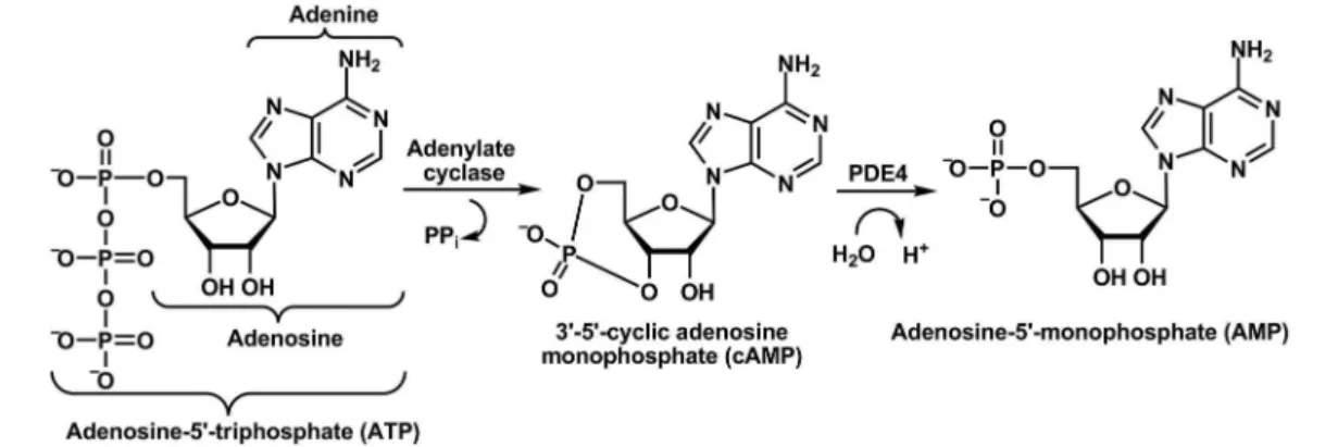
Modulators of intracellular cAMP levels
3’,5’-cyclic adenosine monophosphate (cAMP) and 3’,5’-cyclic guanosine monophosphate (cGMP) are second messengers generated by the action of adenylate cyclase and guanylate cyclase, respectively, which are involved in a vast array of cellular responses to different stimuli (Figure 3) (Lima et al., 2002; Chung, 2006).
channels, resulting in diverse biological effects. In the inlammatory response, the activation of protein kinase A prevents transcriptional factors, such as NF-κB, from promoting the expression of genes encoding cytokines. Thus, agents that induce higher levels of cAMP, either by stimulating its formation or by preventing its degrada-tion, inhibit pro-inlammatory cytokines, e.g. TNF-α and IL-1�, resulting in an immunosuppressive effect (Figure 4) (Chung, 2006).
In this context, new classes of drugs able to modulate intracellular levels of cAMP emerge as useful therapeutic strategies, such as phosphodiesterase inhibitors and modu-lators of the adenosine system (Lipworth, 2005; Chung, 2006; Jacobson, Gao, 2006).
• Phosphodiesterase inhibitors
Phosphodiesterases are a group of eleven families of enzymes (PDEs 1-11) responsible for the hydrolysis of cyclic nucleotides cAMP and cGMP, generating the respec-tive 5'-monophosphates. These families of
phosphodiestera-ses are expressed differently in different tissues and diverge in terms of amino acid sequence, substrate specificity, sensitivity to certain inhibitors, biochemical parameters and modulation of enzyme activity. For the last decade, studies involving the control of cAMP and cGMP levels through the activity of PDEs have intensiied, with their relevance evidenced by the number of clinical trials of selective PDE inhibitors for various inlammatory diseases, e.g. asthma and COPD (Lipworth, 2005; Chung, 2006).
Phosphodiesterase 4 (PDE4) in particular plays a central role in the immune cells involved in inlammatory response and is predominantly expressed in mast cells, eosinophils, neutrophils, T lymphocytes, macrophages and some structural cells, e.g. epithelial cells and neurons. The PDE4 family, speciic for cAMP hydrolysis, is divided into four subtypes, PDE4A, PDE4B, PDE4C and PDE4D (Houslay et al., 2005; Chung, 2006).
Rolipram (41), developed by Schering AG phar-maceutical industry, is a highly selective irst generation PDE4 inhibitor used as a pharmacological tool for inves-tigating the role of this enzyme. This compound has anti-inlammatory and immunomodulatory effects, and inhibits the release of cytokines from activated T lymphocytes, basophils, monocytes, macrophages and airway epithelial cells (Chung, 2006).
In a bid to reduce the adverse effects associated with this class of drugs, the occurrence of nausea and vomiting in particular, several second generation PDE4 inhibitors were described, such as Cilomilast (42, GlaxoSmithKline) and Rolumilast (43, Altana), which reached the stage of phase III clinical trials (Houslay et al., 2005; Lipworth, 2005).
Recent studies describe the involvement of PDE4 subtypes in the onset of the different biological effects of these inhibitors. The PDE4B appears to mediate most of the anti-inlammatory effects, while PDE4D is responsible for the adverse effects reported, mainly emesis (Lipworth,
FIGURE 3 - Biosynthesis of cAMP by adenylate cyclase and its hydrolysis to adenosine-5’-monophosphate (AMP) by the action of phosphodiesterase 4 (PDE4).
2005). Researchers have described that PDE4D knockout mice showed no adverse effects, while studies with PDE4B knockout mice conirmed that this PDE4 subtype is essential for biosynthesis of TNF-α induced by LPS
in vivo (Boswell-Smith et al., 2006). PDE4B knockout animals have shown almost complete suppression of this cytokine (Yamamoto et al., 2007), suggesting that a PDE4B selective inhibitor could potentially be an effective anti-inlammatory agent which does not induce emesis (Boswell-Smith et al., 2006).
Cilomilast (42) is ten times more selective for PDE4D than for other isoenzymes (i.e., PDE4A, PDE4B and PDE4C), while Rolumilast (43) shows no selectivity for the different isoforms of PDE4. The selectivity of 42 for PDE4D, responsible for the induction of nausea in patients, explains why this compound is less tolerated than Rolumilast (43) (Lipworth, 2005).
The search for PDE4 inhibitors without emetic effects led to the discovery of compounds with lower activity against the PDE4D isoform, such as the indole derivative AWD-12-281 (44, GlaxoSmithKline), the inda-zole derivative Toimilast (45, Pizer), and the benzofuran derivative Lirimilast (46, Bayer) (Houslay et al., 2005).
• Modulators of the adenosine system
Adenosine is an endogenous purine nucleoside (Figure 4) released by various cells, which regulates physi-ological processes by activating speciic receptors. There are four known subtypes of adenosine receptors, named A1,
A2A, A2B and A3, all coupled to the G protein, related to a
variety of signal transduction pathways (Haskó, Cronstein, 2004; Zhang et al., 2005).
The release of adenosine is increased in response to tissue injury. An accentuated production of this nucleo-side is described in several pathological conditions, e.g.
inlammation, hypoxia and ischemia. Adenosine can be metabolized by action of the enzyme adenosine
deami-nase, leading to inosine; or can be converted intracellularly to the nucleotide adenosine monophosphate (AMP) by the action of the enzyme adenosine kinase (Fredholm et al., 2001; Zhang et al., 2005).
Each adenosine receptor subtype has its particulari-ties with regard to signaling and inal physiological effects. In the classical description, the signaling triggered by these receptors is mediated by inhibition or stimulation of adenylate cyclase, although there are current indications of the relevance of other mediators and enzymes, e.g.
calcium, phospholipase C and mitogen-activated protein kinases (MAPKs) (Jacobson, Gao, 2006).
The A2A and A2B receptors, coupled to stimulatory
G protein, activate adenylate cyclase and increase intra-cellular cAMP (Figure 4). On the other hand, the A1 and
A3 receptors coupled to inhibitory G protein, reduce the
release of intracellular cAMP. Thus, several studies have shown that agonists of A2A and A2B receptors could act as
anti-inlammatory agents, and this activity was attributed, at least in part, to reduced release of TNF-α. However, recent studies have indicated that activation of A2B
recep-tors is also associated with the release of several allergic and pro-inlammatory mediators, whereas activation of the A2A receptor present in most immune cells including
lymphocytes, monocytes, macrophages and dendritic cells, appears to effectively alleviate inlammation and reperfusion injury in different tissues (Zhang et al., 2005; Jacobson, Gao, 2006).
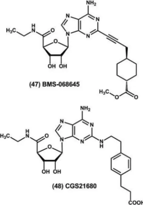
In this context, several A2A receptor agonists have
been reported in the literature as drug candidates for the treatment of inflammatory diseases. For example, the A2A receptor agonist BMS-068645 (ATL-146e) (47),
anti-inlammatory proile associated with the modulation of TNF-α and interferon gamma (INF-γ) release by activated
T lymphocytes, limiting the activation of T cells and mac-rophages in inlamed tissues. Conversely, the prototype CGS21680 (48), produced by Novartis, has pronounced anti-inlammatory activity in different murine models of allergic asthma, emerging as an alternative therapy for the treatment of this disease (Jacobson, Gao, 2006).
Dual inhibitors of TNF-α and PDE4
Aiming to optimize TNF-α modulator activity, dif-α modulator activity, dif- modulator activity, dif-ferent research groups have explored the ability of thalido-mide (3) and derivatives to directly inhibit the biosynthesis
of this cytokine associated with the strategy of indirect modulation, which is based on increasing the intracel-lular cAMP concentration (Lima et al., 2006). Agents that induce higher levels of cAMP, either by stimulating its production or preventing its degradation, cause an inhibition of pro-inlammatory cytokines such as TNF-α, IL-1� and INF-γ, reducing the recruitment and activation
of inlammatory cells resulting in an immunosuppressive effect (Marriott et al., 1997; Gallant et al., 2008).
Muller and colleagues described phthalimidic ana-logues of thalidomide (3) containing the dialcoxy-phenyl core, a pharmacophoric group present in PDE4 inhibitors,
e.g. rolipram (41), cilomilast (42) and rolumilast (43), aiming to obtain dual inhibitors of TNF-α and PDE4. The new phthalimidic dialcoxy-phenyl derivatives (49-52, Figure 5) effectively reduced TNF-α release in peripheral blood mononuclear cells stimulated by LPS, and signii-cantly inhibited the enzymatic activity of PDE4, acting through a different mechanism of action to that described for prototype 3 (Muller et al., 1998; Lima et al., 2006).
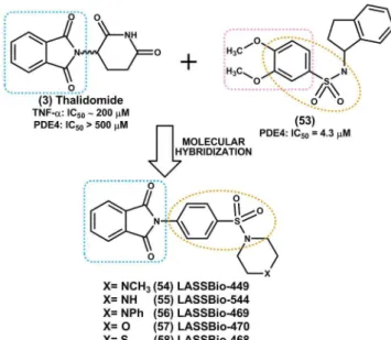
The Laboratório de Avaliação e Síntese de Sub-stâncias Bioativas (LASSBio®), of the Federal University
of Rio de Janeiro, has also made its contribution to the research and development of dual inhibitors of TNF-α and PDE4. In this lab, a congener series of N-phenyl sul-fonamide phthalimidic derivatives designed by molecular hybridization of the prototype thalidomide (3) with the selective PDE-4 inhibitor N -(2,3-dihydro-1H-1-indenil)-3,4-dimethoxy-benzene-sulfonamide (53) was synthesized (Figure 6) (Montana et al., 1998; Lima et al., 2002; Lima
et al., 2006).
FIGURE 5 - Inhibitory activity of thalidomide (3) and analogues (49-52) in the TNF-α release in peripheral blood mononuclear
The determination of the anti-inlammatory proper-ties of the new N-phenyl sulfonamide phthalimidic deriva-tives (54-58) was carried out according to the pharmaco-logical protocol for acute lung inlammation induced by LPS inhalation in mice. The results indicated good overall anti-inlammatory activity for this new series of deriva-tives when administered intraperitoneally, especially the compound LASSBio-468 (58), which showed an ED50 of
2.5 mg/kg in this assay (Lima etal., 2002).
The identified prototype (58) also reduced TNF-α mRNA levels analyzed by reverse transcription polymerase chain reaction (RT-PCR) in mice peritoneal macrophages stimulated by LPS (Alexandre-Moreira et al., 2005); and signiicantly inhibited the NF-κB activation induced by TNF-α at concentrations greater than 257 μM (100 mg/ mL) in U937 cell line, comprising monocytic cells extracted from human histiocytic lymphoma (Abreu, 2007).
In addition to the anti-TNF-α activity previously described, LASSBio-468 (58) showed the ability to inhibit PDE-4 isoform in vitro (IC50 = 80 μM) and was also found
to be inactive against the other isoforms of this enzyme (PDE1, PDE2, PDE3 and PDE5) at concentrations greater than 300 μM (Alexandre-Moreira et al., 2005).
This prototype has a pronounced anti-inlammatory and immunomodulatory proile, possibly resulting from a complex mechanism of action involving, besides the elevation of cAMP levels in immunocompetent cells, other independent biological properties, including the ability to reduce the stability of TNF-α mRNA (Alexandre-Moreira
et al., 2005; Lima et al., 2006).
FINAL CONSIDERATIONS
The anti-TNF biological therapies currently avail-able in the pharmaceutical market have led to a revolution in the treatment of chronic degenerative inflammatory diseases, characterizing this therapeutic approach as ef-fective in improving the quality of life of a significant number of patients. Worldwide, approximately one million individuals are either undergoing treatment or have been treated with TNF inhibitors, comprising indications that include rheumatoid arthritis, psoriatic arthritis, psoriasis and inlammatory bowel diseases. In addition, numerous potential clinical applications are still undergoing evalu-ation. However, these therapies have risks and limitations associated with their continued employment including their high cost, the inconvenience of exclusive administra-tion in hospitals and the incidence of signiicant adverse effects such as a high susceptibility to infections.
In this context, the discovery of small molecules able to modulate TNF activities, preferentially when adminis-tered orally, could represent an alternative to biopharma-ceutical agents. To this end, different mechanisms of TNF modulation have been characterized, e.g. the inhibition of TACE; the inhibition of protein kinases, such as p38 MAPK; the modulation of NF-κB signaling pathway; and the increase in intracellular cAMP levels through the use of phosphodiesterase inhibitors and/or adenosine system modulators; offering a wide range of possible targets for the design and development of new drug candidates with better pharmacotherapeutic proiles.
REFERENCES
ABREU, T. P. Estudo de derivados ftalimídicos análogos
à talidomida: inibição do fator transcricional NF-κB e investigação da ação anti-angiogênica. Rio de Janeiro, 2007. 121 p. [Master’s Degree Thesis in Genetics. Institute of Biology. Federal University of Rio de Janeiro].
ADAMS, J. Potential for proteasome inhibition in the treatment
of cancer. Drug Discov. Today, v.8, n.7, p.307-315, 2003.
ALEXANDRE-MOREIRA, M.S.; TAKIYA, C.M.; ARRUDA, L.B., PASCARELLI, B.; GOMES, R.N.; FARIA NETO, H.C.; LIMA, L. M..; BARREIRO, E. J. LASSBio-468: a new achiral thalidomide analogue with modulates TNF-α and NO production and inhibits endotoxic shock and
arthritis in na animal model. Int. Immunopharmacol., v.5,
n.3, p.485-494, 2005. FIGURE 6 - Structural design of the N-phenyl sulfonamide
phthalimidic derivatives, idealized as dual inhibitors of TNF-α
ARANAPAKAM, V.; DAVIS, J.M.; GROSU, G.T.; BAKER, J.; ELLINGBOE, J.; ZASK, A.; LEVIN, J.I.; SANDANAYAKA, V.P.; DU, M.; SKOTNICKI, J.S.; DIJOSEPH, J.F.; SUNG, A.; SHARR, M.A.; KILLAR, L.M.; WALTER, T.; JIN, G.; COWLING, R.; TILLETT, J.; ZHAO, W.; MCDEVITT, J.; XU, Z.B. Synthesis
and structure-activity relationship of N-substituted
4-arylsulfonylpiperidine-4-hydroxamic acids as novel, orally active matrix metalloproteinase inhibitors for the
treatment of osteoarthritis. J. Med. Chem., v.46, n.12,
p.2376-2396, 2003.
BANNER, D. W.; D’ARCY, A.; JANES, W.; GENTZ, R.; SCHOENFELD, H-J.; BROGER, C.; LOETSCHER, H.; LESSLAUER, W. Crystal structure of the soluble human 55kDa TNF receptor-human TNF� complex: implications
for TNF receptor activation. Cell, v.73, n.3, p.431-445,
1993.
BECK, G.; BOTTOMLEY, G.; BRADSHAW, D.; BREWSTER, M.; BROADHURST, M.; DEVOS, R.; HILL, C.; JOHNSON, W.; KIM, H.J.; KIRTLAND, S.; KNEER, J.; LAD, N.; MACKENZIE, R.; MARTIN, J.; NIXON, J.; PRICE, G.; RODWELL, A.; ROSE, F.; TANG, J.P.;
WALTER, D.S.; WILSON, K.; WORTH, E. (E)-2(R)-[1(S
)- (hydroxycarbamoyl)-4-phenyl-3-butenyl]-2’-isobutyl-2’-(methanesulfonyl)-4-methylvalerohydrazide (Ro 32-7315), a selective and orally active inhibitor of tumor necrosis
factor-α convertase. J. Pharmacol. Exp. Ther., v.302, n.1,
p.390-396, 2002.
BLACK, R. A.; BIRD, T. A., MOHLER, K. M. Agents that block
TNF-α synthesis or activity. Annu. Rep. Med. Chem., v.32,
p.241-249, 1997.
BOEHM, J. C.; SMIETANA, J. M.; SORENSON, M. E.; GARIGIPATI, R. S.; GALLAGHER, T. F.; SHELDRAKE, P. L.; BRADBEER, J.; BADGER, A. M.; LAYDON, J. T.; LEE, J. C.; HILLEGASS, L. M.; GRISWOLD, D. E.; BRETON, J. J.; CHABOT-FLETCHER, M. C.; ADAMS, J. L. 1-Substituted 4-Aryl-5-pyridinylimidazoles: a new class of cytokine suppressive drugs with low 5-lipoxygenase and
cyclooxygenase inhibitory potency. J. Med. Chem., v.39,
n.20, p.3929-3937, 1996.
B O S W E L L - S M I T H , V. ; S P I N A , D . ; PA G E , C . P.
Phosphodiesterase inhibitors. Br. J. Pharmacol., v.147,
n.S1, p.S252-S257, 2006.
BRAKEBUSCH, C.; VARFOLOMEEV, E.E.; BATKIN,M.; WALLACH, D. Structural requirements for inducible
shedding of the p55 tumor necrosis factor receptor. J. Biol.
Chem., v.269, n.51, p.32488-32496, 1994.
BURKE, J.R.; PATTOLI, M.A.; GREGOR, K.R.; BRASSIL, P.J.; MACMASTER, J.F.; MCINTYRE, K.W.; YANG, X.; IOTZOVA, V.S.; CLARKE, W.; STRNAD, J.; QIU, Y.; ZUSI, C. BMS-345541 is a highly selective inhibitor
of IκB kinase that binds at an allosteric site of the enzyme
and blocks NFκB-dependent transcription in mice. J. Biol.
Chem., v.278, n.3, p.1450-1456, 2003.
CAPORALI; R.; PALLAVICINI, F.B.; FILIPPINI, M.; GORLA, R.; MARCHESONI, A.; FAVALLI, E.G.; SARZI-PUTTINI, P.; ATZENI, F.; MONTECUCCO, C. Treatment of rheumatoid arthritis with anti-TNF-alpha agents: A
reappraisal. Autoimmun. Rev., v.8, n.3, p.274-280, 2009.
CAWTHORN, W.P.; SETHI, J.K. TNF-α and adipocyte biology.
FEBS Letters, v.582, n.1, p.117-131, 2008.
CHERNEY, R.J.; KING, B.W.; GILMORE, J.L.; LIU, R.Q.; COVINGTON, M.B.; DUAN, J.J.W.; DECICCO, C.P. Conversion of potent MMP inhibitors into selective TACE
inhibitors. Bioorg. Med. Chem. Lett., v.16, n.4,
p.1028-1031, 2006.
CHUNG, K.F. Phosphodiesterase inhibitors in airways disease.
Eur. J. Pharmacol., v.533, n.1-3, p.110-117, 2006.
CORRAL, L.G.; KAPLAN, G. Immunomodulation by
thalidomide and thalidomide analogues. Ann. Rheum. Dis.,
v.58, suppl.1, p.l107-l113, 1999.
DASGUPTA, S.; MURUMKAR, P.R.; GIRIDHAR, R.; YADAV, M.R. Current perspective of TACE inhibitors: A
review. Bioorg. Med. Chem., v.17, n.2, p.444-459, 2009.
DENG, L.; DING, W.; GRANSTEIN, R.D. Thalidomide inhibits tumor necrosis factor-α production and antigen presentation
by Langerhans cells. J. Invest. Dermatol., v.121, n.5,
p.1060-1065, 2003.
DE-BLANCO, E.J.C.; PANDIT, B.; HU, Z.; SHI, J.; LEWIS,
A.; LI, P.K. Inhibitors of NF-κB derived from thalidomide.
DUAN, J.J.W.; CHEN, L.; WASSERMAN, Z.R.; LU, Z.; LIU, R.Q.; COVINGTON, M.B.; QIAN, M.; HARDMAN, K.D.; MAGOLDA, R.L.; NEWTON, R.C.; CHRIST, D.D.; WEXLER, R.R.; DECICCO, C.P. Discovery of
γ-lactam hydroxamic acids as selective inhibitors of tumor
necrosis factor-α converting enzyme: design, synthesis, and
structure-activity relationships. J. Med. Chem., v.45, n.23,
p.4954-4957, 2002.
EDWARDS, M.R.; BARTLETT, N.W.; CLARKE, D.; BIRRELL, M.; BELVISI, M.; JOHNSTON, S.L. Targeting the
NF-κB pathway in asthma and chronic obstructive pulmonary
disease. Pharmacol. Ther., v.121, n.1, p.1-13, 2009.
FELDMANN, M.; BRENNAN, F.M.; FOXWELL, B.M.J.; TAYLOR, P.C.; WILLIAMS, R.O.; MAINI, R.N. Anti-TNF
therapy: where have we got to in 2005? J. Autoimmun., v.25,
suppl.1, p.26-28, 2005.
FREDHOLM, B.B.; IJZERMAN, A.P.; JACOBSON, K.A.; KLOTZ, K.N.; LINDEN, J. International union of pharmacology. XXV. Nomenclature and classiication of
adenosine receptors. Pharmacol. Rev., v.53, n.4,
p.527-552, 2001.
GALLANT, M.; CHAURET, N.; CLAVEAU, D.; DAY, S.; DESCHÊNES, D.; DUBÉ, D.; HUANG, Z.; LACOMBE, P.; LALIBERTÉ, F.; LÉVESQUE, J-F; LIU, S.; MACDONALD, D.; MANCINI, J.; MASSON, P.; MASTRACCHIO, A.; NICHOLSON, D.; NICOLL-GRIFFITH, D.A.; PERRIER, H.; SALEM, M.; STYHLER, A.; YOUNG, R.N.; GIRARD, Y. Design, synthesis and biological evaluation of 8-biarylquinolines: a novel class
of PDE4 inhibitors. Bioorg. Med. Chem. Lett., v.18, n.4,
p.1407-1412, 2008.
GEARING, A. J. H.; BECKETT, P.; CRISTODOULOU, M.; DRUMMOND, A. H.; GALLOWAY, W. A.;GILBERT, R.; GORDON, J. L.; LEBER, T. M.; MAGAN, M.; MILLER, K.; NAYEE, P.; OWEN, K.; PATEL, S.; THOMAS, G.; WELLS, G.; WOOD, L. M.; WOOLLEY, K. Processing of tumor necrosis factor-α precursor by metalloproteinases.
Nature, v.370, n.6490, p.555-557, 1994.
GRAY, P. W.; AGGARWAL, B. B.; BENTON, C. V.; BRINGMAN, T. S.; HENZEL, W. J.; JARRETT, J. A.; LEUNG, D. W.; MOFFAT, B.; NG, P.; SVEDERSKY, L. P.; PALLADINO, M. A.; NEDWIN, G. E. Cloning and expression of cDNA for human lymphotoxin, a lymphokine
with tumor necrosis activity. Nature, v.312, n.5996,
p.721-724, 1984.
HANADA, T.; YOSHIMURA, A. Regulation of cytokine
signaling and inlammation. Cytokine Growth Factor Rev.,
v.13, n.4, p.413-421, 2002.
HASHIMOTO, Y. Structural development of biological
response modifiers based on thalidomide. Bioorg. Med.
Chem., v.10, n.3, p.461-479, 2002.
HASKÓ, G.; CRONSTEIN, B.C. Adenosine: an endogenous
regulator of innate immunity.Trends Immunol., v.25, n.1,
p.33-39, 2004.
HENRY, J. R.; RUPERT, K. C.; DODD, J. H.; TURCHI, I. J.; WADSWORTH, S. A.; CAVENDER, D. E.; FAHMY, B.; OLINI, G. C.; DAVIS, J. E.; PELLEGRINO-GENSEY, J.; SCHAFER, P. H.; SIEKIERKA,J. J.
6-Amino-2-(4-luorophenyl)-4-methoxy-3-(4-pyridyl)-1H-pyrrolo[2,3-b]
pyridine (RWJ 68354): A potent and selective p38 kinase
inhibitor. J. Med. Chem., v.41, n.22, p.4196-4198, 1998.
HOCHBERG, M.C.; LEBWOHL, M.G.; PLEVY, S.E.; HOBBS, K.F.; YOCUM, D.E. The beneit/risk proile of
TNF-blocking agents: indings of a consensus panel. Sem.
Arthritis Rheum., v.34, n.6, p.819-836, 2005.
HORSSEN, R.V.; HAGEN, T.L.M.; EGGERMONT, A.M.M. TNF-α in cancer treatment: molecular insights, antitumor
effects, and clinical utility. Oncologist, v.11, p.397-408,
2006.
HOUSLAY, M.D.; SCHAFER, P.; ZHANG, K.Y.J. Keynote
review: Phosphodiesterase-4 as a therapeutic target. Drug
Discov. Today, v.10, n.22, p.1503-1519, 2005.
JACOBSON, K.A.; GAO, Z.G. Adenosine receptor as
therapeutic targets. Nat. Rev. Drug Discov., v.5, n.3,
p.247-264, 2006.
KANG, Y.J.; CHEN, J.; OTSUKA, M.; MOLS, J.; REN, S.; WANG, Y.; HAN, J. Macrophage deletion of p38α partially
impairs lipopolysaccharide-induced cellular activation. J.
Immunol., v.180, n.7, p.5075-5082, 2008.
KARIN, M . The IKKbeta subunit of IkappaB kinase (IKK) is essential for nuclear factor kappa B activation and
prevention of apoptosis. J. Exp. Med., v.189, n.11,
p.1839-1845, 1999.
KENNY, P.A. TACE: a new target in epidermal growth factor
receptor depedent tumors. Differentiation, v.75, n.9,
KEIFER, J.; GUTTRIDGE, D.; ASHBURNER, B.; BALDWIN, A. Inhibition of NFκB activity by Thalidomide through
suppression of IκB Kinase activity. J. Biol. Chem., v.276,
n.25, p.22382-22387, 2001.
KIM, Y.S.; KIM, J.S.; JUNG, H.C.; SONG, I.S. The effects of thalidomide on the stimulation of NF-κappaB activity and TNF-alpha production by lipopolysaccharide in human
colonic epithelial cell line. Mol. Cell, v.17, n.2, p.210-216,
2004.
KIM, D.K.; LIM, J.H.; LEE, J.A.; DEWANG, P.M. Synthesis and biological evaluation of trisubstituted imidazole derivatives as inhibitors of p38α mitogen-activated protein
kinase. Bioorg. Med. Chem. Lett., v.18, n.14, p.4006-4010,
2008.
KISHORE, N.; SOMMERS, C.; MATHIALAGAN, S.; GUZOVA, J.; YAO, M.; HAUSER, S.; HUYNH, K.; BONAR, S.; MIELKE, C.; ALBEE, L.; WEIER, R.; GRANETO, M.; HANAU, C.; PERRY, T.; TRIPP, C.S. A
selective IKK-2 inhibitor blocks NFκB-dependent gene
expression in interleukin-1�-stimulated synovial ibroblasts.
J. Biol. Chem., v.278, n.35, p.32861-32871, 2003.
KNOWLTON, A. A. NFκB, heat shock proteins, HSF-1, and
inlammation. Cardiovasc. Res., v.69, n.1, p.7-8, 2006.
KUMAR, S.; BOEHM, J.; LEE, J.C. p38 MAP kinases: key signaling molecules as therapeutic targets for inlammatory
diseases.Nat. Rev. Drug Discov., v.2, n.9, p.717-726, 2003.
LEVIN, J.I.; CHEN, J.M.; LAAKSO, L.M.; DU, M.; SCHMID, J.; XU, W.; CUMMONS, T.; XU, J.; JIN, G.; BARONE, D.; SKOTNICKI, J.S. Acetylenic TACE inhibitors. Part 3:
Thiomorpholine sulfonamide hydroxamates. Bioorg. Med.
Chem. Lett., v.16, n.6, p.1605-1609, 2006.
LIMA, L.M.; CASTRO, P.; MACHADO, A.L.; FRAGA, C.M.A.; LUGNIER, C.; MORAES, V.L.G.; BARREIRO, E.J. Synthesis and anti-inlammatory activity of phthalimide derivatives, designed as new thalidomide analogues.
Bioorg. Med. Chem., v.10, n.9, p.3067-3073, 2002.
LIMA, L.M.; FRAGA, C.A.M.; KOATZ, V.L.G.; BARREIRO, E.J. Thalidomide and analogs as anti-inflammatory and
immunomodulator drug candidates. inlamm.
Anti-allergy Agents Med. Chem., v.5, n.1, p.79-95, 2006.
LIN, J.; ZIRING, D.; DESAI, S.; KIM, S.; WONG, M.; KORIN, Y.; BRAUN, J.; REED, E. GJERTSON, D.; SINGH, R.R. TNFα blockade in human diseases: An overview of eficacy
and safety. Clin. Immunol., v.126, n.1, p.13-30, 2008.
LIPWORTH, B. Phosphodiesterase-4 inhibitors for asthma
and chronic obstructive pulmonary disease. Lancet, v.365,
n.9454, p.167-175, 2005.
LIVERTON, N.J.; BUTCHER, J.W.; CLAIBORNE, C.F.; CLAREMON, D.A.; LIBBY, B.E.; NGUYEN, K.T.; PITZENBERGER, S.M.; SELNICK, H.G.; SMITH, G.R.; TEBBEN, A.; VACCA, J.P.; VARGA, S.L.; AGARWAL, L.; DANCHECK, K.; FORSYTH, A.J.; FLETCHER, D.S.; FRANTZ, B.; HANLON, W.A.; HARPER, C.F.; HOFSESS, S.J.; KOSTURA, M.; LIN, J.; LUELL, S.; O’NEILL, E.A.; OREVILLO, C.J.; PANG, M.; PARSONS, J.; ROLANDO, A.; SAHLY, Y.; VISCO, D.M.; O’KEEFE, S.J. Design and synthesis of potent, selective, and orally bioavailable tetrasubstituted imidazole inhibitors of p38
mitogen-activated protein kinase. J. Med. Chem., v.42, n.12,
p.2180-2190, 1999.
MAN, H.W.; CORRAL, L.G.; STIRLING, D.I.; MULLER,
G.W. α-Fluoro-substituted thalidomide analogues. Bioorg.
Med. Chem. Lett., v.13, n.20, p.3415-3417, 2003.
MARRIOTT, J. B.; WESTBY, M.; DALGLEISH, A. G. Therapeutic potential of TNF-α inhibitors old and new.
Drug Discov. Today, v.2, n.7, p.273-282, 1997.
MATTHEWS, S.J.; McCOY, C. Thalidomide: A review of
approved and investigational uses. Clin. Ther., v.25, n.2,
p.342-395, 2003.
MELCHERT, M.; LIST, A. The thalidomide saga. Int. J.
Biochem. Cell Biol., v.39, n.7-8, p.1489-1499, 2007.
MERCURIO, F.; MANNING, A. M. Multiple signals
converging on NF-κB. Curr. Opin. Cell Biol., v.11, n.2,
p.226-232, 1999.
MOHAN, M.J.; SEATON, T.; MITCHELL, J.; HOWE, A.; BLACKBURN, K.; BURKHART, W.; MOYER, M.; PATEL, I.; WAITT, G.M.; BECHERER, D.; MOSS, M.L.; MILLA, M.E. The tumor necrosis factor alpha converting ezyme (TACE): A unique metalloproteinase with highly
defined substrate selectivity. Biochemistry, v.41, n.30,
MONTALBAN, A.G.; BOMAN, E.; CHANG, C.D.; CEIDE, S.C.; DAHL, R.; DALESANDRO, D.; DELAET, N.G.J.; ERB, E.; ERNST, J.T.; GIBBS, A.; KAHL, J.; KESSLER, L.; LUNDSTRÖM, J.; MILLER, S.; NAKANISHI, H.; ROBERTS, E.; SAIAH, E.; SULLIVAN, R.; WANG, Z.; LARSON, C.J. The design and synthesis of novel
α-ketoamide-based p38 MAP kinase inhibitors. Bioorg.
Med. Chem. Lett., v.18, n.6, p.1772-1777, 2008.
MONTANA, J.G.; BUCKLEY, G.M.; COOPER, N.; DYKE, H.J.; GOWERS, L.; GREGORY, J.P.; HELLEWELL, P.G.; KENDALL, H.J.; LOWE, C.; MAXEY, R.; MIOTLA, J.; NAYLOR, R.J.; RUNCIE, K.A.; TULADHAR, B.; WARNECK, J.B.H. Aryl sulfonamides as selective PDE4
inhibitors. Bioorg. Med. Chem. Lett., v.8, n.19,
p.2635-2640, 1998.
MOREIRA, A.L.; SAMPAIO, E.P.; ZMUIDZINAS, A.; FRINT, P.; SMITH, K.A.; KAPLAN, G. Talidomide exerts its inhibitory action on tumor necrosis factor alpha by
enhancing mRNA degradation. J. Exp. Med., v.177, n.6,
p.1675-1680, 1993.
MOSS, M. L.; JIN, S. L. C.; MILLA, M. E.; BURKHART, W.; CARTER, H.L.; CHEN, W. J.; DIDSBURY, J. R.; HASSIER, D.; HOFFMAN, C. R.; KOST, T. A.; LAMBERT, M. H.; LEESNITZER, M. A.; McCAULEY, P.; McGEEHAN, G.; MITCHELL, J.; MOYERS, M.; PAHEL, G.; ROCQUE, W.; OVERTON, L. K.; SCHOENEN, F.; SEATON, T.; SU, J.L.; WARNER, J.; WILLARD, D.; BECHERE, J. D. Cloning of a disintegrin metalloproteinase that processes precursor tumor necrosis factor-alpha.
Nature, v.385, n.6618, p.733-736, 1997.
MUKHOPADHYAY, S.; HOIDAL, J. R.; MUKHERJEE, T. K.
Role of TNF-α in pulmonary pathophysiology. Respir. Res.,
v.7, n.125, p.1-9, 2006.
MULLER, G.; SHIRE, M.G.; WONG, L.; CORRAL, L.G.; PATTERSON, R.; CHEN, Y.; STIRLING, D. Thalidomide
analogs and PDE4 inhibition. Bioorg. Med. Chem. Lett., v.8,
n.19, p.2669-2674, 1998.
MULLER, G.; CHEN, R.; HUANG, S.; CORRAL, L.; WONG, L.; PATTERSON, R.; CHEN, Y.; KAPLAN, G.; STIRLING, D. Amino-substituted thalidomide analogs:
Potent inhibitors of TNF-α production. Bioorg. Med. Chem.
Lett., v.9, n.11, p.1625-1630, 1999.
MUTALIK, V.K.; VENKATESH, K.V. Effect of the MAPK cascade structure, nuclear translocation and regulation of
transcription factors on gene expression. BioSystems, v.85,
n.2, p.144-157, 2006.
NG, S.S.W.; BROWN, M.; FIGG, W.D. Thalidomide, an antiangiogenic agent with clinical activity in cancer.
Biomed. Pharmacother., v.56, n.4, p.194-199, 2002.
PALLADINO, M. A.; BAHJAT, F. R.; THEODORAKIS, E. A.; MOLDAWER, L. L. Anti-TNF-α therapies: the next
generation. Nat. Rev. Drug Discov., v.2, n.9, p.736-746, 2003.
PAPPAS, D.A.; BATHON, J.M.; HANICQ, D.; YASOTHAN,
U.; KIRKPATRICK, P. Golimumab. Nat. Rev. Drug Discov.,
v.8, n.9, p.695-696, 2009.
PAUL, A.T.; GOHIL, V.M.; BHUTANI, K.K. Modulating
TNF-α signaling with natural products. Drug Discov. Today,
v.11, n.15-16, p.725-732, 2006.
PENNICA, D.; NEDWIN, G. E.; HAYFLICK, J. S.; SEEBURG, P. H.; DERYNCK, R.; PALLADINO, M. A.; KOHR, W. J.; AGGARWAL, B. B.; GOEDDEL, D. V. Human tumor necrosis factor: precursor structure, expression and
homology to lymphotoxin. Nature, v.312, n.5596,
p.724-729, 1984.
PETERSON, P.K.; HU, S.; SHENG, W.S.; KRAVITZ, F.H.; MOLITOR, T.W.; CHATTERJEE, D.; CHAO, CC. Thalidomide inhibits tumor necrosis factor-alpha production by lipopolysaccharide and
lipoarabinomannan-stimulated human microglial cells. J. Infect. Dis., v.172, n.4,
p.1137-1140, 1995.
PIERCE, J.W.; SCHOENLEBER, R.; JESMOK, G.; BEST, J.; MOORE, S.A.; COLLINS, T.; GERRITSEN, M.E. Novel
inhibitors of cytokine-induced IκBα phosphorylation and
endothelial cell adhesion molecule expression show
anti-inlammatory effects in vivo.J. Biol. Chem., v.272, n.34,
p.21096-21103, 1997.
PODOLIN, P.L.; CALLAHAN, J.F.; BOLOGNESE, B.J.; LI, Y.H.; CARLSON, K.; DAVIS, T.G.; MELLOR, G.W.; EVANS, C.; ROSHAK, A.K. Attenuation of murine collagen-induced arthritis by a novel, potent, selective small molecule
inhibitor of IκB kinase 2, TCPA-1 (2-[(aminocarbonyl)
amino]-5-(4-luorophenyl)-3-thiophenecarboxamide), occurs via reduction of proinflammatory cytokines and
antigen-induced T cell proliferation. J. Pharmacol. Exp. Ther., v.312,