SEIZURES OUTCOME AFTER CORTICAL
RESECTIONS INCLUDING THE FACE AND
TONGUE ROLANDIC AREAS IN PATIENTS WITH
REFRACTORY EPILEPSY AND NORMAL MRI
SUBMITTED TO SUBDURAL GRIDS IMPLANTATION
Arthur Cukiert, Jose A. Buratini, Elcio Machado, Alcione Sousa, Joaquim Vieira,
Cassio Forster, Meire Argentoni, Cristine Baldauf, Leila Frayman
ABSTRACT - Purpose: To study the seizures outcome in patients with refractory epilepsy and normal MRI submitted to resections including the rolandic cortex. Methods: Four adult patients were studied. All patients had motor or somatosensory simple partial seizures and normal MRI and were submitted to subdural grids implantation with extensive coverage of the cortical convexity (1 in the non-dominant and 3 in the dominant hemisphere). Results: ECoG was able to define focal areas of seizures onset in every patient. All patients were submitted to resection of the face and tongue motor and sensitive cortex; two patients had resections including the perirolandic cortex and 2 had additional cortical removals. Three patients are seizures free and one had a greater then 90% reduction in seizure frequency. Conclusion: Resections including the face and tongue rolandic cortex can be safely performed even within the dominant hemisphere.
KEY WORDS: epilepsy, surgery, outcome, subdural grids, rolandic cortex.
Resultados cirúrgicos em pacientes portadores de epilepsia refratária e ressonância magnética normal submetidos a ressecções das áreas rolândicas da face e língua investigados por meio de eletrodos subdurais
RESUMO - Objetivo: Estudar o efeito na frequência de crises epilépticas de ressecções de cortex rolândico em pacientes com epilepsia refratária e ressonância normal. Material: Quatro pacientes epilépticos adultos foram estudados. Todos possuíam crises parciais simples motoras ou sensitivas e ressonância normal, e foram submetidos ao implante de eletrodos subdurais cobrindo extensamente a convexidade hemisférica (1 no hemisfério não-dominante e 3 no hemisfério dominante). Resultados: O ECoG foi capaz de definir áreas ictais focais em todos os pacientes. Todos os pacientes foram submetidos à ressecção das áreas da face e língua do cortex rolândico motor e sensitivo; em dois, o cortex perirolândico foi incluído na ressecção e em dois pacientes, outras ressecções corticais foram adicionadas. Três pacientes estão livres de crises e um obteve melhora maior que 90% da frequência de crises. Conclusão: Ressecções corticais envolvendo o cortex rolândico da língua e face podem ser realizadas com segurança mesmo no hemisfério dominante.
PALAVRAS-CHAVE: epilepsia, cirurgia, resultados, placas subdurais, córtex rolândico.
Epilepsy Surgery Program, Hospital Brigadeiro and Clinica de Epilepsia de São Paulo, São Paulo SP, Brazil. Received 28 February 2001, received in final form 6 June 2001. Accepted 13 June 2001.
Dr. Arthur Cukiert - Rua Dr. Alceu Campos Rodrigues 247 / 121 04544-000 São Paulo SP Brasil. FAX: 55 11 384 63273. E-mail: [email protected]
Cortical resections including the rolandic cortex
have been rarely performed in patients with
refrac-tory epilepsy due to potential motor and
speech-related morbidity. This is especially true for patients
without lesions demonstrated by magnetic
resonan-ce and dominant hemisphere epileptic foci
1. Motor
and somatosensitive simple partial seizures may arise
from the rolandic cortex
2-4. The rolandic cortex may
and intraoperative direct cortical stimulation under
local or general anesthesia, have already been used to
map the motor cortex
5-7. Subdural grids implantation
represents a safe and effective procedure allowing
both cortical and neurophysiological mapping.
This paper describes the anatomical and
neuro-physiological data obtained from patients with
re-fractory epilepsy and seizures arising in the rolandic
areas submitted to extensive subdural grids
implan-tation.
CASES
All patients were investigated within the Epilepsy Sur-gery Program and gave formal consent for all surgical pro-cedures.
Patient I. A right-handed 28 years-old man presented with daily motor simple partial seizures (SPS) initiating in the left side of the face and spreading to the left arm, with secondary tonic-clonic generalization, since the age of 4. Interictal electroencephalogram (EEG) showed wide-spread spiking over the right hemisphere. Ictal EEG showed diffuse background attenuation and no focal seizures onset. MRI was normal. He was submitted to subdural grids implantation (96 contacts) covering the whole right fron-toparietooccipital convexity. Interictal electrocorticogram (ECoG) showed diffuse spiking over the right convexity. Ictal ECoG showed seizures onset at the face rolandic mo-tor region with early spread to the posterior frontal , an-terior parietal and superior temporal gyri. Stimulation of the motor face cortex was able to reproduce the patients typical seizure. Cortical resection including both sensory and motor tongue and face rolandic areas, the perirolandic cortex surrounding them and the superior temporal cor-tex was performed. Pathological examination showed mild gliosis. There was a transient (2 weeks) left facial paresis. He has had no SPS and one tonic-clonic seizure every 6 months since surgery (follow-up time = 24 months).
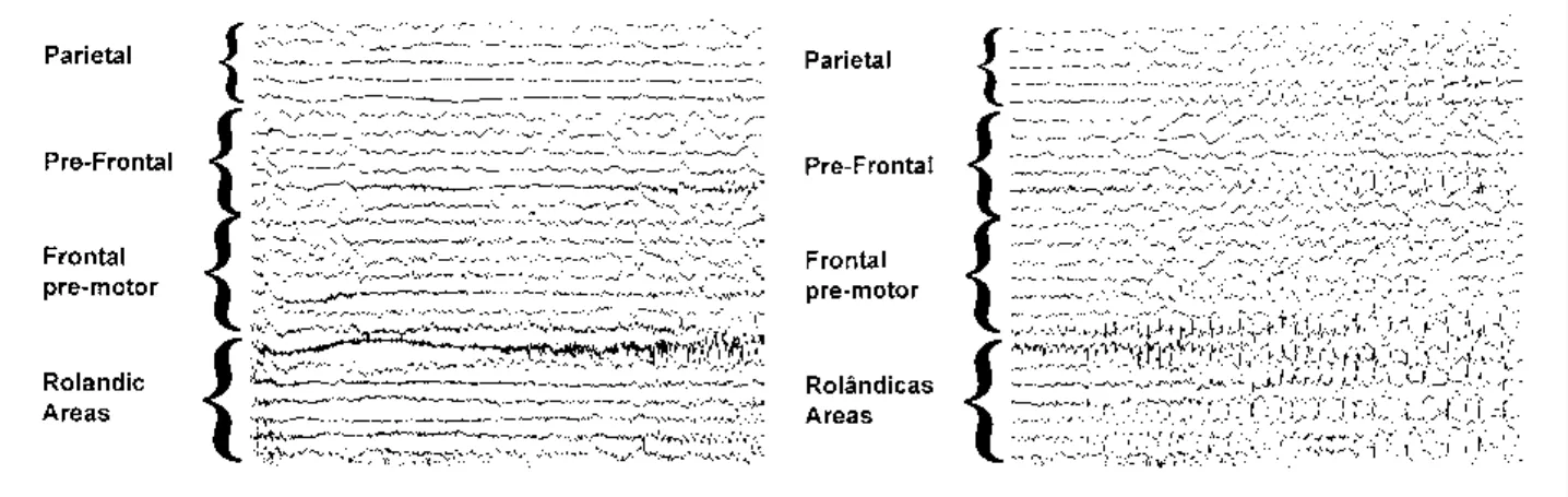
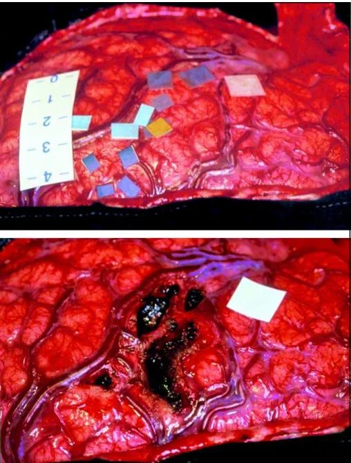
Patient II. A right-handed 23 years-old man presented with daily motor SPS involving the right side of the face associated to aphasic blockage, since the age of 5. Interictal EEG showed widespread left hemispheric spiking and ic-tal EEG showed seizures characterized by diffuse spiking over the frontoparietooccipital convexity. MRI was nor-mal. He was submitted to subdural grids implantation covering the frontoparietooccipital convexity (96 contacts). Interictal ECoG showed diffuse spiking over the left con-vexity. Ictal ECoG showed that seizures started within the left tongue and face motor and sensory areas (Fig 1). Stimulation of the tongue motor area was able to repro-duce the patients typical seizure. He was submitted to resection restricted to the rolandic face and tongue areas (Fig 2). Pathological examination showed severe gliosis. There was a transient (2 weeks) right facial paresis and dysphasia (preserved comprehension, low verbal output and word finding difficulties). He has been seizure-free since surgery (follow-up time = 18 months).
Patient III. A right-handed 24 years-old man, presented with daily motor SPS involving the right side of the face associated to aphasic blockage and eventually evolving to complex partial seizures without automatisms, since the age of 3. Interictal EEG showed diffuse spiking over the left convexity. Ictal EEG showed a large left frontotempo-ral area of seizures onset. MRI was normal. He was submit-ted to subdural grids implantation covering the left con-vexity (96 contacts). Interictal ECoG showed diffuse fronto-parietal spiking. Ictal ECoG showed seizures starting over the left motor and sensory face cortex and from the ante-rior and posteante-rior perirolandic cortex (Fig 3). Stimulation of the face sensory cortex was able to reproduce the pa-tients typical seizure. A cortical resection including the face and tongue sensory-motor areas and perirolandic tex was performed. Pathological examination showed cor-tical dysplasia. There was a transient (1 week) right facial paresis and dysphasia (similar to Patient II). He has been seizure-free since surgery (follow-up time = 15 months).
Patient IV. A right-handed 20 years-old man presented with daily motor SPS involving the right side of the face and aphasic blockage, frequently evolving to complex partial seizures with bimanual automatisms, since the age of 3. Interictal and ictal EEG showed left frontoparietal spi-king and seizures onset, respectively. MRI was normal. He was submitted to subdural grids implantation cover-ing the frontoparietal convexity (96 contacts). Interictal ECoG showed widespread frontoparietal spiking. Ictal ECoG showed seizures onset over the left face and tongue sensorimotor areas with very early spread to the posterior superior frontal gyrus. Stimulation of the face motor area was able to trigger the patients typical seizure. He was submitted to resection of the left rolandic cortex related to the face and tongue and of the posterior two-thirds of the superior frontal gyrus. Pathological examination dis-closed gliosis. There was a transient (2 weeks) right facial paresis and dysphasia (similar to Patient II). He has been
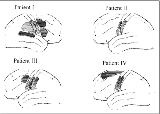
seizure-free since surgery (follow-up time = 20 months). A summary of the resections topography can be found in Figure 4.
DISCUSSION
Three out of the four patients in this series have
been submitted to dominant hemisphere resections.
Despite the potential speech-related morbidity,
pa-tients recovered extremely well after a mean of two
weeks. The absence of long-lasting speech
disturban-ces in these patients might be related to the careful
subpial intragyral resection and preservation of the
intrasulcal vessels and of the deeper fibers
belong-ing to the arcuate and uncinate fasciculi.
All the patients had focal motor seizures and
widespread interictal spiking areas, much larger then
Fig 3. Left: ECoG sample obtained from Patient III showing ictal onset over the somatosensitive gyrus. Right: ECoG sample showing the spread of this activity within the rolandic and perirolandic cortex.
Fig 4. Summary of the topography of the resections performed in this series.
the ictal or symptomatogenic zones. The actual
re-sections included both the ictal onset and
symptoma-togenic zones whenever possible, but large
interic-tally spiking areas were left behind. The latter might
represent a more widespread epileptogenesis and
might correlate to residual seizures, as seen in
Pa-tient I, who had a marked decrease in seizures
fre-quency but still presents sporadic tonic-clonic
sei-zures.
Three of our four patients have been rendered
seizure-free by surgery. Restricted cortical resections
within the rolandic and perirolandic cortex proved
to be effective even in the presence of large
interic-tally spiking areas, as was also noted by Lehman et
al
8. In our series, ictal spread from the rolandic
tex occurred predominantly to the perirolandic
cor-tex although early spreading to the superior
tempo-ral and superior frontal gyri was also seen. Cortical
areas involved in the early spread of ictal activity
should also be included in the resection if feasible.
Resections were limited basically by the hand motor
(not the sensitive) cortex, Brocas and Wernickes
areas and critical draining veins.
Pathological examination showed that one
pa-tient had cortical dysplasia
9not demonstrated
Non-invasive neurophysiological data were
non-localizatory in all patients in this series and invasive
recordings were needed for adequate
neurophysi-ological and anatomical mapping of potentially
re-sectable cortex
10,11. Subdural grids seem to be
supe-rior to depth electrodes for the mapping of large
superficial cortical areas located over the brain
con-vexity.
REFERENCES
1. Holtzman RNN, Mark MH, Wiener LM, Minzer L. Lingual epilepsy: a case report of an unusual expression of focal cerebral discharge. J Neurol Neurosurg Psychiatry 1984;47:317-318.
2. Jabbari B, Coker SB. Paroxyxmal, rhytmic lingual movements and chronic epilepsy. Neurology 1981;31:1364-1367.
3. Neufeld MY, Blumen SC, Nisipeanu P, Korczyn AD. Lingual Seizures. Epilepsia 1988;29:30-33.
4. King RB, Schell MD. Cortical localization and monitoring during cere-bral operations. J Neurosurg 1987;67:210-219.
5. LeRoux PD, Berger MS, Haglund MM, Pilcher WH, Ojemann GA. Re-section of intrinsic tumors from nondominant face motor cortex using stimulation mapping: report of two cases. Surg Neurol 1991;36:44-48. 6. Wood CC, Spencer DD, Allison T, McCerthy G. Williamsin PD, Goff WR. Localization of human sensorimotor cortex during surgery by cortical surface recording of somatosensory evoked potentials. J Neurosurg 1988;68:99-111.
7. Picard C, Olivier A. Sensory cortical tongue representation in man. J Neurosurg 1983;59:781-789.
8. Lehman R, Andermann F, Olivier A, Tandon PN, Quesnay LF, Rasmussen TB. Seizures with onset in the sensorimotor face area: clini-cal patterns and results of surgiclini-cal treatment in 20 patients. Epilepsia 1994;35:1117-1124.
9. Kuzniecky R, Morawetz R, Faught E, Black L. Frontal and central lobe focal dysplasia: clinical, EEG and imaging features. Dev Med Childs Neurol 1995;37:159-166.