CortiCal Stimulation of language
fieldS under loCal aneStheSia
Optimizing removal of brain lesions adjacent to speech areas
Robson Luis Oliveira de Amorim
1, Antônio Nogueira de Almeida
1, Paulo Henrique Pires de Aguiar
1,
Erich Talamoni Fonoff
1, Suely Itshak
1, Daniel Fuentes
2, Manoel Jacobsen Teixeira
1Abstract – Objective: The main objective when resecting benign brain lesions is to minimize risk of postoperative neurological deficits. We have assessed the safety and effectiveness of craniotomy under local anesthesia and monitored conscious sedation for the resection of lesions involving eloquent language cortex. Method: A retrospective review was performed on a consecutive series of 12 patients who underwent craniotomy under local anesthesia between 2001 and 2004. All patients had lesions close to the speech cortex. All resection was verified by post-operative imaging. Six subjects were male and 6 female, and were aged between 14 and 52 years. Results: Lesions comprised 7 tumour lesions, 3 cavernomas and 1 dermoid cyst. Radiological gross total resection was achieved in 66% of patients while remaining cases had greater than 80% resection. Only one patient had a post-operative permanent deficit, whilst another had a transient post-operative deficit. All patients with uncontrollable epilepsy had good outcomes after surgery. None of our cases subsequently needed to be put under general anesthesia. Conclusion: Awake craniotomy with brain mapping is a safe technique and the “gold standard” for resection of lesions involving language areas.
Key WORdS: cortical mapping, awake craniotomy, tumor, speech area.
mapeamento cortical da fala com o paciente acordado: optimização para ressecção de lesões intracranianas localizadas próximas à área da fala
Resumo – Objetivo: O presente estudo visa discutir as vantagens e as limitacões do uso da técnica de mapeamento cortical da área da fala com o paciente acordado. Método: esta é uma revisão retrospectiva dos casos em que foi realizado monitoramento cortical intraoperatório em cirurgias para ressecção de lesões intracranianas localizadas próximas à área da fala. Todos os pacientes foram submetidos a avaliação neuropsicológica no pré e intra-operatório. O grau das ressecções foi verificado através de exames de imagem pós-operatórios. Foram avaliados um total de 12 pacientes. destes, 6 eram do sexo masculino e 6 do feminino. Resultados: 7 lesões eram tumorais. A ressecção total foi atingida em 66% e ressecção subtotal nos remanescentes. Apenas 1 paciente apresentou déficit motor permanente no pós-operatório e todos os pacientes com quadro prévio de epilepsia refratária obtiveram bom controle das crises no pós-operatório. em nenhum caso houve necessidade de conversão da anestesia para geral. Conclusão: O mapeamento funcional intraoperatório na craniotomia com o paciente acordado otimiza a extensão da ressecção da lesão minimizando morbidade permanente. esta é uma técnica eficaz no manejo de lesões em íntimo contato com o córtex eloqüente, que outrora, seriam designadas inoperáveis.
PALAvRAS-ChAve: mapeamento cortical, craniotomia acordado, tumor, área da fala
1department of Neurology, division of Functional Neurosurgery of Clinics hospital, University of São Paulo School of Medicine, São Paulo, Brazil; 2department of Psychology and Psychiatry, Psychiatry Institute of Clinics hospital, University of São Paulo School of Medicine, São Paulo, Brazil.
Received 8 February 2008, received in inal form 15 May 2008. Accepted 11 June 2008.
Dr. Robson Luis Oliveira de Amorim – Rua Oscar Freire 1811 / 511 - 05409-011 São Paulo SP - Brasil. E-mail: [email protected]
Cortical locations corresponding to neurological func-tions can vary signiicantly among individuals and some brain lesions are able to distort the anatomy, hindering the localization of certain key intra-operative points. de-termining the exact functional area is essential for safe and effective resection1,2.
As the extent of tumor lesion resection is directly re-lated to outcome, the surgeon should attempt to max-imize the extent of removal without compromising the patient’s quality of life.
analyze how the application of this technique allows to-tal resections, and to determine to what extent cortical mapping can prevent postoperative deicits.
method
Twelve patients, with brain lesions in close proximity to lan-guage-speciic cortex, operated from 2001 to 2004, were stud-ied. Of these, 6 were female and 6 male. Age varied from 14 to 52 years (mean 35.4 y). The patients were submitted to a pro-tocol where age, clinical symptomatology, the Karnofsky scale (KPS) and radiological indings through computed tomography (CT) and magnetic resonance (MRI) were analyzed.
A neuropsychological evaluation composed by the Boston diagnostic Aphasia examination-III was conducted at bedside to identify patient characteristics in order to optimize subsequent language testing in the operating room.
All were submitted to a left craniotomy under sedation and local anesthesia without muscular relaxation. A three-pin head holder was placed after head block using 0.5% lidocaine, 0.25% bupivacaine with epinephrine (1:200,000) and saline (Fig 1). Sur-gical incision and skin lap base were anesthetized with the same solution. In half of the surgeries, sedation was achieved using
midazolam, fentanil and propofol. The main anesthetic for seda-tion in six procedures was dexmedetomidine at a loading dose of 3 mg/Kg/h over 20 minutes, maintained with 0.5 mg/Kg/h (Almeida et al.). The anesthesist and the prinicpal surgeon were the same in all surgeries (S.I. and A.N.A., restrospectively).
Cortical stimulation was carried out to identify the eloquent areas. Intraoperative stimulation was carried out using a mono-polar probe (Grass Stimulator) in 11 patients (Figs 2 and 3), while in one patient a bipolar probe was applied (Micromar). The cur-rent for stimulation varied from 3–13 mA, with single pulse of 1 millisecond and frequency of 60 hz.The safety limit adopted in this study was the adjacent pia mater of the functional cortex with preservation of the intersulcal vessels. The resection type was graded into total, subtotal (>80%), partial (< 80%) and biop-sy, according to postoperative radiological indings (contrasted CT scan and MRI). The surgeries were accomplished in an onco-logic view when applicable, in a bid to achieve both maximum le-sion removal and treating the epileptic condition, present in the vast majority of patients.
After surgical procedures, all patients were submitted to a CT scan within 6 hours. An MRI was performed after 3 to 9 months to evaluate the resection extent compared to previous exams.
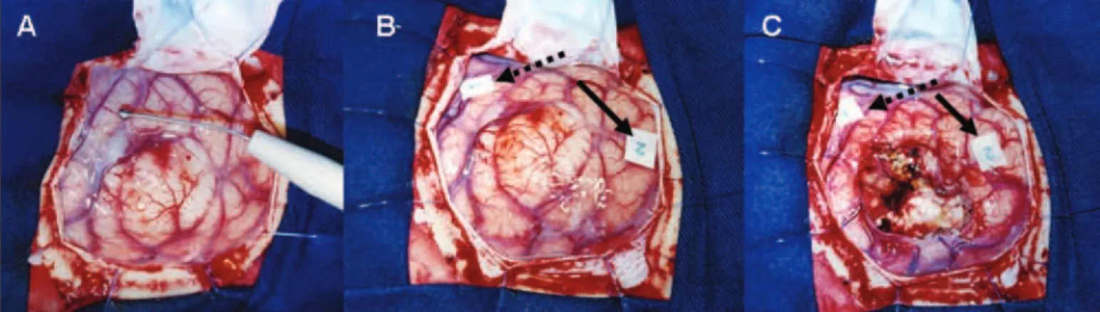
Fig 2. (A) Intraoperative cortical stimulation with electrode monopolar; (B) identiication of 2 speech areas in posterior temporal lobe (black dotted narrow and black narrow) and (C) satisfactory exeresis of the lesion preserving the functional cortex.
The postoperative complications were classiied into neurolog-ical, regional or systemic and median follow up was 6 months.
reSultS
All the patients presented with seizures. half of these presented with refractory seizures non-responsive to clin-ical treatment. Just one patient presented speech distur-bance in the preoperative neurologic exam (luent apha-sia). No patients had motor deicits in the preoperative clinical evaluation. headache was the initial symptom in one patient, who presented a cavernous angioma with
signs of bleeding. All patients harboring tumorous lesions had Karnofsky scale (KPS) 100.
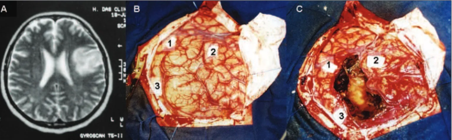
Most lesions were located in the inferior left frontal lobe (8), followed by the left posterior temporal lobe (2) and inferior parietal lobe (2). We identiied the language areas in 11 patients. Patient 1 exhibited 2 areas where stim-ulation provoked aphasia (Fig 3). We were unable to lo-cate the speech area in patient 5.
Of the identiied lesions, 7 were tumorous, 6 of which were low grade tumors. The ive benign lesions were cav-ernous angiomas (3), dermoid cyst (1), and gliosis (1) in a
Table. Patients characteristics included in the study. Sex Age
(y)
Symptoms of presentation
KPS Lesion Localization Resection Postoperative
deicit
Recover Neurological
compl.
Local compl.
Systemic compl.
1 M 50 Refractory crisis 100 Oligodendroglioma Frontal Subtotal Motor aphasia
+ hemyplegia
No Permanent
deicit
Wound infection
No
2 F 38 GTC crisis 100 Oligodendroglioma Medium
frontal gyrus
Total SMA yes Transitory
deicit
No No
3 M 21 Refractory crisis nda Gliosis Frontal-opercular Total No yes No No No
4 M 36 Refracory
tonic crisis
100 dNeT Frontal-opercular Total No yes No No No
5 F 36 Refractory CPC 100 Xantoastrocytoma Posterior
temporal lobe
Total No yes No No No
6 M 40 Refractory GTC nda dermoid cyst Frontal-opercular Total No yes No No No
7 F 14 Refractory CPC 100 Ganglioglioma Medium
temporal gyrus
Subtotal No yes No No No
8 M 28 headache +
GTC crisis
nda Cavernous angioma Supramarginal
gyrus
Total No yes No No No
9 F 45 Comprehension
aphasia + SPC
nda Cavernous angioma Angular gyrus Total No yes No No No
10 M 35 GTC crisis nda Cavernous angioma Frontal-opercular Total No yes No No No
11 F 30 GTC crisis 100 Oligodendroglioma Frontal Subtotal No yes No No No
12 F 52 GTC crisis 100 MGB Frontal Subtotal No yes No No No
GTC, generalized tonic-clonic; SPC, simple partial crisis; CPC, complex partial crisis; KPB, Karnofsky Scale; nda, do not apply; dNeT, disembryoblastic neuroepithelial tumor; MGB, multiform glioblastoma; AMS, supplementary motor area syndrome.
patient who underwent craniotomy due to brain contu-sion and was admitted with refractory epilepsy.
Concerning grade of resection, 8 patients (66%) had total gross resection whereas 4 patients underwent sub-total resection. In patient 2 the number of speech distur-bances decreased postoperatively.
Two out of the six patients submitted to conscious se-dation using dexmetadomidine presented focal seizures during surgery. Patient 1 had two focal and one gener-alized seizure. All of the episodes were transitory and ceased upon application of cold crystaloids (NaCl 0.9%) to the cerebral cortex. Patient 1 presented three seizures during the procedure, one before dural opening and two during cortical stimulation. The other patients had sei-zures during cortical stimulation.
Six patients (50%) underwent sedation with dexme-tomidine (patients 1,2,6,8,9,10) as this was deemed by the anesthesiologist and patients to be the most comfortable anesthetic. however, no its occurred under the other an-esthetic scheme.
One patient, harboring an oligodendroglioma, had a postoperative transitory motor deicit that receded by the second postoperative month, while only 1 patient developed permanent deicit characterized by hemiple-gia and expression aphasia. The same patient presented with meningitis due to cerebrospinal luid leakage and de-ceased 4 months later. Control of epilepsy was achieved in all patients with refractory episodes through the sur-gical procedure. Subsequent reduction of anti-epileptic drugs resulted and 1 patient had total withdrawal of drugs after 6 months (Table).
diSCuSSion
In mapping studies performed by Ojemann et al. on 117 patients2, 67% had more than one distinct essential
language area and 24% had three or more distinct areas subserving the language function in the dominant hemi-sphere peri-Sylvian region. Thus, in lesions that invade the language centers the surgical procedure to achieve total gross resection becomes progressively more difi-cult. The gold standard for identifying cortical function remains the direct cortical stimulation at surgery time in the awake patient3,4, despite the several techniques such
as functional MRI, magnetoencephalography which have been described recently5-7. Given intraoperative cortical
stimulation can identify eloquent areas, many authors routinely adopt this technique to guide brain lesion re-sections and to maintain integrity of the functional path-ways8. vitaz et al.9 compared the use of local anesthesia
with general anesthesia in patients submitted to resection of lesions adjacent to eloquent cortex and reported a higher success rate of stimulation in awake patients (100%
versus 50%) who required a signiicantly lower stimulation current (5 mA versus 13 mA, p<0.0001). A recent random-ized prospective study compared general anesthesia with awake surgery and found that better tumor cytoreduction and neurological improvement was seen in the former, demonstrating that this topic remains controversial10. In
our series, it proved impossible to identify the speech area in patient 1, although this did not affect the postop-erative outcome. This can be explained by the fact that speech area can often be located in different regions and may be as small as 2.5 cm2 . The stimulation current ranged
from 3 mA to 13 mA. Gross total resection was achieved in 66% of our series, similar to the rate to found by other authors ranging from 37.5 to 62%4,11,12. Neurological
wors-ening in the immediate postoperative period followed by recovery within several weeks coincides with other series reported in the literature (27 to 83%)13,14. This worsening
could be related to edema caused by surgical manipula-tion, to the effect of traction on eloquent areas or due to transgression of certain safety margins in mapping. danks et al.4 found a rate of 4% for permanent deicit
postop-eratively, similar to the 8% found in our study.
Surgical procedures performed under local anesthesia allow evaluation of certain intra-operative cerebral func-tions, such as the language area, and to avoid the risks attributed to general anesthesia, besides reducing length of stay in intensive care units3,13. danks et al.4 found some
anesthetic complication in 45% of the procedures, where pain represented the main cause (10%), with epileptic ep-isodes occurring in 7.6%. In their series of 122 patients there was a need to resort to general anesthesia in one patient. In the present study, the high frequency of sei-zures did not represent increased morbidity or length of hospital stay. The seizures lasted some seconds and were controlled quickly using cold isotonic solution on the cor-tex. None of our cases called for general anesthesia.
For a satisfactory procedure we ideally require an awake, cooperative patient capable of undergoing neu-rocognitive tests13-15. different anesthetic combinations,
including neurolept, propofol with or without opioid in-fusions, and asleep-awake-asleep techniques, have been reported for awake craniotomy. In all these techniques, respiratory depression has been reported as a compli-cation14,16. There was no respiratory complication in our
In the majority of series, surgical resection is recom-mended to within 0.5 to 2 cm of the functional cortex. however, the technique used by the authors was subpial resection, adjacent to the sulci that delimits the func-tional cortex. There was no signiicant increase in post-operative deicits employing this technique. We believe that using motor mapping in awake patients the neuro-surgeons can feel safer nearing the functional cortex in this type of resection.
In conclusion, brain mapping by cortical stimulation allows the extent of resection to be optimized thereby minimizing postoperative deicits. Awake surgery is a safe technique that allows the direct physiologic feedback of patients in the operating room. Association with cortical mapping optimizes resections in close contact with elo-quent areas, allowing similar results to those achieved for surgical procedures carried out in less critical areas.
referenCeS
1. Haglund MM, Berger MS, Shamseldin M, Lettich E, Ojemann GA. Cor-tical localization of temporal lobe language sites in patients with glio-mas. Neurosurgery 1994;34:567-576.
2. Ojemann G, Ojemann J, Lettich B, Berger M. Cortical language local-ization in left, dominant hemisphereL an electrical stimulation map-ping investigation in 117 patients. J Neurosurg 1989;71:316-326. 3. Taylor MD, Bernstein M. Awake craniotomy with brain mapping as the
routine surgical approach to treating patients with supratentorial intra-axial tumors: a prospective trial of 200 cases. J Neurosurg 1999;90:35-41. 4. Danks RA, Aglio LS, Gugino LD, Black PM. Craniotomy under local anesthesia and monitored conscious sedation for the resection of tu-mors involving eloquent cortex. J Neurooncol 2000;49:131-139. 5. Berger M. Functional mapping-guided resection of low-grade gliomas.
Clin Neurosurg 1995;42:437-452.
6. Berger MS, Cohen WA, Ojemann GA. Correlation of motor cortex brain mapping data with magnetic resonance imaging. J Neurosurg 1990; 72:383-387.
7. Lurito JT, Lowe MJ, Sartorius C, et al. Comparison of fMRI and intraop-erative direct cortical stimulation in localization of receptive language areas. J Comput Assist Tomogr 2000; 24:99-105.
8. Duffau H, Capelle L, Sichez J. Intra-operative direct electrical stimula-tions of the central nervous system: the Salpetriere experience with 60 patients. Acta Neurochir (Wien) 1999;141:1157-1167.
9. Vitaz T , Marx W, Victor JD, Gutin PH. Comparison of conscious se-dation and general anesthesia for motor mapping and resection of tu-mors located near motor córtex. Neurosurg Focus 2003;15:1-5. 10. Gupta DK, Chandra PS, Ojha BK, Sharma BS, Mahapatra AK, Mehta
VS. Awake craniotomy versus surgery under general anesthesia for re-section of intrinsic lesions of eloquent cortex: a prospective randomised study. Clin Neurol Neurosurg 2007;109:335-343.
11. Brell M, Conesa G, Acebes JJ. Intraoperative cortical mapping in the surgical resection of low-grade gliomas located in eloquent areas. Neu-rocirugia (Astur) 2003;14:491-503.
12. Keles GE, Lundin DA, Lamborn KR, Chang EF, Ojemann G, Berger MS. Intraoperative subcortical stimulation mapping for hemispherical peri-rolandic gliomas located within or adjacent to the descending motor pathways: evaluation of morbidity and assessment of functional out-come in 294 patients. J Neurosurg 2004;100:369-375.
13. Guyotat J, Signorelli F, Isnard J, et al. Perioperative cortical stimulation
of language ields under local anesthesia in preparation to excision of
tumors of the dominant hemisphere. Neurochirurgie 2001;47:523-532. 14. Walsh AR, Schmidt RH, Marsh HT. Cortical mapping and local anes-thetic resection as an aid to surgery of low and intermediate gliomas. Br J Neurosurg 1992;6:119-124.
15. Almeida AN, Tavares C, Tibano A, Sasaki S, Murata KN, Marino R Jr. Dexmedetomidine for awake craniotomy without laryngeal mask. Arq Neuropsiquiatr 2005;63:748-750.
16. Ard JL Jr, Bekker AY, Doyle WK. Dexmedetomidine in awake craniot-omy: a technical note. Surg Neurol 2005;63:114-117.
17. Mirski MA, Rossell LA, McPherson RW, Traystman RJ. Dexmedetomi-dine decreases seizure threshold in a rat model of experimental gener-alized epilepsy. Anesthesiology 1994;81:1422-1428.
18. Miyazaki Y, Adachi T, Kurata J, Utsumi J, Shichino T, Segawa H.
Dex-medetomidine reduces seizure threshold during enlurane anaesthesia