Pediatric Department of University of São Paulo (USP) M edical School, São Paulo SP, Brasil:1Pediatric Neurology Unit;2Neurology Department;3Psychological Department;4Pediatric Department;5Speech Patology and Audiology Department.
Received 8 October 2003, received in final form 9 January 2004. Accepted 11 February 2004.
Dr. Erasmo Barbante Casella - Rua Oscar Freire 1827 - 05409-011 São Paulo SP - Brasil. E-mail:[email protected]
SEQUELAE FROM M ENINGOCOCCAL M ENINGITIS IN
CHIL-DREN
A critical analysis of dexamethasone therapy
Erasmo Barbante Casella
1, Saul Cypel
2, André Alexandre Osmo
3, Yassuhiko Okay
4, Beatriz Helena
Lefèvre
3, Ida Lichtig
5, M aria Joaquina M arques-Dias
1ABSTRACT - Objective:To evaluate the effectiveness of dexamethasone as an adjunctive therapy to antibiotics in children with meningo-coccal meningitis. Method: A total of 81 children diagnosed with meningococcal meningitis hospitalized in sequence were stud-ied at the University Hospital of São Paulo University, with the objective of evaluating the presence of sequelae in four different groups of patients, following the administration of dexamethasone: Group I - 25 patients who received the first dose at least 10 minutes before the introduction of the antibiotic therapy; Group II - 19 patients who received the corticosteroid concomitantly; Group III - 14 patients for which the dexamethasone was administered after beginning the antibiotic scheme; Group IV - 23 patients that did not receive dexamethasone. The groups were evaluated for homogeneity through the prognostic indexes and clinical and laboratory characteristics, based on the records obtained at hospitalization. Results: Some degree of sequelae occurred in 16 (26.22%) of the survivors and 23 patients (28.39%) coursed with sequelae or died. The mean period of neurological attendance was 36.97 months and neurological alterations were detected in 16.17% of the patients. No significant difference was found between the four groups. There was also no statistical difference in the comparison of the neurological sequelae in the children from group IV with the children of groups I and II or even with groups I, II and III analyzed as a whole. The presence of hearing loss occurred in 11.11% of the patients, again there was no significant difference between the four groups. Psychological evaluation was performed using the WPSSI and WISC tests. A mild mental disability was detected in one patient from group I and another in group III. The overall analysis of the sequelae (neurological, auditory and intellectual level) also did not demonstrate any significant difference between the four groups. Comparing the children from groups I and II together and also groups I, II and III as a whole with the children in group IV also failed to detect a significant difference arising from the use or nonuse of the corticosteroid. Conclusion: Dexamethasone was not proven to be effective in decreasing the number of sequelae among patients with meningococcal menin-gitis.
KEY WORDS: sequelae, meningococcal meningitis, dexamethasone.
Seqüelas de meningite meningocócica em crianças: análise crítica da utilização da dexametasona
RESUMO - Objetivo: Avaliar a eficácia da dexametasona como uma terapia adjunta à antibioticoterapia em crianças com menin-gite meningocócica. Método: Foram avaliadas 81 crianças com diagnóstico de meningite meningocócica e hospitalizadas sequen-cialmente no Hospital Universitário da Universidade de São Paulo, com o objetivo de avaliar a presença de sequelas em 4 difer-entes grupos de pacidifer-entes, segundo a administração da dexametasona: Grupo I - 25 pacidifer-entes, que receberam a primeira dose pelo menos 10 minutos antes da introdução da antibioticoterapia; Grupo II - 19 pacientes que receberam o corticosteróide de modo concomitante à antibioticoterapia; Grupo III - 14 pacientes, nos quais a dexametasona foi administrada após o início do esquema antibiótico; Grupo IV - 23 pacientes que não receberam dexametasona. A avaliação das características clínicas e laboratoriais no momento da internação demonstrou que os 4 grupos poderiam ser considerados homogêneos. Resultados: Algum grau de sequela ocorreu em 16 (26,22%) dos sobreviventes e 23 pacientes (28,39%) apresentaram sequelas ou faleceram. O período médio de seguimento neurológico foi 36,97 meses e foram detectadas alterações neurológicas em 16,17% dos pacientes. Não se obser-varam diferenças significativas em relação a presença de seqüelas neurológicas entre os 4 grupos, não havendo também difer-enças entre as crianças do grupo IV em relação ao agrupamento das crianças dos grupos I e II ou mesmo I, II e III. A presença de disacusia ocorreu em 11,11% dos pacientes, também não havendo diferenças entre os 4 grupos. A avaliação psicológica foi real-izada através dos testes WPSSI and WISC. Foi detectada a presença de deficiência mental leve em um paciente do grupo I e em outro do grupo III. A avaliação global das sequelas (neurológicas, auditivas e cognitivas) também não mostrou haver diferenças significativas entre os 4 grupos. A comparação conjunta das crianças dos grupos I e II e também dos grupos I, II e III com as do grupo IV, também não permitiu a verificação da presença de diferenças significativas em relação a utilização ou não do corticosteróide. Conclusão: A dexametasona não foi efetiva na redução de sequelas em crianças com meningite meningocóccica.
Several therapeutic studies have discussed the role of corti-costeroid as a factor responsible for the lower incidence of neu-rological or auditory disturbances among patients with bacter-ial meningitis1-8. Analysis of these studies enables the
conclu-sion that this occurs in meningitis caused by
Haemophilus
in-fluenzae
and possibly also due toStreptococcus
pneumoni-ae
, in children aged over 6 w eeks2,4,5,9,10. How ever, in relationto meningitis caused by
Neisseria meningitidis
, the few stud-ies in the literature have not demonstrated any benefits aris-ing from the use of corticosteroid and this has been the most frequent causal agent among the author’s patients and respon-sible for an epidemic outbreak in São Paulo w hich began in February 1988, w hen its incidence crossed the confidence limits of the control diagram and where it has remained to da-te, with a mean of about 5-8 cases/100,000 inhabitants/year 11-13.With the objective of evaluating the role of dexametha-sone therapy in the treatment of the meningococcal menin-gitis, we studied children with this diagnosis, interned sequen-tially at the University Hospital of the University of São Pau-lo (HU-USP), from December 1987 to July 1994, in order to ana-lyze the sequelae.
M ETHOD
Patients
All children w ere over six w eeks of age and hospitalized at HU-USP with a diagnosis of meningococcal meningitis.This diagnosis was only established in those cases in w hich there was isolation of the bacterium in the cerebrospinal fluid (CSF), or in the blood, and in this last circumstance, only if there w ere more than 20 leukocytes/mm3
in the CSF. All the patients w ere treated w ith conventional antibiot-ic therapy, for a minimum period of seven days.Antibiotantibiot-ics were always administered intravenously, namely ampicillin, 400 mg/kg/day, divid-ed into four daily doses and/or chloramphenicol, 100 mg/kg/day, four times a day, or; ceftriaxone, 100 mg/ kg/ day, in two doses a day (asso-ciated to the ampicillin for those under three months of age), or crys-talline penicillin, 400,000 units/kg/day in four to six administrations. Regarding the use of corticosteroid and the moment of its intro-duction, as an adjunctive therapy for bacterial meningitis, four differ-ent options were studied: Group I - Patidiffer-ents that received dexametha-sone at least 10 minutes before the introduction of intravenous antibi-otic therapy; Group II - Children that received dexamethasone concomi-tant with the start of the antibiotic scheme; Group III - Patients that received dexamethasone after beginning the antibiotic therapy; Group IV - Patients that did not receive dexamethasone as an adjunctive ther-apy.
The dexamethasone was administered at a dose of 0.6mg/kg/day, every 6 hours, for 4 days.
The children admitted after July 1989, received dexamethasone concomitant to or close to the first dose of the intravenous antibi-otic therapy, a procedure at that time justified by the studies of Lebel et al.1and M ustafa et al.14. How ever, during the course of the study,
the cortiscosteroid was introduced at least 10 minutes before the first antibiotic dose, based on new findings concerning the
physiopathol-ogy of the lesion as show ed by Waagner et al.15, Odio et al.16and
Schaad et al.7.
The study started, in a prospective form, in July 1989. In order to create a control group, w ith patients w ho w ere not submitted to corticotherapy, patients hospitalized betw een December 1987 and June 1989 w ith the same diagnosis at HU-USP w ere also studied.
Evaluation of the homogeneity between the four groups, was per-formed in relation to the follow ing three clinical scores of gravity: Herson and Todd16, Turini et al.17and Tesoro and Selbst18. These
eval-uate, at the moment of the diagnosis, the disease duration, presence of petechiae, hemodynamic instability, level of conscience, meningeal signs and epileptic crises.The four groups were also compared in rela-tion to their age, sex, race, nutrirela-tional state and to the previous occurrence of hospitalization due to fever, vomiting and convulsion. Blood cell count, hemoglobin, platelets, CSF leucocyte count, protein and glucose at admission w ere also analysed.
Procedures
Whenever possible, the children w ere submitted to initial neu-rological evaluation, w ithin the first 24 hours after hospitalization and at regular intervals. With the objective of evaluating the neuro-logical, psychological and auditory sequelae, they w ere follow ed-up in outpatient consultations.
The neurological evaluation was always performed by the same neurologist and those children under four years of age, besides the neurological exam, were also assessed using the growth scale of Gesell and Amatruda19, thus enabling the Quotient of Development (QD) to
be established.
Learning difficulties was investigated among the patients older than six years of age. A teacher’s report usually pointed the difficul-ties observed; parents observations on concentration disturbances and the children’s exercise books w ere analyzed. Behavioral disor-der was defined as any change in the child’s usual conduct, charac-terized by aggressiveness, excessive crying or attention deficit. Epileptic crises were considered as sequelae when they occurred after hospital discharge.
Evaluation of auditory acuity was performed in 63 out-patients, using behavioral or tonal audiometry, immitance measurements and brainstem evoked responses, w ith determination of the hearing threshold. Behavioral audiometry was performed in non-collabora-tive patients or those under tw o years of age, according to the methodology of Northern and Dow ns20. The results of hearing
eval-uations w ere classified, according to Davis21as - normal: 0 to 25 dB;
and serious bilateral hearing deficit, consequently this patient was also submitted to the Stanford-Binet Intelligence Scale Tests. The cri-teria of the Diagnostic and Statistical M anual of M ental Disorders (DSM -IV)22w ere used classifying the gravity of the mental
impair-ment, based on the evaluation of IQ, as follow s: deficiency - IQ from 55 to 69; moderate deficiency - IQ betw een 40 and 55; severe defi-ciency - IQ of 25 to 40; and profound defidefi-ciency - IQ below 25. Again the psychologists responsible for the cognitive assessment w ere not informed w hich patients had been administered corticosteroid.
According to the relatively small number of sequelae in each indi-vidual section (neurological, auditory and intellectual), and to achieve a better comparison of the therapeutic groups, regarding the use of dexamethasone, a modified criterion of Sell et al.23was used and
pa-tients w ere grouped in four categories according to their long term course as follow s:a. w ithout sequelae: IQ > 70; w ithout significant neurological or auditory alterations; w ithout epileptic crises or seri-ous behavioral disturbances. b. slight sequelae: slight alteration in hearing or speech, or; behavioral problems (w ith IQ > 70) or; learn-ing difficulty. c. moderate sequelae: IQ between 50 and 69, or; epilep-tic crises requiring medication, or; moderate auditory deficit, or; motor deficit, or; partial alterations in vision. d. severe sequelae: IQ < 50, or patient totally dependent. The patients with preexistent con-ditions, such as epileptic crises and congenital anomalies associat-ed to developmental impairments w ere excludassociat-ed from this analysis. The patients included in the study w ere classified according to the criteria of the Brazilian Association of M arket Research (five socioeconomic classes) and also in relation to the level of maternal education.
Statistical analysis was accomplished using the Statistical Packages for Social Sciences software (SPSSPC+ ,-5.0) and LOTUS 123 and FOX-PRO-2.0 database software. For comparison of the categorical variables, the non-corrected chi-square test was used for the contingency tables with Yates correction whenever the expected frequencies were less than five, or when the chi square was only slightly significant. In the test for difference between groups with continuous variables and scores, the Kruskal-Wallis test was applied. For all tests, the significance lev-el was set at p = 0.05 for rejection of the null hypothesis.
RESULTS
A total of 81 patients with a diagnosis of meningococcal meningitis was analyzed, of these 40 (49.38%) were male and 41(50.62%) females. The patients’ age varied from two to 150 months, with a mean of 44.17 and median of 33 months. The patients’ distribution in the groups was as follows: Group I - 25 children; Group II - 19 children; Group III - 14 children, and Group IV - 23 children.
There was a prevalence of children aged under 48 months (59.25%), but there was no significant difference in the dis-t ribudis-t ion by age group in dis-t he four dis-t herapeudis-t ic groups. Homogeneity was also verified in the four therapeutic groups in relation to the level of severity of symptoms assessed by the three clinical scores of gravity (Table 1), level of mothers’ education and their distribution according to socioeconomic class.
The four groups of patients w ere homogeneous in terms
of the findings from the blood count (hemoglobin and leuko-cytes) and in the CSF (cells, glucose and protein) (Table 2).
There w ere seven deaths (8.64%), all of w hich occurred in the first 24 hours of hospitalization and w ere related to a picture of septicemia, with cardiovascular and coagulation dis-turbances. Five children presented a normal neurological exam at hospital discharge, but failed to return for reevaluation by the neurologist and consequently w ere not included in the analysis of the sequelae. Hearing evaluation was not per-formed in another six children. The IQ tests were not perper-formed in three patients, besides the children that did not return for any consultation and in those one under three years and 11 months of age.
The presence of some form of sequel was detected in 16 patients corresponding to 26.22% of the 61 patients that completed the neurological, psychometric and auditory eval-uations. Of the 81 children included initially, at least 28.39% coursed w ith sequelae or died.
The neurological follow up was completed by 69 patients, w ith a mean out-patient attendance of 36.97 months (medi-an, 34.50 months). One patient was excluded from the analy-ses, a patient with delayed development identified prior to the onset of meningeal infection. The alterations detected are summarized in Table 3. Due to the small number of neurolog-ical sequelae it was not possible to identify significant differ-ences betw een the four therapeutic groups. Furthermore, no statistical differences were found between the age at the mo-ment of diagnosis or a given serologic group and the presence of sequelae.
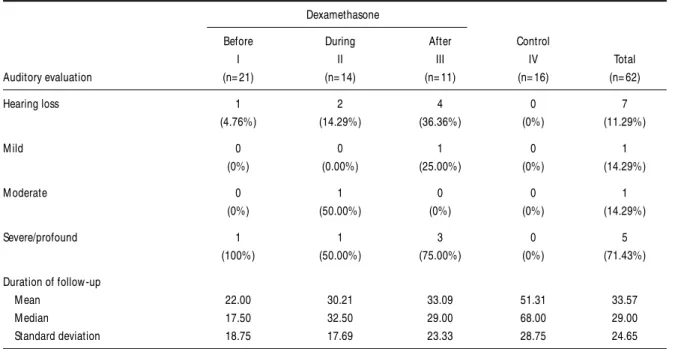
The presence of hearing impairment related to the four ther-apeutic groups is show n in Table 4. Auditory evaluation was performed in 63 children, w ith a mean audiological follow -up of 33.57 months (mean, 29 months); one patient, w ith a pro-found hearing loss in the left ear was excluded from the analy-sis as this problem was already present before hospitalization to treat the meningitis. The statistical analysis did not demon-strate a significant difference between the incidence of dysacou-sia and any therapeutic group.
IQ tests w ere performed in 44 patients. The presence of mental deficiency was found in only two patients, one belong-ing to group I and the other to group III.
Table 5 analyzes the presence of all researched sequelae (neurological, mental and auditory impairment) in the patients studied. From the 74 survivors, 61 children w ere evaluated in the three areas and the presence of some type of alteration was detected in 16 children (26.22%).
con-trol group (group IV), w ith 31.25% presenting sequelae. The evaluation of only the most intense sequelae, more eas-ily attributed to the meningitis and classified according to the method of Sell et al.23as moderate (there w ere no cases of
serious sequelae in this population), occurring in 15% of the patients in group I, 15.38% of the children in group II, 25% of the patients in group III and 12.50% of those in group IV, also did not demonstrate any significant differences betw een the groups. Analyzing the children that received dexametha-sone concomitantly, before or close to the beginning of the
antibiotic therapy (groups I and II), of w hich 18.51% present-ed moderate sequelae and groups I, II and III (23.52% w ith moderate sequelae) also failed to detect any significant dif-ferences from those patients that did not receive dexametha-sone.
DISCUSSION
Despite the experimental demonstration of the therapeu-tic action of dexamethasone in bacterial meningitis, clinical
expe-Table 1. Clinical characteristics at admission of 81 patients with meningococcal meningitis, according to therapeutic group. Dexamethasone
Before During After Control
I II III IV Total
(n= 25) (n= 19) (n= 14) (n= 23) (n= 81)
Feve 24 18 14 22 78
(96.00%) (94.74%) (100%) (95.65%) (96.30%)
M eningeal signs 20 16 12 20 68
(80.00%) (84.21%) (85.71%) (86.96%) (83.95%)
Duration of disease: > 3 days 1 3 2 1 7
(4.00%) (15.79%) (14.29%) (4.35%) (8.64%)
< 1 day 0 0 0 1 1
(0%) (0%) (0%) (4.35%) (1.23%)
Seizures 5 2 3 4 14
(20.00%) (10.53%) (21.43%) (17.39%) (17.28%)
M otor disturbances 0 3 2 1 6
(0.%) (15.79%) (14.29%) (4.35%) (7.41%)
Prostration 13 13 8 11 45
(52.00%) (68.42%) (57.14%) (47.83%) (55.56%)
Coma 0 1 0 1 2
(0.%) (5.26%) (0%) (4.35%) (2.47%)
Hemodynamic instability 3 4 0 8 15
(12.00%) (21.05%) (0%) (34.78%) (18.52%)
Cutaneous signs 18 9 5 17 49
(72.00%) (47.37%) (35.71%) (73.91%) (60.49%)
Herson and Todd gravity score
M ean 1.84 2.08 2.43 2.37 2.15
M edian 2.00 1.75 2.25 2.00 2.00
Standard deviation 1.31 1.78 1.30 1.54 1.48
Turini gravity score
M ean 1.60 1.28 1.86 1.65 1.59
M edian 1.00 1.00 2.00 1.00 1.00
Standard deviation 1.04 1.07 0.77 1.03 1.00
Tesoro and Selbst gravity score
M ean 1.32 1.28 1.00 1.52 1.31
M edian 1.00 1.00 1.00 1.00 1.00
rience in human beings has presented limited results. In spite of the beneficial results reported by some studies in childhood meningitis due to
S. pneumoniae
and mainlyH. influenzae
1,2,4,5,9,there are still no conclusive answers in relation to the action of corticosteroid in decreasing the degree of sequelae in menin-gitis caused by
N. meningitidis
9.The main limitation of the present study lies in the fact that a randomized and blind distribution was not effected. Nevertheless, the patients analyzed came from the same area of the city of São Paulo, w ith a similar distribution among the different therapeutic groups in relation to several parameters, such as nutritional state, age group, socioeconomic class and level of maternal education.Analysis of the laboratory and clin-ical parameters, including indexes for the prognosis of grav-ity, at the moment of hospitalization also demonstrated a homogeneity betw een the various therapeutics groups. Thus, the sample of patients was an appropriate population for evaluating the therapeutic effectiveness of dexamethasone.
Out of the 74 survivors, it was possible to evaluate 69 chil-dren (93.24%) as out-patients for a mean period of 36.97 months after hospital discharge (median = 34.50 months); 63
patients (85.13%) underw ent an audiological assessment for a mean time of 33.57 months (median = 29 months) and 66 children (89.18%) w ere appraised using the IQ tests (WPPSI and WISC) or QD test (Gesell and Amatruda).
At the end of the research, some degree of alteration was detected in relation to motor deficit, epileptic seizures, delayed neuropsychomotor development, behavioral disturbance, learn-ing disturbance, language disorder, mental or hearlearn-ing im-pairments in 16 of the 61 (26.22%) children that w ere sub-mitted to the complete evaluation, including the audiological, psychological and neurological tests. Hence, considering the total sample of 81 patients, 24 (29.62%) coursed w ith some of the above mentioned disturbances or died.
Hearing loss was confirmed in seven patients (11.29%) of the 62 patients assessed. This incidence is in agreement w ith the findings of other studies, in relation to meningococcal meningitis treated conventionally without the use of corticos-teroid24-26.
In the present study, hearing loss was most frequent in group III, in w hich four patients (36.36%) presented dysacousia. On
Table 2. Laboratorial characteristics of the 81 patients, at their admission and according to the therapeutic group. Dexamethasone
Before During After Control
I II III IV Total
(n= 25) (n= 19) (n= 14) (n= 23) (n= 81)
I - Hemogram Hb (g%)
M ean 10.8 9.6 10.3 10.3 10.3
M edian 11.0 10.1 10.3 10.2 10.4
Standard deviation 2.2 2.7 0.9 1.4 2.0
Leukocytes (/mm3)
M ean 15392.0 16508.2 11850.0 14921.7 14867.6
M edian 13600.0 13700.0 8300.0 15000.0 13700.0
Standard deviation 7657.9 8832.8 7959.0 6795.0 7742.0
II – CSF Leukocytes (/mm3)
M ean 6925.3 5620.3 9653.5 3100.8 6041.1
M edian 3820.0 4181.0 2463.5 866.5 2503.0
Standard deviation 11731.4 6417.4 17968.2 6359.1 10958.2
Protein (mg/dl)
M ean 239.5 412.6 265.0 167.8 262.3
M edian 198.0 180.0 259.0 53.0 199.0
Standard deviation 152.0 822.2 146.1 185.8 416.6
Glucose (mg/dl)
M ean 30.0 26.7 20.4 45.5 32.0
M edian 25.0 16.0 7.5 54.0 21.0
comparing the incidence of cases of hearing alteration among the children from group III with those of group I: 4.76%, group II: 14.29%, and group IV: no case, no statistically significant difference was detected at the 5% confidence limit.
Analyzing all the sequelae as a w hole (neurological, audi-tory and cognitive), as show n in Table 5, also did not allow the characterization of any effect from the dexamethasone in the therapeutics for meningococcal meningitis in these patients.
These data demonstrate the absence of benefit from the corticosteroid in terms of decreasing the sequelae, suggest-ing that w ith regard to mensuggest-ingococcal mensuggest-ingitis adjunctive therapy w ith dexamethasone is not so effective as the results of the research by Lebel et al.1, Odio et al.5demonstrated in
relation to
H. influenzae
.Consequently, based on this study and also the absence of proven benefits from the use of corticosteroid in meningo-coccal meningitis in other researches, w e suggest that dex-amethasone should be removed from the therapeutics of patients w ith meningitis, w hen this causal agent is identified. We consider that the pathophysiology of meningitis caused by
N. meningitidis
is similar to that due toS. pneumoniae
andH. influenzae
, how ever w ith a much greater rapidity in trig-gering the chain reaction that eventually causes lesions of the nervous system. Thus, even with precocious therapeutic there is no longer sufficient time available for the action of dexam-ethasone in the sense of reducing the liberation of cytokinesand their consequences, such as: the expression of adhesion glycoproteins betw een the polymorphonuclear and endothe-lial cells; the action of phospholipase A2 and nitric oxide syn-thase, among others27,28. This must be the same reason that
explains the lesser effect of corticosteroid in the clinical trials (even in meningitis caused by
S. pneumoniae
andH.
influen-zae
)1,2,5, in relation to studies and experiments with animalmod-els, in w hich there is greater control of the onset of the infec-tion and administrainfec-tion of dexamethasone29-32.
REFERENCES
1. Lebel MH, Freji GJ, Syrogiannopoulos GA, et al. Dexamethasone ther-apy for bacterial meningitis: results of two double-blind, placebo-con-trolled trials. N Engl J Med. 1988;319:964-971.
2. Girgis NI, Farid Z, Mikhail IA, et al. Dexamethasone treatment for bacte-rial meningitis in children and adults. Pediatr Infect Dis J 1989;8:848-851. 3. Kaplan SL. Dexamethasone for children with bacterial meningitis
should it be routine therapy?AJDC. 1989;143:290-292.
4. Kennedy WA, Hoyt MJ, Mccracken GH Jr. The role of corticosteroid ther-apy in children with pneumococcal meningitis. AJDC.1991;145:1374-1378. 5. Odio CM, Faingezicht I, Paris M, et al. The beneficial effects of early dexamethasone administration in infants and children with bacterial meningitis. N Engl J Med 1991;324:1525-1531.
6. Marguet C, Mallet E. Intérêt de la dexaméthasone au cours des ménin-gites purulentes de lenfants a propos d`une étude comparative chez 85 enfants. Arch Fr Pediatr 1993;50:111-117.
7. Schaad UB, Lips U, Gnehm HE, Blumberg A, Heinzer I, Wedgwood. Dexamethasone therapy for bacterial meningitis in children. Lancet. 1993;342:457-461.
8. Syrogiannopoulos GA, Lourida NA, Theodoridou MC, et al. Dexamethasone therapy for bacterial meningitis in children: 2-versus 4-days regimen. J Infect Dis 1994;169:853-858.
9. Mcintyre PB, Berkey CS, King SM, et al. Dexamethasone as adjunctive
Table 3. Presence of neurological sequelae, according to therapeutic group. Dexamethasone
Before During After Control
I II III IV Total
Neurological sequelae (n= 23) (n= 14) (n= 14) (n= 17) (n= 68)
M otor disabilities 0 1 0 1 2
(0%) (7.14%) (0%) (5.88%) (2.94%)
Epileptic seizures 0 0 0 1 1
(0%) (0%) (0%) (5.88%) (1.47%)
Behavioral problems 1 1 1 1 5
(4.35%) (7.14%) (7.14%) (5.88%) (7.35%)
Learning disabilities 1 1 0 2 4
(4.35%) (7.14%) (0%) (11.76%) (5.88%)
Developmental delay 1 2 1 1 5
(4.35%) (14.29%) (7.14%) (5.88%) (7.35%)
Language disorders 1 1 1 2 5
(4.54%) (8.33%) (11.11%) (11.76%) (7.81%)
Duration of follow -up
M ean 24.83 36.57 33.71 56.41 36.97
M edian 20.00 39.50 34.00 70.00 34.50
therapy in bacterial meningitis: a meta-analysis of randomized clini-cal trials since 1988. JAMA 1997;278:925-931.
10. Coyle PK. Glucocorticoids in central nervous system bacterial infection. Arch Neurol 1999;56:796-801.
11. Sacchi CT, Zanella RC, Caugant DA, et al. Emergence of a new clone of serogroup C Neisseria meningitidisin São Paulo, Brazil. J Clin Microbiol 1992;30:1282-1286.
12. Sacchi CT, Tondella MLC, Lemos APS, et al. Characterization of epi-demic Neisseria meningitidisserogroup C strains in several Brazilian States. J Clin Microbiol. 1994;32:1-5.
13. Centro de Vigilância Epidemiológica. A doença meningocócica: situ-ação atual. http://www.cve.saude.sp.gov.br/htm/inf_meni0711.htm 14. Mustafa MM, Ramilo O, Mertsola J, et al. Modulation of inflammation and cachectin activity in relation to treatment of experimental Haemophilus influenzaetype b meningitis. J Infect Dis 1989;160:818-25. 15. Waagner DC, Hoyt MJ, Finitzo T, Mccracken GH Jr. Administration of dexamethasone before antibiotic therapy in bacterial meningitis in children. N Engl J Med. 1990;322:141-147.
16. Herson VC, Todd JK. Prediction of morbidity in Haemophilus influen-zaemeningitis. Pediatrics 1977;59:35-39.
17. Turini TL, Baldy JLS, Passos JN, Takata PK. Fatores prognósticos da doença meningocócica: estudo relativo a 254 casos. Rev Saúde Públ S Paulo.1979;13:173-182.
18. Tesoro LJ, Selbst SM. Factors affecting outcome in meningococcal infec-tions. AJDC. 1991;145:218-220.
19. Gesell A, Amatruda C. Diagnostico del desarollo normal y anormal del nino. Buenos Aires: Paidos, 1962.
20. Northern JL, Downs NP. Hearing in children. 3.Ed. Baltimore: Willians & Wilkins, 1984.
21. Davis, H. Audiometric: pure tone and simple speech tests. In Hearing and deafness. New York, 3.Ed. Holt 1970:179-220.
22. American Psychiatric Association. Diagnostic and statistical manual of mental disorders (DSM-IV) 4.Ed. Washington, DC: APA Press, 1994. 23. Sell SHW, Merrill RE, Doyne EO, Zimsky EP Jr. Long-term sequelae of
Hemophilus influenzae meningitis. Pediatrics 1972;49:206-211. 24. Edwards MS, Baker CJ. Complications and sequelae of meningococcal
infections in children. J Pediatr 1981;99:540-545.
25. Dodge PR, Davis H, Feigin RD, et al. Prospective evaluation of hear-ing impairment as a sequel of acute bacterial menhear-ingitis. N Engl J Med 1984;311:869-874.
26. Baraff LJ, Lee SI, Schriger DL. Outcomes of bacterial meningitis in chil-dren: a meta-analysis. Pediatr Infect Dis 1993;12:389-394.
27. Rosenstein NE, Perkins BA, Stephens DS, Popovic T, Hughes JM. Meningococcal disease. N Engl J Med 2001;344:1378-1388.
28. Hackett SJ, Thomson APJ, Hart CA. Cytokines, chemokines and other
Table 4. Presence of auditory sequelae, according to therapeutic group.
Dexamethasone
Before During After Control
I II III IV Total
Auditory evaluation (n= 21) (n= 14) (n= 11) (n= 16) (n= 62)
Hearing loss 1 2 4 0 7
(4.76%) (14.29%) (36.36%) (0%) (11.29%)
M ild 0 0 1 0 1
(0%) (0.00%) (25.00%) (0%) (14.29%)
M oderate 0 1 0 0 1
(0%) (50.00%) (0%) (0%) (14.29%)
Severe/profound 1 1 3 0 5
(100%) (50.00%) (75.00%) (0%) (71.43%)
Duration of follow -up
M ean 22.00 30.21 33.09 51.31 33.57
M edian 17.50 32.50 29.00 68.00 29.00
Standard deviation 18.75 17.69 23.33 28.75 24.65
Hearing loss: mild 26 to 40 dB, moderate 41 to 70 dB, severe 71 to 90 dB, profound above 91 dB.
Table 5. Presence of sequelae, according to the modified scale of Sell, according to therapeutic group. Dexamethasone
Before During After Control
I II III IV Total
(n= 20) (n= 13) (n= 12) (n= 16) (n= 61)
Normal 17 10 7 11 45
(85.00%) (76.92%) (58.33%) (68.75%) (73.77%)
Sequelae M ild 0 1 2 3 6
(0%) (7.69%) (16.67%) (18.75%) (9.84%)
M oderate 3 2 3 2 10
effector molecules involved in meningococcal disease. J Med Microbiol 2001;50:847-859.
29. Syrogiannopoulus GA, Olsen KD, Reisch JS, Mccracken GH Jr. Dexamethasone in the treatment of experimental Haemophilus influen-zae type b meningitis. J Infect Dis. 1987;155:213-219.
30. Tuomanen E. Molecular mechanisms of inflammation in experimental pnemococcal meningitis. Pediatr Infect Dis 1987;6:1146-1149.