Clinical features and outcomes of diffuse endocapillary
proliferation Henoch-Scho¨nlein purpura nephritis
in children
Haidong Fu, Jianhua Mao,* Yanping Xu, Weizhong Gu, Xiujuan Zhu, Aimin Liu The Children’s Hospital of Zhejiang University School of Medicine, Department of Nephrology, Hangzhou 310003, China.
OBJECTIVE: To investigate the outcomes of childhood diffuse endocapillary proliferation Henoch-Scho¨nlein purpura nephritis (DEP-HSPN) in response to early diagnosis and prompt treatment.
METHODS:Eleven cases of DEP-HSPN in children were investigated in comparison to HSPN without diffuse endocapillary proliferation (non-DEP-HSPN).
RESULTS: DEP-HSPN had a higher prevalence of nephrotic syndrome but a lower prevalence of hematuria compared to non-DEP-HSPN. IgA, IgG and IgM antibody deposition was found in DEP-HSPN by histopatho-logical examination. Proteinuria cleared in all 11 cases through treatment with steroids and/or immuno-suppressive drugs. However, half of the DEP-HSPN patients continuously had hematuria after treatment.
CONCLUSION: The early diagnosis and prompt initiation of immunosuppressive treatment based on renal biopsy are important for achieving favorable outcomes.
KEYWORDS: Henoch-Scho¨nlein Purpura Nephritis; Diffuse Endocapillary Proliferation; Renal Biopsy.
Fu H, Mao J, Xu Y, Gu W, Zhu X, Liu A. Clinical features and outcomes of diffuse endocapillary proliferation Henoch-Scho¨nlein purpura nephritis in children. Clinics. 2016;71(9):550-554
Received for publication onMarch 11, 2016;First review completed onApril 16, 2016;Accepted for publication onApril 29, 2016 *Corresponding author. E-mail: [email protected]
’ INTRODUCTION
Henoch-Schönlein purpura (HSP) is an immunoglobulin A (IgA)-mediated, systemic small-vessel vasculitis that occurs in children (1). The deposition of IgA antibodies in vessel walls leads to symptoms involving the skin, joints, intestines and kidneys (2). Henoch-Schönlein purpura nephritis (HSPN), which is defined as HSP for 6 months with kidney involve-ment, including hematuria and/or proteinuria, is the most serious complication and often determines the prognosis of an HSP patient (1,2).
The progression of renal involvement is important in evaluating HSPN prognosis and in selecting individualized therapeutic strategies. The clinical manifestations of HSPN are miscellaneous and the pathological features are divergent (3,4). In addition, the severity of the clinical manifestations of HSPN in children does not always correlate with the severity of the pathological renal biopsy findings (3). HSPN with diffuse endocapillary proliferation (DEP-HSPN) may have a different prognosis than HSPN without diffuse endocapillary proliferation (non-DEP-HSPN). Therefore, in the current study,
the pathological features and corresponding clinical mani-festations of DEP-HSPN were compared with those of non-DEP-HSPN in children.
’ METHODS
Subjects
From December 2007 to November 2013, 4212 HSP patients were hospitalized in the Children’s Hospital, School of Medicine, Zhejiang University. Of these patients, 1823 were diagnosed with HSPN and 503 cases were further confirmed by renal biopsy with indications of the following diseases or manifestations: 1) nephritic syndrome; 2) moderate or severe proteinuria; 3) acute nephritis; or 4) rapidly progressive glomerulonephritis. The 503 patients were enrolled into the current study and grouped as DEP-HSPN (n=11) and non-DEP-HSPN (n=492). A written consent form was obtained from each patient’s parents. The study protocol was approved by the Ethics Committee of Zhejiang University. The diagnosis of HSP fulfills the criteria for the classification of childhood vasculitis (5). The inclusion criteria for this prospective study were as follows: 1) patients werep16 years old; 2) patients presented with
typical palpable purpura or petechiae and at least one of the following: arthralgia or arthritis, abdominal pain or renal involvement. The following diseases were excluded: thrombo-cytopenia, post-streptococcus-infection glomerulonephritis, Parvovirus B19 infection, lupus nephritis, ANCA-associated nephritis, hepatitis B-induced nephritis, and coagulopathy.
DOI:10.6061/clinics/2016(09)11
Copyright&2016CLINICS–This is an Open Access article distributed under the terms of the Creative Commons License (http://creativecommons.org/licenses/by/ 4.0/) which permits unrestricted use, distribution, and reproduction in any medium or format, provided the original work is properly cited.
HSPN was defined as the presence of gross or microscopic hematuria (45 erythrocytes per high-power field) with or
without proteinuria (44 mg/kg/d), nephrotic syndrome
(urine protein 450 mg/kg/d, serum albumin o2.5 g/dL,
edema and hyperlipidemia), or acute nephritis (hematuria plus at least one of the following: increased serum creatinine, hypertension, or oliguria).
Histopathology
Kidney biopsies were obtained from all 503 patients and were stained with hematoxylin and eosin (H&E) and for immunofluorescence (IF). IF staining was performed on 3-mm cryostat sections using polyclonal
fluorescein-isothio-cyanate-conjugated antibodies to IgG, IgA, IgM, C3, C4, C1q and fibrinogen according to the manufacturer’s instruc-tions (DakoCytomation, Glostrup, Denmark). The intensity of IF staining was subjectively scored on scale of 0-3. The International Study of Kidney Disease in Children classifica-tion was used to classify HSPN (6).
Statistical analysis
Chi-squared tests or student’s two-tailed t tests were performed.P values less than 0.05 were considered signifi-cant. All data analysis was carried out using the SPSS soft-ware for windows (version 13.0; SPSS, Inc., Chicago, IL).
’ RESULTS
A total of 11 (4 girls and 7 boys) out of the 503 HSPN patients were given a confirmative diagnosis of DEP-HSPN, and the remaining 492 patients were diagnosed with non-DEP-HSPN. All DEP-HSPN patients had typical manifesta-tions of HSP during the clinical visit, including skin rash, abdominal pain and joint symptoms. As shown in Table 1, of the 11 patients, 36.36% (4/11) had edema, 45.45% (5/11) had hypertension, 27.27% had gross hematuria, 72.73% had severe proteinuria (^50 mg/kg/d), 18.18% (2/11) had moderate proteinuria (^25 mg/kg/d, but o50 mg/kg/d),
9.09% (1/11) had mild proteinuria (o25 mg/kg/d), 27.27%
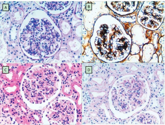
(3/11) had albumin deficiency, and 9.09% (1/11) had acute renal dysfunction. The diagnosis of DEP-HSPN was patho-logically confirmed by kidney biopsy in all 11 patients, and diffuse endocapillary proliferation was easily observed in the cases of DEP-HSPN via H&E staining (Figure 1A) and periodic acid-Schiff (PAS) staining (Figure 1B). In contrast, non-DEP-HSPN was characterized by the significant pro-liferation of mesangial cells, as indicated by H&E staining (Figure 1C) and PAS staining (Figure 1D).
As shown in Table 2, crescent formation was found in 2 of the 11 specimens and affected an average of 1.06% glomeruli (range: 0-7.69%). The clinical impact of crescent formation was not analyzed due to the limited number of cases. Of the 11 cases of DEP-HSPN, 9 were class IIb and 2 were class IIIb.
The IF staining indicated that 3 patients (27.27%) were positive for IgA, 4 cases (36.36%) were positive for IgA and IgG, 2 cases (18.18%) were positive for IgA and IgM, and 2 cases (18.18%) were positive for IgA, IgM, and IgG (Table 2). In addition, C3 deposits were found in 10 out the 11 patients (90.90%) (Table 2).
Compared to non-DEP-HSPN at the IIb stage (43 cases), DEP-HSPN (9 cases) had a higher prevalence of nephrotic syndrome (32.6% of non-DEP-HSPNvs77.8% of DEP-HSPN,
p=0.012) but a lower prevalence of hematuria (60.5% of non-DEP-HSPNvs11.1% of DEP-HSPN,p=0.007, Table 3).
Of the 11 DEP-HSPN patients, 3 patients received methylprednisolone pulse therapy followed by prednisone and cyclophosphamide (CTX), 2 patients received predni-sone plus mycophenolate mofetil (MMF), 3 patients were treated with prednisone plus Tripterygium, 2 patients were treated only with Tripterygium, and one patient was treated only with prednisone. In addition, all 11 patients were given angiotensin-converting enzyme inhibitors. As shown in Table 4, 6 patients still had hematuria after 13-20 months of treatment with MMF alone (3 cases), prednisone alone (1 case), Tripterygium alone (1 case), or methylprednisolone, prednisone, and CTX (1 case). The remaining 5 patients’
urine test results were normal after 7-17 months of treatment with Tripterygium alone (3 cases) or methylprednisolone, prednisone, and CTX (2 cases).
’ DISCUSSION
The histopathological feature of HSP is the deposition of immune complexes on organs such as the skin and glomeruli (7). Glomerular nephritis in HSP patients, known as HSPN, occurs in approximately 33% of pediatric cases and approxi-mately 63% of adult cases (8). The current study reviewed 11 cases of DEP-HSPN and 492 cases of non-DEP-HSPN. Compared to non-DEP-HSPN, DEP-HSPN had a higher prevalence of nephrotic syndrome and IgA, IgG and IgM antibody deposition but a lower prevalence of hematuria. After pulse steroid therapy followed by standard therapy with steroids with or without immunosuppressive drugs, proteinuria disappeared in all 11 cases. However, half of the
Table 1-Clinical presentation of DEP-HSPN patients.
ID # Age (yr) Sex RBT (w) Symptoms HP ED GH PU AD RD
1 5.9 M 4 Severe
2 13.4 M 3 Arthralgia Mild
3 5.4 M 6 + + Severe
4 5.4 M 3 Severe
5 10.8 F 3 Abdominal pain + Moderate
6 10.6 M 9 + Severe +
7 7.9 F 3 + Severe +
8 12.0 F 6 + + Severe +
9 7.7 F 12 Abdominal pain, arthralgia, + Moderate
10 9.1 M 3 Abdominal pain, arthralgia, + + + Severe +
11 9.2 M 2 Arthralgia + Severe
DEP-HSPN patients continuously had hematuria, suggest-ing that hematuria in DEP-HSPN requires a more effective treatment and a longer follow-up period.
Steroid therapy is recommended for HSP patients with severe renal damage (9,10). Patients refractory to steroids may be successfully treated with immunosuppressive agents (11,12). However, there are few reports on the findings of histopathological improvement that demonstrate the effec-tiveness of immunosuppressive therapy for HSPN. In this regard, while 93.9% of pediatric HSPN patients and 89.2% of adult HSPN patients achieve short-term remission (13), the long-term prognosis is not always encouraging, especially in cases of adult onset. A survival rate of 74% and a complete remission rate of only 20% over a median observation period of 15 years were reported by Sherestha et al. (14). Consi-stently, we found that only half of the 11 DEP-HSPN cases
became negative for hematuria in response to the steroid treatment with or without immunosuppressive drugs, suggesting that a longer follow-up period is required in DEP-HSPN patients.
The degrees of crescent formation, interstitial fibrosis and diffuse endothelial cell proliferation are important in predicting HSPN prognosis (14). In this context, chronic proteinuria greater than 1 g/day and/or accompanying nephrotic syndrome predict a poor HSPN prognosis (15). Therefore, it has been recommended that the treatment plan should be determined based on the clearance of or at least a reduction in proteinuria to less than 1 g/day. In the present study, proteinuria was completely cleared in DEP-HSPN patients after treatment with a single reagent or a com-bination of steroids and immunosuppressive drugs, sug-gesting that the strategies used in the current study,
Table 2-Histopathological examination in DEP-HSPN patients.
ID# Immunostaining Stage Crescent Time for clearance of proteinuria (Months)
1 IgA+++, IgM+, C3++ IIIb 1/25 4
2 IgA+++, C3++ IIb 0/11 2
3 IgG+, IgA+++, IgM+-, C3+++ IIb 0/9 4
4 IgG+, IgA++, C3++ IIIb 1/13 5
5 IgA++, C3++ IIb 0/30 1
6 IgA+++, IgM+, C3++ IIb 0/17 5
7 IgG+, IgA+++, C3++ IIb 0/21 4
8 IgG+, IgA+++, IgM+, C3+ IIb 0/5 2
9 IgA++ IIb 0/25 4
10 IgG+-, IgA++, C3+ IIb 0/31 4
11 IgG+, IgA+++, C3++ IIb 0/18 4
especially Tripterygium alone, were effective in reducing proteinuria.
In clinical practice, renal biopsies are rarely used to eval-uate therapeutic efficacy. Furthermore, reducing or dis-continuing an effective therapy in a patient is often carried out without confirming whether the nephritis has improved pathologically. As a result, relapse often occurs in patients who appear to have had a remission, but there is no improvement in their renal histopathology. In this study, repeat renal biopsies were not performed in the 11 cases of DEP-HSPN to monitor renal histopathological improve-ments and guide the therapeutic strategy, which was one of the major limitations of the current study.
The current study describes 11 cases of DEP-HSPN in comparison to cases of non-DEP-HSPN. DEP-HSPN had a higher prevalence of nephrotic syndrome but a lower prevalence of hematuria. Histopathologically, IgA, IgG and IgM antibody deposition was found in DEP-HSPN. After standard treatment with steroids and/or immunosuppressive drugs, proteinuria cleared in all 11
cases. However, half of the DEP-HSPN patients continu-ously had hematuria for as long as 20 months; thus, hematuria in DEP-HSPN needs to be monitored for a longer period.
’ AUTHOR CONTRIBUTIONS
Fu H and Liu A contributed to the conception and design of the study. Mao J and Xu Y contributed to the data acquisition. Gu W and Zhu X contributed to the data analysis. Fu H wrote the manuscript. All of the authors reviewed and approved thefinal version of the manuscript.
’ REFERENCES
1. Rostoker G. Schonlein-henoch purpura in children and adults: diagnosis, pathophysiology and management. BioDrugs. 2001;15(2):99-138, http:// dx.doi.org/10.2165/00063030-200115020-00004.
2. Ting TV. Diagnosis and management of cutaneous vasculitis in children. Pediatr Clin North Am. 2014;61(2):321-46, http://dx.doi.org/10.1016/ j.pcl.2013.11.007.
3. Davin JC, Coppo R. Henoch-Schonlein purpura nephritis in children. Nat Rev Nephrol. 2014;10(10):563-73, http://dx.doi.org/10.1038/nrneph. 2014.126.
4. Mao S, Huang S. Association of AGT M235T gene polymorphism with HSP/HSPN risk. Ren Fail. 2015;37(1):16-21, http://dx.doi.org/10.3109/ 0886022X.2014.977142.
5. Ozen S, Ruperto N, Dillon MJ, Bagga A, Barron K, Davin JC, et al. EULAR/PReS endorsed consensus criteria for the classification of child-hood vasculitides. Ann Rheum Dis. 2006;65(7):936-41, http://dx.doi.org/ 10.1136/ard.2005.046300.
6. Counahan R, Winterborn MH, White RH, Heaton JM, Meadow SR, Bluett NH, et al. Prognosis of Henoch-Schonlein nephritis in children. Br Med J. 1977;2(6078):11-4, http://dx.doi.org/10.1136/bmj.2.6078.11.
7. Kauffmann RH, Herrmann WA, Meyer CJ, Daha MR, Van Es LA. Circulating IgA-immune complexes in Henoch-Schonlein purpura. A longitudinal study of their relationship to disease activity and vascular deposition of IgA. Am J Med. 1980;69(6):859-66, http://dx.doi.org/ 10.1016/S0002-9343(80)80011-8.
8. Rieu P, Noel LH. Henoch-Schonlein nephritis in children and adults. Morphological features and clinicopathological correlations. Ann Med Interne (Paris). 1999;150(2):151-9.
9. Ronkainen J, Koskimies O, Ala-Houhala M, Antikainen M, Merenmies J, Rajantie J, et al. Early prednisone therapy in Henoch-Schonlein purpura: a randomized, double-blind, placebo-controlled trial. J Pediatr. 2006; 149(2):241-7, http://dx.doi.org/10.1016/j.jpeds.2006.03.024.
10. Bluman J, Goldman RD. Henoch-Schonlein purpura in children: limited benefit of corticosteroids. Can Fam Physician. 2014;60(11): 1007-10.
11. Tanaka H, Suzuki K, Nakahata T, Ito E, Waga S. Early treatment with oral immunosuppressants in severe proteinuric purpura nephritis. Pediatr Nephrol. 2003;18(4):347-50, http://dx.doi.org/10.1007/s00467-003-1094-4.
12. Shin JI, Park JM, Shin YH, Kim JH, Kim PK, Lee JS, et al. Cyclosporin A therapy for severe Henoch-Schonlein nephritis with nephrotic syndrome.
Table 4-Treatment and outcome.
Case MP Pulse Therapy Prednisone Others Proteinuria (month) Outcome
1 + MMF 7 RBC, 14/HP after 15 months
2 Tripterygium 2 Normal urine test after 13 months
3 + MMF 4 RBC, 14/HP after 20 months
4 + + CTX 5 Normal urine test after 17 months
5 + Tripterygium 1 Normal urine test after 7 months
6 + + CTX 5 RBC, 10/HP after 15 months
7 + 4 RBC, 8/HP after 19 months
8 + Tripterygium 2 RBC, 6/HP after 17 months
9 Tripterygium 4 Normal urine test after 13 months
10 + + CTX 4 Normal urine test after 15 months
11 + MMF 4 RBC, 15/HP after 13 months
HP: hypertension; MP: methylprednisolone; PN: prednisone; MMF: mycophenolate mofetil; CTX: cyclophosphamide; RBC: red blood cell; HPF: high-power field.
Table 3-Comparison of clinical and pathological presentation between DEP- HSPN (class IIb) and non-DEP-HSPN (class IIb).
DEP-HSPN (n=9)
Non-DEP-HSPN (n=43)
pvalue
Age (years) 9.57±0.81 9.44±0.40 0.896
Gender (M/F) 5/4 31/12 0.328
Abdominal pain 3/9 15/43 0.929
Arthralgia 4/9 19/43 0.989
Simplified proteinuria 0/9 1/43 0.644
Hematuria & proteinuria 1/9 26/43 0.007
Acute nephritis 1/9 2/43 0.450
Nephrotic syndrome 7/9 14/43 0.012
Serum IgA (g/L) 1.47±0.59 1.88±0.72 0.119 Serum C3 (g/L) 1.22±0.17 1.18±0.30 0.707
IgA deposits 3/9 12/43 0.744
IgA+IgG deposits 3/9 7/43 0.238
IgA+IgM deposits 1/9 13/43 0.240
IgA+IgM+IgG deposits 2/9 11/43 0.832
Simplified proteinuria: urine protein44 mg/kg/d, buto50 mg/kg/d. Hematuria and proteinuria:45 RBCs/HPF with urine protein44 mg/kg/d,
buto50 mg/kg/d.
Acute nephritis: hematuria plus at least one of the following: increased serum creatinine, hypertension, or oliguria.
Nephrotic syndrome: urine protein450 mg/kg/d, serum albumino2.5 g/dL,
Pediatr Nephrol. 2005;20(8):1093-7, http://dx.doi.org/10.1007/s00467-005-1864-2.
13. Blanco R, Martinez-Taboada VM, Rodriguez-Valverde V, Garcia-Fuentes M, Gonzalez-Gay MA. Henoch-Schonlein purpura in adulthood and childhood: two different expressions of the same syndrome. Arthritis Rheum. 1997; 40(5):859-64, http://dx.doi.org/10.1002/art.1780400513.
14. Shrestha S, Sumingan N, Tan J, Alhous H, McWilliam L, Ballardie F. Henoch Schonlein purpura with nephritis in adults: adverse prognostic indicators in a UK population. QJM. 2006;99(4):253-65, http://dx.doi.org/10.1093/qjmed/hcl034. 15. Bogdanovic R. Henoch-Schonlein purpura nephritis in children: risk