Arq Neuropsiquiatr 2003;61(3-B):842-847
LEPTOMENINGEAL DISSEMINATION OF PILOCYTIC
ASTROCYTOMA AT DIAGNOSIS IN CHILDHOOD
Two cases report
Eberval Gadelha Figueiredo, Hamilton Matushita, André Guelman G. Machado,
José Píndaro P. Plese, Sérgio Rosemberg, Raul Marino Jr
ABSTRACT - Pilocytic astrocytoma (PA) is a benign tumor that rarely spread along the neuraxis. At the moment there are no more than five cases of leptomeningeal dissemination (LD) from PA at diagnosis described in the literature. Different patterns of presentation or recurrence may be noted: local recurrence, malignant transformation, multicentric disease or metastatic disease. LD and multicentric disease can be distinct pathological entities. We report two cases and analyse literature, emphasizing leptomeningeal spread at presentation. Hydrocephalus, biopsy and parcial ressection are likely to be favorable factors to the occurrence of LD. Otherwise, LD may be part of natural history of PA, as evidenced by its ocurrence in non-treated cases. KEY WORDS: pilocytic astrocytoma, leptomeningeal dissemination, multicentric disease, cerebrospinal fluid, metastasis, radiation therapy, chemotherapy, hydrocephalus.
Disseminação leptomeníngea de astrocitoma pilocítico ao diagnóstico: relato de dois casos. Disseminação leptomeníngea de astrocitoma pilocítico ao diagnóstico: relato de dois casos.Disseminação leptomeníngea de astrocitoma pilocítico ao diagnóstico: relato de dois casos. Disseminação leptomeníngea de astrocitoma pilocítico ao diagnóstico: relato de dois casos.Disseminação leptomeníngea de astrocitoma pilocítico ao diagnóstico: relato de dois casos.
RESUMO - Astrocitoma pilocítico (AP) é tumor benigno que raramente se dissemina ao longo do neuroeixo. Até o momento não há mais que cinco casos de AP que se tenham apresentado com disseminação leptomeníngea (DL) descritos na literatura. Diferentes padrões de apresentação ou recorrência podem ser observados: recorrência local, transformação maligna, doença multicêntrica ou doença metastática. DL e doença multicêntrica podem ser entidades diferentes. Relatamos dois casos e analisamos a literatura. Hidrocefalia, biópsia e ressecção parcial são provavelmente fatores predisponentes à DL. Por outro lado, DL pode ser parte da história natural de AP, como pode ser evidenciado pela sua ocorrência em casos não tratados. PALAVRAS-CHAVE: astrocitoma pilocítico, disseminação leptomeníngea, doença multicêntrica, líquor, metástases, quimioterapia, radioterapia.
Neurosurgery Division, Clinics Hospital, School Of Medicine, São Paulo University, São Paulo SP, Brazil. Received 10 February 2003, received in final form 14 April 2003. Accepted 12 May 2003.
Dr. Eberval Gadelha Figueiredo - Rua Alves Guimarães, 485/141 - 05410-000 São Paulo SP - Brasil. E-mail: [email protected]
Pilocytic astrocytoma (PA) is a benign subset of gliomas with an excelent prognosis1 and that rarely spread along the neuraxis. Leptomeningeal dissemi-nation (LD) of primary central nervous system (CNS) tumors in children has been reported mainly in ependymomas, germ-cell tumors, primitive neu-roectodermal tumors, high grade gliomas2-9. Low grade gliomas dissemination has been documented in few cases1,2,5,6,9. Leptomeningeal dissemination of pilocytic astrocytoma in children is much more uncommon with 32 cases reported in the litera-ture1,2,6,7,9-24. These include only five with LD at diag-nosis13,17-19. Dissemination patterns, clinical picture, treatment and outcome are still poorly understood. We reported two cases evaluated by the child neurosurgery team at Hospital das Clínicas of São
Paulo University Medical School that presented LD at diagnosis, enhancing clinical, diagnoses, manage-ment and prognosis features.
CASES
dischar-Arq Neuropsiquiatr 2003;61(3-B) 843
ged without symptoms. Radiotherapy was proposed but it was refused by her relatives. Four months after discharge, the patient returned with poor general appereance, cons-ciousness impairement, fever, stiff neck and generalized spasticity. CT scan showed diffuse sub-arachnoid enha-cement (Fig 3). CSF culture was negative and cytologic examination revealed important pleocytosis with neoplastic cells. Death occured in a few weeks.
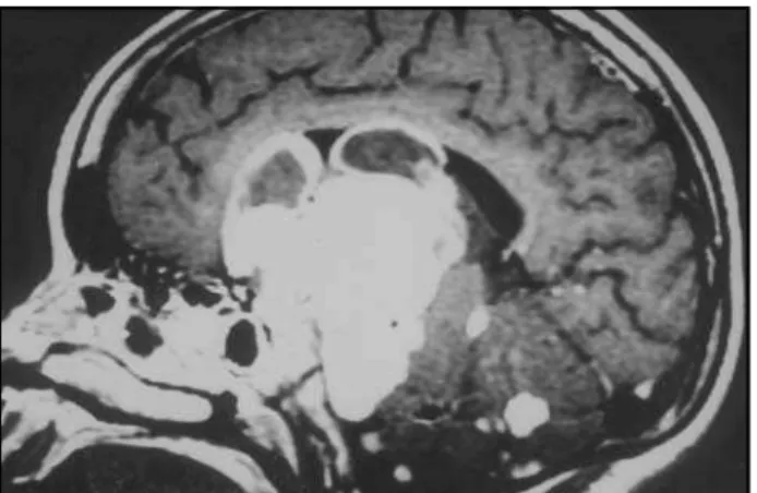
Case 2. In February 1994, an eight years old girl was admitted with a four years history of headache, vomiting and visual acuity deterioration. At that period she mentioned stiff neck and occipital pain; neurologic exa-mination showed axial and apendicular cerebellar symp-toms, amaurosis and meningismus. The optic disks were atrophic. A hypothalamic syndrome ( Russell syndrome ) was present and nutritional status was precarious.
Mag-netic resonance image (MRI) with gadolinium revealed a lesion with cystic and solid parts at the optic nerve and chiasma with multiple tumoral implants on the subarach-noid space and hydrocephalus (Fig 4). Ventriculoperitoneal shunting and stereotactic puncture of the cyst were perfor-med. Histological examination revealed similar features to previous case. Patient underwent radiation therapy (RDT) and was discharged. Surgery was delayed due to poor clinical conditions. Infectious complications caused death after months.
DISCUSSION
Although PA has usually a benign course, it can eventually show a malignant behavior. Different pat-terns of presentation or recurrence may be noted: local recurrence, malignant transformation, multi-centric disease or metastatic spread1,6,25-30. Contro-versy exists about multicentric disease definition. Ma-melak defined it as either diffuse subependymal or leptomeningeal dissemination beyond the margins of the primary tumor or as discrete nodular disease separate from the primary tumor mass1. However, multiplicity of PAs might be due to the multiple ge-nesis of the tumor instead of tumor spread to the subarachnoid space12. LD and multicentric disease can be distinct pathological entities with different pathogenesis and perhaps different clinical features.
LD of PA is uncommon with only few cases repor-ted in the literature. At the moment we can find just
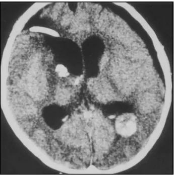
Fig 1. Contrast-enhanced CT scan shows two lesions in ventricular spaces, at right frontal and at left horns, and hydrocephalus.
Fig 2. Histological appearance of pilocytic astrocytoma showing fusiform cells with wavy fibrillary processes and Rosenthal fibers.
8
4
4
Arq Neuropsiquiatr 2003;61(3-B)
TABLE 1. LEPTOMENINGEAL DISSEMINATION IN CHILDREN WITH PILOCYTIC ASTROCYTOMA
AUTHOR AGE AT SITE OF PREDISSEMINATION TIMING THERAPY OUTCOME INITIAL PRIMARY PROCEDURE OF LD FOR LD
DIAGNOSIS TUMOR
MCLAUGHIN (1976) 08 Ys III VENTRICLE - - - DIED
13 Ys III VENTRICLE - - - DIED
6,5 Ys III VENTRICLE - - - DIED
18 Ys III VENTRICLE - - - DIED
AUER (1981) 8 Ys CEREBELLUM SURGERY + LOCAL XRT 10 Ds - DIED CIVITELLO (1988) 8 Ys CEREBELLUM SURGERY 6 Ys CHT + CS XRT ALIVE 1 Ys 5 Mos
4 Ys 9 Mos CHIASM BPS + LOCAL XRT 3Ys 5Mos SPINAL XRT DIED 1 Ys 9 Mos KOCKS (1989) 10 Ys CHIASM SURGERY 2 Ys XRT ALIVE 1Ys BRUGGERS (1991) Mos CHIASM SURGERY AT DIAGNOSIS CHT ALIVE OBANA (1991) 16Mos CHIASM SURGERY 8, 5 Ys CHT ALIVE 1,5Ys MISHINA 6 Ys CEREBELLUM SURGERY+ LOCAL XRT 6 Ys CS XRT ALIVE 2Ys VERSARI 7 Ys CHIASM SURGERY+LOCAL XRT 3 Ys PARCIAL RES. DIED 48 Mos POLLACK (1994) 5 ½ Ys CEREBELLUM AT DIAGNOSIS XRT ALIVE MAMELAK (1994) 3 Ys HYPOTHALAMIC SURGERY +CHT - CS XRT ALIVE 34 Mos
9 Mos HYPOTHALAMIC SURGERY +CHT - CHT ALIVE 20 Mos 16 Mos HYPOTHALAMIC SURGERY+ XRT - CHT ALIVE 4 Ys
Arq Neuropsiquiatr 2003;61(3-B)
8
4
5
GAJJAR (1995) 3 Ys HYPOTHALAMIC BPS - CHT
-1 6 Ys HYPOTHALAMIC BPS - RT
-11 Ys TEMPORAL LOBE SURGERY - PARCIAL RES. -MCCOWAGE (1996) 8Ys III VENTRICLE SURGERY + XRT 2 Ys CHT ALIVE 27 Mos
5 Ys CEREBELLUM SURGERY 3 Ys CHT ALIVE 10 Mos 2 ½ Ys III VENTRICLE SURGERY 8 Mos CHT ALIVE 3 Ys
7 Ys CHIASM BIOPSY AT DIAGNOSIS CHT ALIVE 6 Mos
MORIKAWA (1997) 4 Ys CEREBELLUM BPS AT DIAGNOSIS SURGERY ALIVE
JAMJOOM (1998) - CEREBELLUM VPS 2Ys -
-TAMURA M (1998) 6Ys CEREBELLUM SURGERY 4Ys SURGERY+ CS XRT ALIVE 5 Ys 4Ys CHIASM SURGERY+XRT 4Ys SURGERY+CHT+CS XRT -PATT S (1999) 16 Ys - SURGERY 4Ys CHT TUMOR STABLE
AKAR (2000) - CHIASM - AT DIAGNOSIS -
-FELLGIEBEL (2001) 14Ys - SURGERY 8Ys - -FIGUEIREDO (2003) 11Ys FRONTAL AND OCCIPITAL - AT DIAGNOSIS SURGERY DIED 06 Mos
VENTRICULAR CORNS
08 Ys CHIASM - AT DIAGNOSIS SP + BPS DIED 05 Mos
846 Arq Neuropsiquiatr 2003;61(3-B)
32 cases reported. Incidence of LD of PA is not still determined. Since the use of MRI has become routine in surveying of patients with brain tumors, a sharp increased in number of cases has been noted. Ma-melak, analising his series with 90 cases of PA, found an incidence of 12%, including cases of multicentric disease1. Gajjar, reviewing low-grade astrocytomas found an incidence of 5,3%2. Civitello and associates described 162 patients with low-grade gliomas; six patients (3.7%) had leptomeningeal spread6. Pollack found an incidence of 4% of dissemination of low grade intracranial astrocytomas17.
Spread mechanisms are not well understood. Tu-mors of any grade arising nearby to CSF pathways may disseminate. Tumoral mass located in the floor of the third ventricle may breach the ependyma and invade ventricular cavity, resulting in to ependymal or leptomeningeal seeding. This is not, however, the only important factor. Tumor cells are able of crossing piamater and invade CSF pathways; their degree of adhesiveness, their metabolic capacities (adhesion molecule production, protease secretion and growth factor pathway activation) and their antigenic factors also may aid leptomeningeal spread1,2,3,5,27. A study with CD44 suggests that this adhesion molecule may play a role in astrocytic invasion and adherence31. Probably hydrocephalus, biopsy and parcial ressec-tion may also be addiressec-tional favorable factors, although this remains unproven1. When primitive tumor does not arise nearby ventricular spaces, disaggregation of tumoral cells and their entry into the Virchow-Robin space is likely to be the responsible mechanism15.
LD can occur after a long postoperative period as well as at diagnosis. In fact, LD may be the first sign of disease or of relapse. Clinically, LD can be presented by hidrocephalus, meningismus,
worse-ning of focal deficits, other neurologic deficits and onset of seizures. However, some cases are asymptomatic1,23.
CSF examination provides unreliable results. In subarachnoid metastatic disease in children, if a single CSF sample is employed, nearly 50% of the patients will not be diagnosed32,33. In evaluating 17 children with primary intracranial neoplams for suba-rachnoid metastatic disease, Kramer found CSF cyto-logic examination positive in only 29% of the ca-ses34. The use of CSF tissue culture can improve stan-dard cytology techniques and almost double the rate of successful diagnosis35. Myelography with CT follow-up and MRI with gadolinium was positive in 47% and 65% of the cases, respectively34. Thus, MRI with ga-dolinium seems provide better sensibility and accuracy for LD diagnosis than other diagnostic methods.
Table 1 summarizes related cases. The average age at initial diagnosis of the primary tumor was six years and nine months. Most of the patients were males. Location of primary tumor was in hypothala-mus, optic pathways and third ventricle (n=22), cere-bellum (n=8) and only one case in the temporal lobe and another in lateral ventricles. Seven patients pre-sented LD at diagnosis (including our patients), what shows that LD may be part of natural history of this tumor. Surgical removal of the primary tumor was total subtotal in most of the cases. Biopsy was employed in 7 patients and RDT and/or chemothe-rapy were used in 8 and 3, respectively. Only two patients who were submitted to total removal before LD diagnosis and developed subarachnoid spread staied alive during follow-up. Thus total removal may be favorable factor in avoiding dissemination and improving prognosis. However, adjuvant therapy seemed not to change the odds for tumor spread. At histologic examination, there was no pathologic caracteristic sign that allowed prediction of lepto-meningeal spread.
LD was treated with chemotherapy (n=14), cra-niospinal RDT (n= 8) , subtotal resection of primary masses (n=7). There were nine deaths during follow-up, of whose only 2 underwent chemotherapy. McCowage reported treatment of four patients with high dose of cyclofosfamide, including intrathecal therapy, demonstrating three significant tumor responses and one prolonged disease estabilization18. These data, however, are not conclusive . RDT seemed not affect prognosis.
We are not certain about which factors are pre-dictive to leptomeningeal spread. Mamelak et al.
Arq Neuropsiquiatr 2003;61(3-B) 847
found that hypothalamic tumors have a high-risk. However, it seems impossible to distinguish the role played by the site of tumor from that played by the extent of surgical resection, tumor location, hydro-cephalus, patient age at diagnosis and type of therapy. Nonetheless, it was possible to draw a pro-file of the patient at highest risk to developing LD: a child under 4 years old with a primary PA in the hy-pothalamic region treated with subtotal resection or biopsy followed by adjuvant therapy1.
The outcome of patients with LD is not well known. Nevertheless, it is likely to be not as good as that of patients with localized recurrence or totaly resected primary disease. However, it is not as bad as the leptomeningeal spread in high-grade gliomas.
In summary, at this time is not possible to predict patients whom will be diseminated disease. More cases are necessary to define optimal treatment and prognosis of this entity. In our opinion, total resection must be performed as often as possible and no adju-vant therapy should be carried out. Children with hypothalamic tumors, subtotal resection, hydroce-phalus or those submitted to adjuvant therapy should be followed with cytologic examination of the CSF, which can be also obtained before surgical resection of primary tumor. MRI with gadolinium should also be used during follow-up to allow early diagnosis of dissemination. Radiotherapy and che-motherapy can be used for LD treatment, the second with better support from the literature18,23,24. Further controlled studies will define the risks factors, opti-mal treatment of primary and disseminated disease and prognosis of this rare pathological entity.
REFERENCES
1. Mamelak NA, Prados MD, Obana WG, Cogen PH, Edwards MSB. Treatment options and prognosis for multicentric juvenile pilocytic astrocytoma. J Neurosurg 1984;81:24-30.
2 Gajjar A, Bhargava R, Jenkins JJ, et al. Low-grade astrocytoma with neuraxis dissemination at diagnosis. J Neurosurg 1995;83:67-71. 3 Kellie SJ, Kovnar EH, Kun LE, et al. Neuraxis dissemination in pediatric
brain tumors:response to preirradiation chemotherapy. Cancer 1992;69:1061-1066.
4. Pezeshhkpour GH, Henry JM, Armbrustmacher VW. Spinal matastases: a rare mode of presentation of brain tumors. Cancer 1984;54:353-356. 5. Ushio Y, Arita N, Hayakawa T, et al. Leptomeningeal dissemination of
primary brain tumors in children: clinical and experimental studies. Prog Exp Tumor Res 1987;30:194-205.
6. Civitello LA, Packer RJ, Rorke LB, Siegel K, Sutton LN, Schut L. Leptomeningeal dissemination of low-grade gliomas in childhood. Neurology 1988;38:562-566.
7. Bell WO, Packer RJ, Seigel K, et al. Leptomeningeal spread of intramedullary spinal cord tumors: report of three cases. J Neurosurg 1988;69:295-300.
8. Arseni C, Horvath L, Carp N, et al. Spinal dissemination following operation on cerebral oligodendroglioma. Acta Neurochir (Wien)1977;37:125-137.
9. Packer RJ, Siegel KR, Sutton LN, Litmann P, Bruce DA, Schut L. Leptomeningeal dissemination of primary central nervous system tumors of childhood. Ann Neurol 1985;18:217-221.
10. McLaughlin JE. Juvenile astrocytomas with subarachnoid spread. J Pathol 1976;118:101-107.
11 Auer RN, Rice GPA, Hinton GG, Amacher AL, Gilbert JJ. Cerebellar astrocytoma with benign histology and malignant clinical course: case report. J Neurosurg 1981;54:128-132.
12. Kocks W, Kalff R, Reinhardt V, Grote W, Hilke J. Spinal metastasis of pilocytic astrocytoma of the chiasma opticum. Childs Nerv Syst 1989;5:110-118.
13. Bruggers CS, Friedman HS, Phillips PC, et al. Leptomeningeal dissemination of optic pathway gliomas in three children. Am J Ophthalmol 1991;111:719-723.
14. Obana WG, Cogen PH, Davis RL, Edwards MS. Metastatic juvenile pilocytic astrocytoma: case report. J Neurosurg 1991;75:972-975. 15. Mishina K, Nakamura M, Nakamura H, Nakamura O, Funata N, Shitara
N. Leptomeningeal dissemination of cerebellar pilocytic astrocytoma: case report. J Neurosurg 1992;77:788-791.
16. Versari P, Talamonti G, D’Aliberti G, Fontana R, Colombo N, Cascadei G. Leptomeningeal dissemination of juvenile pilocytic astrocytoma: case report. Surg Neurol 1994;41:318-321.
17. Pollack IF, Hurtt M, Pang D, Albright AL. Dissemination of low grade intracranial astrocytomas in children. Cancer 1994;73:2869-2878. 18. Mccowage G, Tien R, McLendon R, et al. Successful treatment of
childhood pilocytic astrocytomas metastatic to the leptomeninges with high-dose cyclophosphamide. Med Ped Oncol 1996;27:32-39. 19. Morikawa M, Tamaki N, Kokunai T, et al. Cerebellar pilocytic
astrocytoma with leptomeningeal dissemination: case report. Surg Neurol 1997;48:49-52.
20. Fellgiebel A, Erbguth F, Neundorfer B. Polyradiculopathy and ataxia: clinical manifestation of late recurrence of pilocytic astrocytoma with cerebral and spinal dissemination. Nervenarzt 2001;72:143-146. 21. Jamjoom AB, Jamjoom ZA, al-Rayess M. Intraventricular and
leptomeningeal dissemination of a pilocytic cerebellar astrocytoma in a child with a ventriculoperitoneal shunt: case report.Br J Neurosurg 1998 ;12:56-58.
22. Akar Z, Tanriover N, Kafadar AM, Gazioglu N, Oz B, Kuday C. Chiasmatic low-grade glioma presenting with sacral intradural spinal metastasis. Childs Nerv Syst 2000;16:309-311.
23. Patt S, Haberland N, Graupner H, Schreiber D, Kalff R. 16 years old male with an unexpected MRI finding. Brain Pathol 1999;9:743-744. 24. Tamura M, Zama A, Kurihara H, et al. Management of recurrent
pilocytic astrocytoma with leptomeningeal dissemination in childhood. Childs Nerv Syst 1998;14:617-622.
25. Russell DS, Rubinstein LJ. Pathology of tumors of the central nervous system 5.Ed. London : Butleeer & Tanner, 1989:421-428.
26. Shapiro K, Shulman K. Spinal cord seeding from cerebellar astrocytomas. Childs Brain 1976;2:177-186.
27. Garcia DM, Fulling KH . Juvenile pilocytic astrocytoma of the cerebrum in adults: a distinctive neoplasm with favorable prognosis. J Neurosurg 1985;63:382-386.
28. Gjerris F, Klinken L. Long-term prognosis in children with cerebellar astrocytoma. J Neurosurg 1978;49:179-184.
29. Haddad SF, Menezes AH, Bell WE , Godersky JC, Afifi AK, Bale JF . Brain tumors occuring before 1 year of age: a retrospective review of 22 cases in an 11-year period (1977-1987). Neurosurgery 1991;29:8-13. 30. Wallner KE, Gonzales MF, Edwards MSB, Wara WM, Sheline GE. Treatment results of juvenile pilocytic astrocytoma. J Neurosurg 1988;69:171-176.
31. Cooper DL, Dougherty GJ To metastasize or not? Selection of CD44 splice slices. Nat Med 1995; 1: 635-637.
32. Wilkins RH, Odom GL. Cytological changes in cerebrospinal fluid associated with resections of intracranial neoplasms. J Neurosurg 1966;25:24-34.
33. Balhizen JC, Bots GTAM, Schaberg A, Bosman FT. Value of cerebrospinal fluid cytology for the diagnosis of malignancies in the central nervous system. J Neurosurg 1978;48:747-753.