Arq Neuropsiquiatr 2003;61(2-B):459-462
CAVERNOUS CAROTID ARTERY PSEUDO-ANEURYSM
TREATED BY STENTING IN ACROMEGALIC PATIENT
Jorge Marcondes de Souza¹, Flavio S. Domingues¹,
Gaudencio Espinosa², Monica Gadelha³
ABSTRACT - We report on a case of endovascular management of pseudoaneurysm of the cavernous segment of the internal carotid artery with covered stent reconstruction. A 36 years-old woman with a history of previous transsphenoidal approach for pituitary macroadenoma and false aneurysma formation was studied in a protocol that included balloon test occlusion and cerebral blood flow evaluation. An endovascular covered stent deployment in the area of the carotid laceration was performed with isolation of the aneurysm from the circulation and maintenance of the carotid flow. Helical angio-CT and cerebral digital subtraction angiography showed the carotid preservation without stenosis in the stented area. In conclusion, endovascular stent reconstruction for post-transsphenoidal carotid artery laceration and false aneurysm is demonstrated as useful technical adjunct in the management strategy and with the potential for carotid sacrifice morbidity avoidance.
KEY WORDS: cerebral aneurysms, intravascular stents, transsphenoidal surgery, pituitary tumor.
P PP
PPseudoaneurisma da artéria carótida cavernosa tratado com “stent” em paciente acromegálicaseudoaneurisma da artéria carótida cavernosa tratado com “stent” em paciente acromegálicaseudoaneurisma da artéria carótida cavernosa tratado com “stent” em paciente acromegálicaseudoaneurisma da artéria carótida cavernosa tratado com “stent” em paciente acromegálicaseudoaneurisma da artéria carótida cavernosa tratado com “stent” em paciente acromegálica
RESUMO - Relatamos um caso de manuseio com “stent” recoberto por pseudoaneurisma do segmento cavernoso da artéria carótida interna. A paciente de 36 anos, tinha história de cirurgia trans-esfenoidal para macroadenoma de hipófise e desenvolvimento de falso aneurisma na região cavernosa da ACI, foi estudado com protocolo para avaliação de reserva circulatória carotídea com teste de oclusão por balão e estudo de fluxo sanguíneo cerebral com tomografia computorizada de emissão de fóton único (SPECT). Instalação de “stent” recoberto no segmento lesado isolou o aneurisma da circulação, com manutenção do fluxo carotídeo. Angio-tomografia helicoidal e angiografia digital por subtração demonstraram a reconstrução carotídea sem estenose local. Em conclusão, reconstrução carotídea com “stent” recoberto é possível na estratégia para manuseio de pseudo-aneurisma com potencial para prevenção da morbidade do sacrifício terapêutico carotídeo.
PALAVRAS-CHAVE: stent intravascular, cirurgia trans-esfenoidal, pseudo-aneurisma, tumor hipofisário.
¹Service of Neurosurgery, Department of Surgery, Hospital Universitário Clementino Fraga Filho / Faculdade de Medicina / Universidade Federal do Rio de Janeiro (HUCFF/FM/UFRJ), Rio de Janeiro RJ, Brazil; ²Service of Vascular Surgery, Department of Surgery, HUCFF/FM/ UFRJ; ³Service of Endocrinology, Department of Internal Medicine, HUCFF / FM / UFRJ.
Received 4 October 2002, received in final form 6 January 2003. Accepted 18 January 2003.
Dr. Jorge Marcondes de Souza - Serviço de Neurocirurgia / Hospital Universitário Clementino Fraga Filho - Avenida Brigadeiro Trompowski s /n - 21941-590 Rio de Janeiro RJ - Brasil. E-mail: [email protected]
Vascular injury can be one of the most serious
com-plications associated to the surgery of the
sphenoid-sellar region. Cavernous internal carotid artery (ICA)
injury is uncommon during transsphenoidal surgery
and may result in carotid-cavernous fistula or
pseudo-aneurysm with a reported incidence of 0 to 1.2%
1.
The most common presentation is massive
blee-ding during the operation, usually controlled by
na-sal packing. The resulting false aneurysm does not
have a real wall and its limits are formed by organized
clot. Its natural history is not entirelly defined, some
authors show expansion of those lesions without a
timing pattern
2,3. Most pseudoaneursms following
transsphenoidal operation described in the literature
were associated to growth hormone (GH) secreting
tumors and it is intriguing that acromegalic patients
also have tendency to develop intracranial elongated
and ectatic arteries
4-6.
CASE
hyper-460 Arq Neuropsiquiatr 2003;61(2-B)
tension or cardiac symptoms. There was no headache. The neurological examination was essencially normal as well as her campimetric test. Laboratory tests including her hormonal profile and coagulation studies were unremar-kable except for increased serum level (13 ng/dl) of the GH and abnormal glucose tolerance curve.
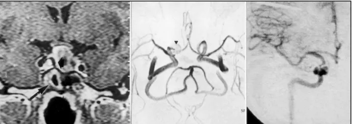
Magnetic resonance imaging (MRI) revealed a para-sellar mass next to the right cavernous ICA, invading the sellar compartment and with a of flow void rim. A macroa-denoma with supra-sellar extension and optic chiasm dis-tortion was clearly demonstrated close to the lesion sur-rounded by a layer of hyperintense signal. There was also enhancement of the adjacent sphenoid sinus wall (Fig 1.A). Magnetic ressonance angiography (MRA) showed an ima-ge sugima-gestive of an aneurysmal dilatation at the level of the right cavernous ICA ( Fig 1.B). A digital subtraction angiography (DSA) of the right ICA confirmed the right
cavernous carotid irregular aneurysm and a stenotic su-pra-aneurysmal segment (Fig 1.C).
After discussion of risks, treatment alternatives and potential complications, the patient consented with the right internal carotid artery colateral flow evaluation with balloon test occlusion (BTO) SPECT in order to stablish the most apropriate management for the aneurysm.
Procedures
She was neurologically intact during the BTO but sho-wed moderate assymetry on the right side on SPECT ima-ging. It was decided for the stenting of the arterial segment harboring the false aneurysm as the best option for her case as opposed to the carotid definitive occlusion. Under general endotracheal anesthesia and systemic heparini-zation, a 7-French sheath (Cordis Endovascular, Miami Lakes, FL) was advanced into the ICA and an extra-stiff 14
Fig 2. A. Helical Angio-CT showing the stent at the level of the cavernous ICA in the coronal section. B. Sagittal section of the helical angio-CT with the stent in place. C. Control digital angiography with normal carotid flow and without disappearance of the pseudoaneurysm.
Arq Neuropsiquiatr 2003;61(2-B) 461
microguidewire (Guidant, Santa Clara, USA) was posi-tioned beyond the lesion. A 4 x 9 mm coronary stent-graft (JOMED, Helsingberg, Germany) mounted on its 4 x 15 mm delivery balloon (Guidant, Santa Clara, USA) was navigated across the region of the aneurysm. The balloon was inflated and the stent deployed with complete exclu-sion of the aneurysm from the circulation.
Heparin was not reversed and the patient was asym-ptomatic afterwards. She was placed on ticlopidine (Ticlid; Roche Pharmaceuticals, Nutley, NJ), 250 mg twice daily for a 4-week period. Follow-up helical-CT angiogram and DSA (Fig 2 A, B and C) one month later showed good flow in the stented area, without recanalization of the aneurysm. After ticlopidine interruption she was submitted to a sublabial transsphenoidal reoperation, when became obvious that the previous operation was a transnasal paramedian transs-phenoidal approach with a right lateral entry site into the sphenoid sinus. We ressected the midline bony bridge that divided her sphenoid sinus and completed the adenoma ressection uneventfully. She was discharged on the fifth post-operative day without intercorrence.
DISCUSSION
Transsphenoidal approach is a safe and accepted
procedure for pituitary lesions, being critical to be
aware of the relationships of the carotid artery with
the sphenoid sinus walls as well as the sela turcica
boundaries.During the operation, the midline
orien-tation remains one of the cornerstones to the route
toward the selar floor
7-9. A computerized
tomogra-phy with bone window may be important in case of
any doubt about variations in the septation of the
sphenoid sinus. False aneurysm from carotid lesion
during transsphenoidal access for pituitary lesions
can be a life-threatening complication. After arterial
bleeding controlled, those patients should undergo
DSA in order to understand the magnitude of the
lesion and to plan the treatment strategy.
Endovascu-lar balloon occlusion of the ICA has been offered as
advantageous over trapping procedures minimizing
the chances of thromboembolic accidents and the
morbidity associated with intracranial
procedu-res
4,7,10. Bavinski et al. described seven cases of
trau-matic false aneurysm of the intracavernous ICA
ma-naged by balloon occlusion at the level or just bellow
to the lesion and advocate it as a very effective and
safe method of treatment
10. Long-term
complica-tions of the permanent carotid artery occlusion
sho-uld be considered. Several studies found the 5 to
10% of delayed infarction after therapeutic carotid
occlusion in spite of normal BTO results
11,12. Awad
et. al showed de novo appearance of subcortical
hyperintense lesions on MRI after therapeutic ICA
occlusion even in patients that tolerated ICA
occlusion with maximal prophilaxis against
thrombo-embolism
13. Link et al. described the experience of
total follow up for sixty patients of 468 patient-years
with four delayed infarcts after permanent carotid
occlusion
14. Endovascular treatment using electrolytic
detachable coils have been described and Lempert
et al had two good outcome but one neck refilling
afterwards
15. The MRI and DSA evaluation of our case
showed two well known disadvantages of using coils
alone: 1) a wide neck aneurysm, due to the risk of
bulging of them into the parent artery with occlusion
of the vessel; 2) the possibilities of dislodging
intra-lesional thrombus and promote distal embolization.
The current endovascular technology includes the
use of endovascular constructive approaches that
could preserve the parent artery either by stent
deploy-ment or concurrent use of stents and detachable coils
for complex intracranial vascular diseases
16-19.
The placement of a covered intravascular stent
within the parent artery across an aneurysm opening
promotes immediate stasis and thrombosis inside
the lesion. The endoluminal reconstruction avoids
the carotid sacrifice and the chances of distal
embo-lization. Long term effects of stenting of the
intra-cranial arterial segments are currently unknown and
myointimal hyperplasia and stenosis remains as a
concern for possible future hemodynamic
compro-mise of the distal circulation, although vessels with
larger diameters have a lower rate of significant
stenosis than smaller ones
17,20.
In conclusion, endovascular covered stent
re-construction for post-transphenoidal carotid artery
laceration and false aneurysm is demonstrated as
useful technical adjunct in the management strategy
and with the potential for carotid sacrifice morbidity
avoidance. To our knowledge, there has been no
pre-vious description of isolated carotid covered stenting
for this clinical situation.
REFERENCES
1. Ciric I, Ragin A, Baumgartner C, Pierce D. Complications of transsphenoidal surgery: results of a national survey, review of the literature and personal experience. Neurosurgery 1997;40:225-237. 2. Amirjamshidi A, Rahmat H, Abbassioun K. Traumatic aneurysms and
arteriovenous fistulas of intracranial vessels associated with penetrating head injuries ocurring during war: principles and pitfalls in diagnosis and management. J Neurosurg 1996;84:769-780.
3. Tokunaga K, Kusaka N, Nakashima H, Date I, Ohmoto T. Coil embolization of intradural pseudoaneurysms caused by arterial injury during surgery: report of two cases. AJNR Am J Neuroradiol 2001;22:35-39.
4. Cabezudo JM, Carrilo R, Vaquero J, Areitio E, Martinez R. Intracavernous aneurysm of the carotid following transsphenoidal surgery. J Neurosurg 1981;54:118-121.
5. Wilson CB, Dempsey LC. Transsphenoidal microsurgical removal of 250 pituitary adenomas.J Neurosurg 1978;48:13-22.
462 Arq Neuropsiquiatr 2003;61(2-B)
7. Ahuja A, Guterman LR, Hopkins LN. Carotid cavernous fistula and false aneurysm of the cavernous carotid artery: complications of transsphenoidal surgery. Neurosurgery1992;31:774-779.
8. Paullus WS Jr, Norwood CW, Morgan HW. False aneurysm of the cavernous carotid artery and progressive external ophtalmoplegia after transsphenoidal hypophysectomy. J Neurosurg 1979;51:707-709. 9. Wright DC. Transsphenoidal approach to sellar and sphenoidal regions.
In Sekhar LN, de Oliveira E, (eds). Cranial microsurgery - approaches and techniques. New York: Thieme, 1999:246-259.
10. Bavinzki G, Killer M, Knosp E, Ferraz-Leite H, Gruber A, Richling B. False aneurysm of the intracavernous carotid artery: report of 7 cases. Acta Neurochir (Wien) 1997;139:37-43.
11. Lee S, Awad AI. Therapeutic carotid occlusion: current management paradigmas. Clin Neurosurg 1998;46:363-391.
12. Segal DH, Sen C, Bederson JB, et al. Predictive value of balloon test occlusion of the internal carotid artery. Skull Base Surg 1995;5:97-107. 13. Awad IA, Masaryk T, Magdinec M. Pathogenesis of subcortical hyperintense lesions on magnetic resonance imaging of the brain: observations in patients undergoing controlled therapeutic internal carotid artery occlusion. Stroke 1993;24:1339-1346.
14. Link MJ, Tomsick TA, Tew JM. Honored guest presentation: therapeutic carotid artery occlusion. Clin Neurosurg 1998;46:326-338.
15. Lempert TE, Halbach VV, Higashida RT, et al. Endovascular treatment of pseudoaneurysms with electrolytically detachable coils. AJNR Am J Neuroradiol 1998;19:907-911.
16. Klein GE, Szolar DH, Raith J, Fruhwirth H, Pascher O, Hausegger KA. Posttraumatic extracranial aneurysm of the internal carotid artery: combined endovascular treatment wiht coils and stents. AJNR Am J Neuroradiol 1997;18:1261-1264.
17. Lanzino G, Wakhloo AK, Fessler RD, Hartney ML, GutermanLR, Hopkins N Efficacy and current limitations of intravascular stents for intracranial internal carotid,vertebral and basilar artery aneurysms. J Neurosurg 1999; 91:538-546.
18. Lavine SD, Larsen DW, Giannota SL, Teitelbaum GP. Parent vessel Guglielmi detachable coil herniation during wide-necked aneurysm embolization: treatment with intracranial stent placement: two technical case reports. Neurosurgery 2000;46:1013-1017.
19. Phatouros CC, Sasaki TYJ, Higashida RT, et al. Stent-supported coil embolization: the treatment of fusiform and wide-neck aneurysms and pseudoaneurysms. Neurosurgery 2000;47:107-115.