96
Correspondence: Ulisses Alexandre Croti
Hospital de Base – FAMERP – Avenida Brigadeiro Faria Lima, 5544 CEP 15090-000 – São José do Rio Preto – São Paulo
E-mail: uacroti@uol.com.br
Clinical-Surgical Correlation
Braz J Cardiovasc Surg 2005; 20(1): 96-97
CLINIC DATA
A male white 10-year-old patient of 34.6 kg was referred to our service. He was asymptomatic even at 6 years of age, when a heart murmur was observed and he was sent to a cardiologist. Since then, he was accompanied clinically without the use of medications. He evolved with dyspnea during exercise and another echocardiogram was taken and subsequently referred for appropriate diagnosis. The patient was in a good general state, ruddy, acyanotic and eupneic. His thorax was asymmetric with slight bulging to the left. The Ictus cordis was palpable in the 6th intercostal space on the left hemiclavicular line. Rhythmic and normophonetic heart sounds were auscultated with a diastolic murmur at ++++/6 at the high left sternal border. The liver was located on the right costal border. Peripheral pulses of the four limbs were easily palpable. The arterial blood pressure was divergent at 110/30 mmHg.
ELECTROCARDIOGRAM
Sinusal rhythm with a heart rate of 75 beats per minute was evidenced. The SÂP was +30º, SÂQRS was + 60º, PR interval was 0.16 seconds, QRS was 0.08 s and QTc was 0.42 s. There was no evidence of left ventricular systolic or diastolic overload.
RADIOGRAM
The radiogram identified visceral situs solitus in levocardia. The cardiac area was at the upper limit and the cardiothoracic index was 0.51 with a slight predominance of the left ventricle. The lungs showed no alterations.
RBCCV 44205-739
Article received in January, 2005 Article accepted in February, 2005
Case 2/2005 – Pediatric Heart Surgery Service – Hospital de Base,
Medical School, São José do Rio Preto
Ulisses Alexandre CROTI, Domingo Marcolino BRAILE, André Luis de Andrade BODINI, Lílian GORAIEB
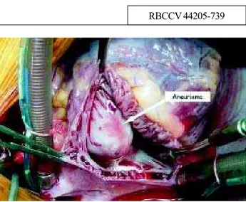
Fig. 1 – Aneurysmal bulging of the right coronary Valsalva sinus observed after opening the right atrium
97 ECHOCARDIOGRAM
The echocardiogram demonstrated situs solitus in levocardia and concordant venoatrial, atrioventricular and ventriculoarterial connections. Significant aortic regurgitation and an aneurysmal dilation at the right coronary Valsalva sinus were seen by Doppler echocardiography. Also a slight increase in the thickness of the left ventricular cavity was seen but with the ejection fraction preserved.
DIFFERENTIAL DIAGNOSIS
Some disease should be considered such as congenital or rheumatic aortic valve regurgitation, spontaneous rupture of a leaflet of the aortic valve, myxomatous degeneration of the aortic valve, left ventricle-aortic shunt and Takayasu disease.
DIAGNOSIS
The echocardiogram generally is sufficient to reliably diagnose but coronary cineangiography can be used in situations where other lesions are associated such as interventricular shunts. The identification of an aneurysm in the right coronary artery and aortic regurgitation, which
are usually silent, requires immediate surgical correction as there is a high risk of rupture.
OPERATION
Median transsternal thoracotomy with the establishment of conventional cardiopulmonary bypass, hypothermia at 25ºC intermittent anterograde sanguineous cardioplegia at 4ºC directly through the coronary ostia. The reperfusion and myocardial ischemia times were 85 and 63 minutes, respectively. The orifice of the right coronary cusp and significant aneurysmal dilation (calcified) of the right coronary sinus which bulged to the right atrium were evidenced (Figure 1). After opening this region, resection of the calcification was performed, followed by direct suturing of the orifice of the coronary cusp and of the aortic wall (Figure 2). The echocardiogram in the intra-operative period was important to guide the conduct in respect to preservation of the aortic valve, which was also submitted to plasty with plicature between the right coronary and non-coronary cusps. In the postoperative period the patient evolved well and was released on the 8th day after hospitalization. The echocardiogram demonstrated a slight regurgitation and absence of the aneurysm.
