online | memorias.ioc.fiocruz.br
Heterologous expression and biochemical characterization of
an α1,2-mannosidase encoded by the
Candida albicans MNS1
gene
Héctor M Mora-Montes, Everardo López-Romero, Samuel Zinker1, Patricia Ponce-Noyola,
Arturo Flores-Carreón/+
Instituto de Investigación en Biología Experimental, Facultad de Química, Universidad de Guanajuato, Apartado postal 187, Guanajuato, Gto. CP 36000, México 1Departamento de Genética y Biología Molecular, CINVESTAV del IPN, México, DF, México
Protein glycosylation pathways, commonly found in fungal pathogens, offer an attractive new area of study for the discovery of antifungal targets. In particular, these post-translational modifications are required for virulence and proper cell wall assembly in Candida albicans, an opportunistic human pathogen. The C. albicansMNS1 gene is predicted to encode a member of the glycosyl hydrolase family 47, with α1,2-mannosidase activity. In order to char-acterise its activity, we first cloned the C. albicans MNS1 gene into Escherichia coli, then expressed and purified the enzyme. The recombinant Mns1 was capable of converting a Man9GlcNAc2N-glycan core into Man
8GlcNAc2 isomer B, but failed to process a Man5GlcNAc2-Asn N-oligosaccharide. These properties are similar to those displayed by Mns1 purified from C. albicans membranes and strongly suggest that the enzyme is an α1,2-mannosidase that is localised to the endoplasmic reticulum and involved in the processing of N-linked mannans. Polyclonal antibodies specifically raised against recombinant Mns1 also immunoreacted with the soluble α1,2-mannosidases E-I and E-II, indicating that Mns1 could share structural similarities with both soluble enzymes. Due to the high degree of simi-larity between the members of family 47, it is conceivable that these antibodies may recognise α1,2-mannosidases in other biological systems as well.
Key words: α1,2-mannosidase - heterologous expression - glycosyl hydrolase family 47 - Candida albicans -
polyclonal antibodies - N-glycosylation
The α1,2-mannosidases that participate in the N -glycan trimming and ER-associated degradation of glycoproteins (Herscovics 1999a, b, Herscovics 2001, Helenius & Aebi 2004) belong to family 47 of glycosyl hydrolases. There are two subgroups of enzymes within family 47: the ER α1,2-mannosidases, that remove only one mannose residue from Man9GlcNAc2 (M9), generat-ing the Man8GlcNAc2 isomer B (M8B) (Ziegler &
Trim-ble 1991, Tremblay & Herscovics 1999, Mora-Montes et al. 2004, Movsichoff et al. 2005), and the Golgi α1,2-mannosidases, which after the action of ER enzyme, trim three α1,2-linked mannose residues to generate the Man
5GlcNAc2 (M5) intermediate required for the forma-tion of the complex and hybrid N-glycans found in mam-malian cells and filamentous fungi (Ichishima et al. 1999, Tremblay & Herscovics 2000, Akao et al. 2006). Golgi α-mannosidases are absent in lower eukaryotes, such as Saccharomyces cerevisiae, and thus no further process-ing of N-glycans is carried out. Instead, the N-glycan core is modified by Golgi mannosyltransferases to gen-erate high mannose N-glycans (Herscovics 1999a).
We have previously demonstrated that roughly 80% of the total α-mannosidase activity in Candida albicans is present as a soluble enzyme, while the remaining activ-ity is associated with the membrane (Vázquez-Reyna et al. 1993). Further work with the soluble fraction revealed the presence of two soluble α-mannosidase isoforms, named E-I and E-II (Vázquez-Reyna et al. 1999). Both enzymes were capable of removing the α1,2-mannose residues from a M9N-linked mannan core, indicating that they belong to family 47 of glycosyl hydrolases
(Mora-Financial support: Consejo Nacional de Ciencia y Tecnología (Grant n° CONACyT-2002-CO1-39528/A-1), Dirección de Investigación y Posgrado, Universidad de Guanajuato, México
HM Mora-Montes’ present address: School of Medical Sciences, In-stitute of Medical Sciences, University of Aberdeen, Foresterhill, Ab-erdeen AB25 2ZD, United Kingdom.
+Corresponding author: [email protected] Received 10 July 2008
Accepted 23 October 2008
Glycosylation is the most common covalent modifi-cation of proteins found in eukaryotic cells. The glycans attached to proteins are categorised into different classes based on the type of glycosidic linkage formed between the sugar and the protein, such as N-linked, O-linked glycans and glycosylphosphatidylinositol anchors. In eukaryotic cells, N-glycosylation of proteins is initi-ated in the endoplasmic reticulum (ER) and completed in the Golgi complex. Formation of the N-glycosidic bond requires a lipid-linked oligosaccharide (Glc3Man
Montes et al. 2004, 2006). Previously, we have shown that the α1,2-mannosidase E-II enzyme is the product of limited proteolysis of either enzyme E-I or a membrane-bound α-mannosidase (Mora-Montes et al. 2006, 2008), suggesting that the three α-mannosidase isoforms are re-lated each other. Indeed, this is supported by our recent report indicating that a C. albicansmns1∆ null mutant lacks both soluble and membrane-bound α-mannosidase activity (Mora-Montes et al. 2007). In order to learn more about the membrane-bound α-mannosidase activ-ity, we expressed the C. albicansMNS1 gene in Escheri-chia coli and characterised the enzyme product.
MATERIALS AND METHODS
Organism and culture conditions - C. albicans ATCC 26555 was used in this study. It was maintained and propagated in YPD medium [1% (w/v) yeast extract, 2% (w/v) mycological peptone, 2% (w/v) glucose] as pre-viously described (Mora-Montes et al. 2004). E. coli One Shot® TOP10 and BL21 StarTM (DE3) were from
Invitro-gen (Carlsbad, CA). Selection of transformants was per-formed at 37°C in LB medium [1% (w/v) tryptone, 0.5% yeast extract (w/v), 1% NaCl (w/v), pH 7.0] supplemented with 100 μg mL-1 carbenicillin.
Construction of pMNS1c - The C. albicans MNS1 open reading frame (GenBank/EBI accession AY167027) was amplified by PCR using genomic DNA from C. albicans ATCC 26555 and the nucleotide primers 5’ - CAC CAT TCT CAT GCT ATT AAA -3’ and 5’ - TTG TCA ATT ACC CAG CAA -3’ (the adaptor nucleotide sequence added for directional cloning is underlined). The pMNS1c construct was generated by ligation of the roughly 1.7 kbp PCR product into a pET100/D-TOPO®
expression vector (Invitrogen, Carlsbad, CA). The con-struct was propagated and maintained in E. coli One Shot® TOP10 cells, while BL21 StarTM (DE3) cells were
used for Mns1 expression, as indicated by the provider. Briefly, 10 mL of carbenicillin-containing LB broth was inoculated with 500 μL aliquots of transformed BL21 StarTM (DE3) cells grown overnight and incubated at
37°C until the culture reached an OD600nm ~ 0.6. Then, isopropyl β-D-thiogalactoside (IPTG) was added to the cultures, at 1mM final concentration, followed by a 5 h incubation at 37°C with shakig (200 rpm).
Extraction and partial purification of recombi-nant Mns1 -E. coli cells were washed three times with 50 mM MES-Tris buffer, pH 6.0 (buffer A), resuspend-ed in 1 mL of the same buffer containing 50 mg mL-1 lysozyme and incubated overnight at 37°C. Then, the cells were lysed by three rounds of sonication for 5 min alternating with freezing at -70°C for 20 min. The ho-mogenate was centrifuged at 4,830 g for 4 min and the supernatant was saved and kept at -20°C until use. For enzyme purification, the cellular preparation was first centrifuged at 21,500 g for 10 min and then the superna-tant was collected and fractionated on a DEAE Bio-Gel A column (0.9 x 2.0 cm) equilibrated with buffer A. The column was first washed with 5 mL of buffer A followed by a discontinuous gradient from 0 to 0.5 M NaCl, in the same buffer. One-mL fractions were collected, with
protein quantification and enzyme activity determined as described below. The most active fractions (usually 6-10) were pooled and kept at -20°C until use.
α-mannosidase assay and protein determination - En-zyme activity was determined using 4-methylumbelliferyl-α-D-mannopyranoside (MUαMan), M9 or Man5GlcNAc2 -Asn (M5-Asn) N-oligosaccharides as described previously (Mora-Montes et al. 2004). Elution of protein during chro-matography was monitored by absorbance at 280 nm and the Bradford method was used for protein quantification (Bradford 1976).
Electrophoresis - Sodium dodecyl sulphate-poly-acrylamide gel electrophoresis (SDS-PAGE) was done in 10% gels and the proteins were stained with Coomassie Blue following standard protocols. For in situ detec-tion of α-mannosidase activity, analytical zymograms revealed with MUαMan were carried out as previously described (Mora-Montes et al. 2004).
Generation of polyclonal anti-Mns1 antibodies - Par-tially purified Mns1 was separated by SDS-PAGE, the protein band was sliced out of the gel, placed in a separa-tion tube (a device consisting of an Eppendorf tube with a small hole at the bottom and one third of its volume filled with sterile glass fibres) and incubated at -70°C for 2 h. The separation tube was placed onto the top of another Eppendorf tube and the protein was eluted from the polyacrylamide gel by centrifugation at 5,200 g for 20 min at 4°C. Antibodies were raised in a New Zealand male rabbit after intramuscular injection of 150 μg of recombinant protein emulsified with complete Freund’s adjuvant (day 0). Booster injections, containing 150 μg of protein emulsified with incomplete Freund’s adjuvant, were given on days 15, 30, 45 and 60 and the animal was bled to death on day 75. The animal was handled and sacrificed following the approved guide from the Ethical Committee of Universidad de Guanajuato. The γ-globulin fraction from both pre and post-immunization sera was purified by precipitation with 50% (w/v) am-monium sulphate and the anti-Mns1 antibody titre was determined by ELISA, as described by Goers (1993).
Purification of α-mannosidases from C. albicans - The soluble enzymes were purified as previously de-scribed (Mora-Montes et al. 2004, 2006). Membrane-bound α-mannosidase activity was purified as follows. Yeast cells were harvested by low-speed centrifugation, resuspended in ice-cold buffer A containing 1 μM pep-statin A, and disrupted with glass beads (0.45 mm in di-ameter) in a MSK cell homogeniser (Braun, Melsungen, Germany) for 3 min, using a CO2 stream to cool the ho-mogenizing chamber. The homogenate was centrifuged at 1,000 g for 10 min at 4°C and the resulting supernatant was collected and further centrifuged at 105,000 g for 1 h at 4oC (ultracentrifugation). The pellet, consisting of a
mixed membrane fraction (MMF), was homogenised in 2-3 ml of buffer A and used as the starting material for enzyme purification. To this purpose, the MMF was suc-cessively subjected to the following treatments and the pellet was recovered by ultracentrifugation after each step: (1) incubation for 1 h at 48ºC and 30 min at -20ºC; (2) resuspension in 1 mL of 3 mM EDTA in buffer A and incubated for 1 h at rt; (3) resuspension in 1 mL of buffer A containing 0.5 M NaCl and incubated for 1 h at rt; (4) resuspension in 1 mL of buffer A containing Triton X-100 in a protein: detergent ratio of 1:1 and incubated for 1 h at rt. Finally, the pellet was resuspended in 1 mL of buffer A and subjected to ion-exchange chromatogra-phy on a DEAE Bio-Gel A column (0.9 x 2.0 cm), which was washed and eluted as described above to purify the recombinant enzyme. The most active fractions (usually 6-10) were pooled, freeze-dried, resuspended in 1 mL buffer A and then subjected to size exclusion chromatog-raphy on a Sephadex G-25 gel filtration column (0.5 x 21 cm) equilibrated with the buffer A. Twenty 1 mL frac-tions were eluted with buffer A and the enzyme activity and protein elution were monitored as described above. The most active fractions (usually 2-8) were pooled, freeze-dried, and kept at -20°C until use.
Chemicals - M9, M5-Asn, MUαMan, pepstatin A, 1-deoxymannojirimycin (1-DMJ), swainsonine (SWN), lysozyme, 3,3’-diaminobenzidine, bovine serum albu-min and Triton X-100 were obtained from Sigma Che-mical Company (St. Louis, MO). The Sephadex G-25, DEAE Bio-Gel A, and all electrophoresis reagents, were purchased from Bio-Rad Laboratories (Hercules, CA). The anti-rabbit IgG-horseradish peroxidase an-tibody was from Amersham Biosciences (Piscataway, NJ). All other chemicals were of the highest purity commercially available.
RESULTS AND DISCUSSION
Heterologous expression of MNS1 - C. albicansMNS1 is the ortholog of S. cerevisiae MNS1, which encodes the ER α1,2-mannosidase (Camirand et al. 1991). Accord-ingly, we have recently demonstrated that a C. albicans mns1∆ null mutant failed to express α-mannosidase activ-ity, as measured with the fluorogenic substrate MUαMan (Mora-Montes et al. 2007). With this background knowl-edge, we decided to express MNS1 and characterise the enzyme product. To this purpose, C. albicansMNS1 was cloned into pET100/D-TOPO® vector and the construct
generated, pMNS1c, was used to transform E. coli BL21 StarTM (DE3) cells, as described in the material and
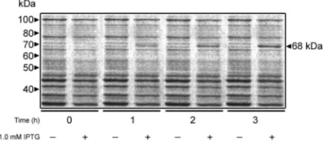
meth-ods. Cells grown in the presence of 1.0 mM IPTG ex-pressed a 68 kDa polypeptide whose intensity increased as a function of the induction time (Fig. 1), although no further increase in induction was observed after 3 h of incubation with IPTG (data not shown). When the crude homogenates were incubated with MUαMan, homoge-nate from cells expressing MNS1 exhibited a 491-fold in-crease in α-mannosidase specific activity with respect to control preparations (Table I). The molecular mass of the recombinant Mns1 was 3 kDa higher than that predicted for the MNS1 product, which was expected due to the addition of affinity tags from the expression vector. The vector utilised to express MNS1 adds a six-histidine tag at the N-terminus of the recombinant protein to allow for protein purification by nickel affinity chromatography. However, this approach could not be used because the enzyme activity was irreversibly inhibited by imidazole, even at concentrations as low as 5 mM (data not shown). Instead, the recombinant α-mannosidase was partially purified by ion-exchange chromatography on a DEAE Bio-Gel A column. The enzyme activity co-eluted with a protein peak at 0.1 M NaCl, well apart from the other protein peaks (Fig. 2A). Analysis of the purified sample by SDS-PAGE revealed protein bands with
molecu-Fig. 1: expression of Candida albicans Mns1 in Escherichia coli. BL21 StarTM (DE3) cells transformed with pMNS1c were grown in the absence or presence of 1 mM IPTG, and 500 μL from each culture was removed at the times indicated. Cells were collected by centrifu-gation, resuspended in 100 μL SDS-PAGE loading buffer, disrupted by incubation at 100°C for 5 min and 20 μL from each sample was analysed by SDS-PAGE on 10% gels. Protein bands were revealed with Coomassie Blue staining.
TABLE I
α-mannosidase activity in cell homogenates from Escherichia coli
Specific Increase
Homogenate source activitya (n-folds)
Non-transformed cells 1.1 ± 2.0 1.0
Non-transformed cells + 1.0 mM IPTG 0.9 ± 1.8 0.8
Cells transformed with pMNS1c 1.0 ± 2.6 0.9
Cells transformed with pMNS1c
+ 1.0 mM IPTG 540 ± 50 491.0
a: expressed as pmoles of 4-methylumbelliferone min-1 mg of
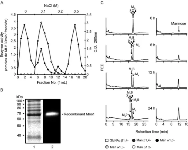
Fig. 2: A: purification of recombinant Mns1 by ion-exchange chromatography. The homogenate from cells expressing Candida albicans Mns1 was applied onto a DEAE Bio-Gel A column (0.9 x 2.0 cm) equilibrated with buffer A. The column was washed with the same buffer and bound proteins were eluted with a discontinuous gradient of 0-0.5 M NaCl in buffer A. Protein elution was monitored by O.D.280 nm (closed symbols) and enzyme
activity was measured with MUαMan (open symbols); B: protein profile and analytical zymogram of recombinant Mns1. Aliquots containing 10 μg of protein were heat-denatured (Lane 1) and separated by Sodium dodecyl sulphate-polyacrylamide gel electrophoresis on 10% gels that were stained with Coomassie Blue (Lane 1). In Lane 2, the sample was not heat-denatured and enzyme activity was revealed with MUαMan; C: hydroly-sis of the M9 N-mannan core by partially purified Mns1. Samples (5 μg) of the enzyme were incubated at 37°C with 2 μg of the M9N-mannan core
substrate and the reactions were stopped by heating after the indicated times. The products were analysed by High-performance anion exchange chromatography as described in materials and methods. Elution of oligosaccharides and mannose is indicated in the left and right panels, respec-tively. M9, Man9GlcNAc2; M8B, Man8GlcNAc2 isomer B; M7B, Man7GlcNAc2 isomer B. M6 is most probably Man6GlcNAc2.
lar masses between 38 and 100 kDa and a clearly en-riched polypeptide of 68 kDa (Fig. 2B, Lane 1) that had α-mannosidase activity (Fig. 2B, Lane 2). These results demonstrated that the C. albicansMNS1 gene product was successfully expressed in E. coli.
Hydrolysis of a M9 N-linked mannan core by recom-binant Mns1 - In order to determine the ability of the recombinant enzyme to hydrolyse natural oligosaccha-rides, a time-course assay was carried out using a M9 N-linked mannan core as substrate. After 12 h of incuba-tion, the M8B N-oligosaccharide (Fig. 2C, left panels) and
mannose (Fig. 2C, right panels) were detected as prod-ucts of the enzymatic reaction. Both prodprod-ucts increased as a function of the incubation time. These results in-dicate that the recombinant enzyme removes only one α1,2-mannose moiety from M9, behaving as a typical ER α1,2-mannosidase of family 47. After 24 h of incubation, two other products, M7B and possibly the M6 N
-oligo-saccharide, were also detected. Similar observations have been reported for ER α1,2-mannosidases from S. cerevisiae and human cells (Herscovics et al. 2002) and also for the soluble α1,2-mannosidases E-I and E-II from C. albicans (Mora-Montes et al. 2004, 2006). Recombi-nant Mns1 failed to trim the M5-Asn N-oligosaccharide (data not shown), which confirmed the specificity of the enzyme for α1,2-linked mannose residues, as M5 con -tains α1,3- and α1,6-linked mannose units.
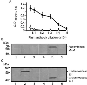
recog-nition band was observed when extracts from non-trans-formed cells (Fig. 3B, Lanes 1, 2) or cells transnon-trans-formed with pMNS1c growing under non-inductive conditions (data not shown) were incubated with anti-Mns1 or pre-immune sera. In extracts prepared from cells expressing Mns1, the anti-Mns1 antibody recognised a polypeptide with a molecular mass of 68 kDa (Fig. 3B, Lane 5). No recognition was observed in mock incubations contain-ing pre-immune serum (Fig. 3B, Lane 4) or lackcontain-ing the primary antibody (Fig. 3B, Lanes 3, 6). Following this initial validation of our antibody, we then decided to de-termine whether the antiserum immunoreacted with oth-er purified soluble α-mannosidases as well. The results indicated that the anti-Mns1 serum immunodetected both α-mannosidases E-I (Fig. 3C, Lane 2) and E-II (Fig. 3C, Lane 5). Control Lanes using pre-immune serum (Fig. 3C, Lanes 1, 4) or lacking the primary antibody (Fig. 3C, Lanes 3, 6) did not give any signal. When simi-lar blot assays were conducted using the high-speed sol-uble fraction from C. albicans, three protein bands with molecular masses of 65, 52 and 43 kDa were detected
(Fig. 4, Lane 2). The 52 and 43 kDa proteins correspond to enzymes E-I and E-II, as previously reported (Mora-Montes et al. 2004, 2006). The 65 kDa protein band may correspond to Mns1, since this is the predicted molecular mass for the MNS1 gene product. Bioinformatic analysis indicated that, like other mannosidases from family 47 (Herscovics 2001), Mns1 has a membrane domain at the N-terminus and is predicted to be a type II membrane bound protein. In order to find evidence supporting this model, immunodetection assays were conducted with the MMF. The anti-Mns1 antibody detected only one protein band with a molecular weight of 65 kDa (Fig. 4, Lane 1). These data suggests that the 65 kDa protein is Mns1 and also strongly indicates that the soluble and membrane-bound enzymes are immunologically related. This is in accordance with our proposal that the E-I and membrane-bound α-mannosidase are precursors to the α1,2-mannosidase E-II (Mora-Montes et al. 2006, 2008). Furthermore, unpublished data from our laboratory sug-gests that the membrane-bound activity is a precursor of E-I, thus confirming a close relationship between the three α-mannosidases.
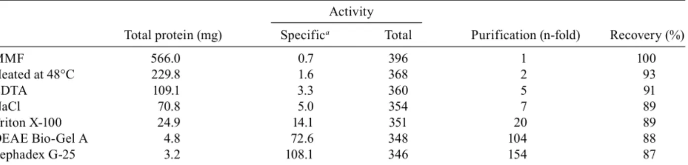
Purification of Mns1 - Our results suggested that the membrane-bound activity previously reported by Vázquez-Reyna et al. (1993) may correspond to Mns1. To further investigate this, we purified the membrane-bound enzyme and compared its biochemical character-istics with those of the recombinant enzyme obtained here. For reasons not fully understood, detergents such as Triton X-100, Igepal CA-630 or Lubrol, at different con-centrations, failed to solubilise the particulate activity (data not shown). Therefore, we used a high-temperature extraction method that was a successful for the solubili-sation of the membrane-bound α-mannosidase from Spo-rothrix schenckii (HM Mora-Montes et al., unpublished observations). The procedure solubilised about 50% of the spurious non-enzymatic protein present in the MMF, without a significant effect on the total α-mannosidase activity remaining in the pellet. Nearly 90% of the α-mannosidase activity was recovered after sequen-tial extractions with EDTA, NaCl and Triton X-100
Fig. 3: A: titration of Mns1p antiserum by ELISA. Anti-Mns1 (closed symbols) or rabbit pre-immune (open symbols) sera were diluted in phosphate buffered saline solution as indicated and added to ELISA 96-well plates coated with 20 μg mL-1 recombinant Mns1. Error bars
indicate the mean ± the standard deviation (n= 4). The results encom-pass pooled data from triplicate experiments; B: immunodetection of recombinant Mns1 in homogenates from Escherichia coli. Samples containing 10 μg of protein from BL21 StarTM (DE3) cells (Lanes 1-3)
or E. coli cells expressing recombinant Mns1 (Lanes 4, 6) were
sepa-rated by Sodium dodecyl sulphate-polyacrylamide gel electrophoresis, transferred to nitrocellulose membranes and immunoblotted with anti-Mns1 (Lanes 2, 5) or rabbit pre-immune (Lanes 1, 4) sera. Control samples lacking the primary antibody are shown in Lanes 3 and 6; C: immunodetection of the purified α1,2-mannosidases E-I and E-II by the Mns1p antibody. Same legend as in panel B, except that aliquots (10 μg) of enzyme E-I (Lanes 1-3) or E-II (Lanes 4-6) were analysed.
(Table II). The enzyme was further purified on DEAE Bio-Gel A and Sephadex G-25 columns, as described in the materials and methods (data not shown). At the end of the purification protocol, the α-mannosidase was puri-fied 154-fold with a recovery of 87% with respect to the starting material (Table II). Analytical SDS-PAGE re-vealed three minor protein bands with molecular masses of 53, 59 and 72 kDa, along with a major polypeptide at 65 kDa (Fig. 5, Lane 1) that was active on MUαMan (Fig. 5, Lane 2) and was also recognised by the Mns1 an-tiserum (Fig. 5, Lane 4). Mock incubations containing the pre-immune serum (Fig. 5, Lane 3) or lacking the primary antibody (Fig. 5, Lane 5) did not reveal any signal.
Biochemical characterization of partially purified particulate α-mannosidase - Purified α-mannosidase exhibited maximum activity at pH 6.0 and at 37oC, re
-spectively. Hydrolysis of MUαMan followed a hyper-bolic kinetic curve and Lineweaver-Burk plots revealed K
m and Vmax values of 0.07 mM and 48.2 nmoles of methylumbelliferone min-1 mg of protein-1, respectively. Specific inhibitors of α-mannosidase, such as
1-deoxy-TABLE II
Purification of membrane-bound α-mannosidase from Candida albicans
Activity
Total protein (mg) Specifica Total Purification (n-fold) Recovery (%)
MMF 566.0 0.7 396 1 100
Heated at 48°C 229.8 1.6 368 2 93
EDTA 109.1 3.3 360 5 91
NaCl 70.8 5.0 354 7 89
Triton X-100 24.9 14.1 351 20 89
DEAE Bio-Gel A 4.8 72.6 348 104 88
Sephadex G-25 3.2 108.1 346 154 87
a: expressed as nmoles of 4-methylumbelliferone min-1 mg of protein-1;mixed membrane fraction (MMF) was sequentially
treated as described in material and methods followed by anionic and gel filtration column chromatographies.
mannojirimycin (1-DMJ) and SWN inhibited the hydrol-ysis of MUαMan with IC50 values of 0.19 mM and 0.62 mM, respectively (data not shown). The optimum pH of 6.0 confirmed that this enzyme is not an acidic vacu-olar α-mannosidase belonging to family 38 (Herscovics 1999a). In addition, the partially purified enzyme showed an increased sensitivity to 1-DMJ over SWN, suggesting that it belongs to family 47 (Herscovics 1999a). This was further confirmed by the hydrolysis of the M9N-linked mannan core, which exhibited the same pattern also ob-served for Mns1 (data not shown). Therefore, our results strongly suggest the 65 kDa α1,2-mannosidase present in the MMF of C. albicans is Mns1.
Overall, results presented here indicate that we have heterologously expressed the C. albicans MNS1 gene en-coding an ER α1,2-mannosidase and that the polyclonal anti-Mns1 serum can be a useful tool for the study of α1,2-mannosidases in C. albicans. We also confirmed a close relationship between the soluble and membrane-associated α1,2-mannosidases. Due to the high degree of similarity among the members of family 47, it is conceiv-able that this antibody may recognise α1,2-mannosidases from other organisms as well.
REFERENCES
Akao T, Yamaguchi M, Yahara A, Yoshiuchi K, Fujita H, Yamada O, Akita O, Ohmachi T, Asada YT, Yoshida T 2006. Cloning and ex-pression of 1,2-α-mannosidase gene (fmanIB) from filamentous fungus Aspergillus oryzae: in vivo visualization of the FmanIBp-GFP fusion protein. Biosci Biotechnol Biochem70: 471-479. Bradford MM 1976. A rapid and sensitive method for the
quantita-tion of microgram quantities of protein utilizing the principle of protein-dye binding. Anal Biochem 72: 248-254.
Camirand A, Heysen A, Grondin B, Herscovics A 1991. Glycopro-tein biosynthesis in Saccharomyces cerevisiae. Isolation and characterization of the gene encoding a specific processing α-mannosidase. J Biol Chem 266: 15120-15127.
Goers J 1993. Immunochemical techniques laboratory manual, Aca-demic Press, San Diego, p. 15-18, 119-135.
Helenius A, Aebi M 2004. Roles of N-linked glycans in the endoplas-mic reticulum. Annu Rev Biochem73: 1019-1049.
Herscovics A 1999a. Importance of glycosidases in mammalian gly-coprotein biosynthesis. Biochim Biophys Acta 1473: 96-107. Fig. 5: analytical electrophoresis, zymogram analysis and
immuno-blotting of partially purified membrane-bound α-mannosidase from
Candida albicans; Lane 1: the protein profile of partially purified
Herscovics A 1999b. Processing glycosidases of Saccharomyces
cerevisiae. Biochim Biophys Acta 1426: 275-285.
Herscovics A 2001. Structure and function of Class I α1,2-mannosidases involved in glycoprotein synthesis and endoplas-mic reticulum quality control. Biochimie 83: 757-762.
Herscovics A, Romero PA, Tremblay LO 2002. The specificity of the yeast and human class I ER α1,2-mannosidases involved in ER quality control is not as strict previously reported. Glycobiology 12: 14G-15G.
Ichishima I, Taya N, Ikeguchi M, Chiba Y, Nakamura M, Kawabata C, Inoue T, Takahashi K, Minetoki T, Ozeki K, Kumagai C, Gomi K, Yoshida T, Nakajima T 1999. Molecular and enzymic properties of recombinant 1,2-α-mannosidase from Aspergillus saitoi overex-pressed in Aspergillus oryzae cells. Biochem J 339: 589-597. Kornfeld R, Kornfeld S 1985. Assembly of asparagine-linked
oli-gosaccharides. Annu Rev Biochem54: 631-664.
Mora-Montes HM, Bates S, Netea MG, Diaz-Jiménez DF, López-Ro-mero E, Zinker S, Ponce-Noyola P, Kullberg BJ, Brown AJ, Odds FC, Flores-Carreón A, Gow NA 2007. Endoplasmic reticulum α-glycosidases of Candida albicans are required for N -glycosyla-tion, cell wall integrity, and normal host-fungus interaction.
Eu-karyot Cell 6: 2184-2193.
Mora-Montes HM, López-Romero E, Zinker S, Ponce-Noyola P, Flores-Carreón A 2004. Hydrolysis of Man9GlcNAc2 and Man8GlcNAc2
oligosaccharides by a purified α-mannosidase from Candida
albi-cans. Glycobiology 14: 593-598.
Mora-Montes HM, López-Romero E, Zinker S, Ponce-Noyola P, Flores-Carreón A 2006. Purification of soluble α1,2-mannosidase from Candida albicans CAI-4. FEMS Microbiol Lett 256: 50-56. Mora-Montes HM, Lopez-Romero E, Zinker S, Ponce-Noyola P,
Flores-Carreón A 2008. Conversion of α1,2-mannosidase E-I from Candida albicans to α1,2-mannosidase E-II by limited pro-teolysis. Antonie Van Leeuwenhoek 93: 61-69.
Movsichoff F, Castro OA, Parodi AJ 2005. Characterization of
Schizosaccharomyces pombe ER α-mannosidase: a reevaluation
of the role of the enzyme on ER-associated degradation. Mol Biol Cell16: 4714-4724.
Towbin H, Staehelin T, Gordon J 1979. Electrophoretic transfer of pro-teins from polyacrylamide gels to nitrocellulose sheets: procedure and some applications. Proc Natl Acad Sci USA 76: 4350-4354. Tremblay LO, Herscovics A 1999. Cloning and expression of a
specific human α1,2-mannosidase that trims Man9GlcNAc2 to
Man8GlcNAc2 isomer B during N-glycan biosynthesis.
Glyco-biology 9: 1073-1078.
Tremblay LO, Herscovics A 2000. Characterization of a cDNA en-coding a novel human Golgi α1,2-mannosidase (IC) involved in N-glycans biosynthesis. JBiol Chem 275: 31655-31660.
Trombetta ES 2003. The contribution of N-glycans and their process-ing in the endoplasmic reticulum to glycoprotein biosynthesis.
Glycobiology13: 77R-91R.
Vázquez-Reyna AB, Balcázar-Orozco R, Flores-Carreón A 1993. Biosynthesis of glycoproteins in Candida albicans: biochemical characterization of a soluble α-mannosidase. FEMS Microbiol
Lett 106: 321-325.
Vázquez-Reyna AB, Ponce-Noyola P, Calvo-Méndez C, López-Romero E, Flores-Carreón A 1999. Purification and biochemical characterization of two soluble α-mannosidases from Candida
albicans. Glycobiology 9: 533-537.