Evolution of flatworm central nervous systems: Insights from polyclads
Sigmer Y. Quiroga
1,§, E. Carolina Bonilla
3,§, D. Marcela Bolaños
2,3,§, Fernando Carbayo
4,5,
Marian K. Litvaitis
2and Federico D. Brown
3,5,6,71
Programa de Biología, Facultad de Ciencias Básicas, Universidad del Magdalena, Santa Marta, Colombia.
2
Department of Natural Resources and the Environment, University of New Hampshire, Durham, NH, USA.
3Laboratorio de Biología del Desarrollo, Departamento de Ciencias Biológicas, Universidad de los Andes,
Bogotá, Colombia.
4
Laboratório de Ecologia e Evolução, Escola de Artes, Ciências e Humanidades,
Universidade de São Paulo, São Paulo, SP, Brazil.
5Programa de Pós-Graduação em Zoologia, Departamento de Zoologia, Instituto de Biociências,
Universidade de São Paulo, São Paulo, SP, Brazil.
6
Departamento de Zoologia, Instituto de Biociências, Universidade de São Paulo, São Paulo, Brazil.
7Centro de Biologia Marinha, Universidade de São Paulo, São Sebastião, Brazil.
Abstract
The nervous systems of flatworms have diversified extensively as a consequence of the broad range of adaptations in the group. Here we examined the central nervous system (CNS) of 12 species of polyclad flatworms belonging to 11 different families by morphological and histological studies. These comparisons revealed that the overall organi-zation and architecture of polyclad central nervous systems can be classified into three categories (I, II, and III) based on the presence of globuli cell masses -ganglion cells of granular appearance-, the cross-sectional shape of the main nerve cords, and the tissue type surrounding the nerve cords. In addition, four different cell types were iden-tified in polyclad brains based on location and size. We also characterize the serotonergic and FMRFamidergic ner-vous systems in the cotyleanBoninia divae by immunocytochemistry. Although both neurotransmitters were broadly expressed, expression of serotonin was particularly strong in the sucker, whereas FMRFamide was particularly strong in the pharynx. Finally, we test some of the major hypothesized trends during the evolution of the CNS in the phylum by a character state reconstruction based on current understanding of the nervous system across different species of Platyhelminthes and on up-to-date molecular phylogenies.
Keywords: brain, FMRFamide, globuli cell masses, orthogon, serotonin. Received: January 13, 2015; Accepted: April 19, 2015.
Introduction
Current phylogenetic views of Platyhelminthes di-vide the phylum into Catenulida and Rhabditophora (Lau-mer and Giribet, 2014; Lau(Lau-meret al,.2015; Eggeret al., 2015). Within the diverse Rhabditophora, strong morpho-logical and molecular evidence suggests Macrostomorpha as the basal clade; whereas Polycladida, often considered a basal clade (for a review see Baguñà and Riutort, 2004; Karling, 1967, 1974; Carranzaet al,. 1997, Litvaitis and Rhode, 1999; Laumer and Giribet, 2014), currently groups together with the Prorhynchida in a more derived position based on recent phylogenomic and transcriptomic analyses
(Laumeret al., 2015; Eggeret al., 2015). The main charac-teristic of the Polycladida is their highly branched intestine (Hyman, 1951), from which they derive their name. Poly-clads have few external traits; however, the presence or ab-sence of clusters of eyespots and either true tentacles or pseudotentacles, which form by folds of the anterior body margin, can be used as systematic characters (Newman and Cannon, 1994). The initial division of the order is based on the presence or absence of a ventral sucker. This character divides the polyclads into the two suborders Acotylea (without sucker) and Cotylea (with sucker) (Lang, 1884).
Among invertebrates, nervous systems exhibit a wide variety of organization, which can range from an unorga-nized diffuse nerve net (cnidarians) to complex systems with highly specialized sensory receptors (arthropods, some mollusks) (Ruppert and Barnes, 1994). The bilater-ally flattened body of flatworms preserves a common
orga-DOI: http://dx.doi.org/10.1590/S1415-475738320150013
Send correspondence to Federico D. Brown. Departamento de Zoologia, Instituto de Biociências, Universidade de São Paulo, Rua do Matão, Travessa 14, No. 101, Cidade Universitária, 05508-090 São Paulo, SP, Brazil. E-mail: [email protected].
§
These authors contributed equally to the present work.
nization of the central nervous system (CNS). The CNS of flatworms consists of: (i) theorthogon,composed of main longitudinal nerve cords and transverse commissures that form a ladder-like network. The main cords have direct connections to the brain, are generally multifibrillar, are more strongly developed on the ventral side, and express serotonin or cholinergic neuropeptides (Reuter and Gus-tafsson, 1995). The number, position, and arrangement of cords vary drastically from one taxonomic group to the next; (ii) an anterior brainconsisting of two lobes con-nected by one or several commissures. The lobes may be ei-ther loosely aggregated or encapsulated by a sheath of extracellular matrix (Reuter and Gustafsson, 1995); and (iii) theplexus,composed of a network of sub- and infra-epidermal, or submuscular nerves that expand throughout the body of the worm. In addition, in free-living flatworms the presence of pharyngeal and stomatogastric plexi, con-sisting of either one or two nerve rings, innervated by sub-and infra-epithelial nerve nets (Reuter sub-and Gustafsson, 1995; Halton and Gustafsson, 1996), are considered plesio-morphic characters (Ehlers, 1985).
Reisinger (1925) defined a standard orthogonal pat-tern for many free-living forms. This patpat-tern consists of several pairs of ventral, lateral, and dorsal longitudinal nerve cords that are connected at right angles along their lengths by transverse commissures, forming a ladder-like arrangement. Although the orthogon organization and oc-currence does not show clear evolutionary trends (Reuter and Gustafsson, 1995), a common origin of the CNS in flatworms has been suggested with convergent evolution of similar orthogonal organizations in different taxa (Koti-kova, 1991). The orthogon varies in position and number of cords, different points of contact with the brain, and thick-ness of cords. Reuteret al.(1998) determined that the main cords derive from strong roots in the brain, are composed of numerous neurons within wide fiber bundles, and generally show immunoreactivity for serotonin and catecholamines. It has been hypothesized that more derived clades often have fewer but thicker nerve cords than more basal taxa (Reisinger, 1972; Reuter and Gustafsson, 1995), and that longitudinal cords may have evolved from the fusion of plexal fibers of ancestral taxa (Joffe and Reuter, 1993).
The architecture of the polyclad nervous system re-flects components of the anatomically diffuse cnidarian system and of a centralized and more condensed nervous system found in the higher invertebrates (Koopowitz, 1974). They have been characterized as being among the most advanced in flatworms with complexities comparable to those of segmented marine worms (Bullock and Hor-ridge, 1965). At the same time, the peripheral nervous sys-tem, and especially a nerve plexus located at the base of the epithelium (infraepithelial plexus) exhibits an organization similar to that of the nerve net found in hydra (Chien and Koopowitz, 1977). Additionally, the polyclad nervous sys-tem lacks ganglia other than those found in the brain.
As part of a comprehensive and comparative study of ca. 20 species of polyclads from the Gulf of Naples, Lang (1884) unified early descriptions of the main morphologi-cal features in polyclad nervous systems. Detailed histo-logical descriptions of the CNS are available for the coty-lean,Thysanozoon brocchiiRisso, 1818 (Lang, 1884) and more recently for the acotylean,Notoplana acticolaBoone, 1929 (Koopowitz, 1973, 1986; Chien and Koopowitz, 1972, 1977; Koopowitz and Chien, 1974, 1975). The CNS consists of an anterior, bilobed brain that functions in coor-dinating locomotion and peripheral reflex activity (Koopo-witz, 1973). In both species, this brain is enclosed by a dis-tinct capsule and is composed of an outer rind of cells, formed by a variety of neuronal types (e.g., multipolar, de-cussating, coupled), and an inner core of thin fibers and a complete lack of nuclei or cell bodies (Lang, 1884; Koopo-witz, 1986). InT. brocchii,the largest, multipolar ganglion cells are mostly confined to the dorsal, ventral, and poste-rior parts of the brain. Thin fibers in the core of the brain form a complex anastomosing network, which gives rise to the nerve cords exiting the brain (Lang, 1884). At the front of each brain lobe and located outside the capsule are two groups of sensory ganglion cells (globuli cell masses, or Körnerhaufenin the original German terminology) that are of granular appearance. Furthermore, Stylochoplana
maculataQuatrefage, 1845,N. acticola, PlanoceraLang,
1879, andGnesiocerosDiesing, 1862 showed six pairs of nerve cords radiating from the brain and forming a typical radial network pattern for the group (Koopowitz, 1973). The first two pairs are directed forward (anterior and ante-rior dorsal nerves), the third and fourth pairs (lateral and lat-eral dorsal nerves) are directed latlat-erally from the brain, and the last two pairs (posterior and posterior dorsal nerves) are directed posteriorly. These nerves soon branch repeatedly towards the margin, becoming more delicate and forming anastomosing plexi that contain multi- and bipolar neurons (Koopowitz, 1974, 1986).
Neuroactive molecules can be classified broadly into neurotransmitters (e.g., acetylcholine, serotonin (5-HT), GABA, histamine), neuropeptides (e.g., FMRF-amide) and nitric oxide, a dissolved gas with nervous and endocrine function. In flatworms, immunocytochemistry (ICC) has demonstrated the presence of several neuroactive mole-cules, including typical neurotransmitters, nitric oxide syn-thase, and also a number of regulatory neuropeptides (Reu-ter and Halton, 2001). Among the neuropeptides, four native flatworm FMRFamide-related peptides have been described (FaRPs), plus two neuropeptide F substances (Mauleet al., 1993, Johnstonet al., 1996). The specific role of neuropeptides is still unclear although a myoexitatory function is likely (Day et al., 1997; Marks et al., 1996, Monneypennyet al., 1997).
Schmidt, 1848), triclads (Dendrocoelum Ørsted, 1844, PolycelisEherenberg, 1831), and catenulids (Stenostomum Schmidt, 1848) all exhibit immunoreactivity to serotonin, as do representatives of monogeneans, trematodes, and cestodes (Wikgren and Reuter, 1985; Reuter, 1988; Reuter et al., 1986, 1996; Ladurneret al., 1997). Functionally, se-rotonin is an excitatory neurotransmitter stimulating mus-cle contraction and it also plays a role in controlling growth, asexual reproduction and regeneration (Villar and Schaef-fer, 1993). Serotonin immunoreactivity has been shown in the submuscular and subepidermal plexi, in the main nerve cords (Reuteret al., 1995), and in positive fibers that encir-cle the pharynx. Nerve fibers but no cell bodies have been visualized in the peripheral nervous system (Reuteret al., 1986). In general, the distribution pattern of serotonin is distinct from cholinergic and peptidergic pathways, how-ever, it parallels catecholamine staining patterns (Shishov, 1991).
Serotonin and FMRFamide related peptides (FaRPs) play important roles in myoexcitatory activities, including motility of the gut and pharynx related to feeding behavior (Forest and Lindsay, 2008), or in the contraction of loco-motory muscles of different species of Platyhelminthes (Mousley et al., 2005, Cebrià, 2008, Kreshchenko and Tolstenkov, 2012). In the freshwater planarianSchmidtea mediterraneaBenazziet al., 1975, both, the CNS and PNS assemble a loose 5-HT nerve network that is expressed throughout the animal from head to tail (Cebrià, 2008). Fur-thermore, FMRFamide was expressed in both the CNS and PNS of the turbellarian species Polycelis tenuis Ijima, 1884, andGirardia tigrinaGirard, 1850 (Kreshchenko and Tolstenkov, 2012). Although both of these two neuro-transmitters may be expressed throughout the nervous sys-tems of flatworms, they often show distinctively different staining patterns. For example, in the basal platyhelminth Macrostomum lignanoLadurner,et al., 2005, serotonergic nerve cells are densely clustered in the pharyngeal region and are relatively sparse in the brain neuropile, whereas FMRFaminergic cells are concentrated in ventro-lateral and centro-lateral compartments of the brain neuropile (Morriset al., 2007).
Hence, the major goals of our study were to compare the gross and fine morphology of the CNS of 12 different polyclad species at the light-microscopic level, to examine serotonin (5-HT) and FMRFamide expression in the ner-vous system ofBoninia divaeMarcus and Marcus, 1968 us-ing immunocytochemistry (ICC), and to review our results in light of the newest phylogenetic understanding of platy-helminth relationships (Laumer and Giribet, 2014; Eggeret al., 2015; Laumeret al., 2015) in an attempt to place the di-versity of flatworm nervous systems into an evolutionary context.
Materials and Methods
Animals
Specimens representing 12 species of polyclads were collected from different sites in the Caribbean. In addition, a deep-sea specimen of Anocellidus profundus Quiroga, Bolaños and Litvaitis, 2006, from the North Pacific was also included in the analysis (specimen courtesy of Dr. Janet Voight, Field Museum of Natural History, Chicago, Illinois, USA). For details of collection, fixation, and spe-cies identification see Litvaitiset al.(2010).
Histological studies of polyclad nervous systems
A specimen of each species was embedded in paraffin in its entirety, longitudinally sectioned at 7-10 mm, and stained with Milligan Trichrome technique which allows differentiating well among connective tissue, muscles, and nervous fibers (Presnell and Schreibman, 1997). Additional cross and sagittal sections were also prepared. De-paraffi-nized sections were treated with 3% potassium dichro-mate-hydrochloric acid solution for 5 min. Following a distilled water rinse, sections were stained in acid fuchsin for 8 min. After a second distilled water rinse, they were placed into 1% phosphomolybdic acid for 2 min and then stained with 2% solution of Orange G for 5 min. After a fi-nal rinse with distilled water, the sections were treated with 1% hydrochloric acid solution for 2 min stained in Fast Green for 8 min, treated with 1% acetic acid for 3 min and then rinsed in 95% alcohol and dehydrated. Finally, the sec-tions were cleared with Histoclear (National Diagnostics) and mounted in Permount (Fisher Scientific). Slides were observed and photographed under an Axiostar Plus (Zeiss, Thornwood, New York) light microscope. We applied the morphological criteria of Reuteret al.(1998) to distinguish main nerve cords from secondary nerve cords for the exam-ination of the NS. Cross and sagittal sections were used for interpretation. In addition, whole mounts of all species were prepared, by dehydrating the specimens in a graded alcohol series, cleared with Histoclear, and mounted in Permount.
Immunocytochemistry (ICC) inBoninia divae
Boninia divae specimens were anaesthetized with
7.14% magnesium chloride hexahydrate before fixation with freshly made 4% paraformaldehyde for 4 h at 4 °C. Then the specimens were washed in BSA-T: PBS-T (PBS containing 0.3% Triton X 100) + BSA 1%. Overnight bleaching with light was carried out with 6% H2O2in
College London). After incubation for 72 h at 4 °C in the primary antibody, the samples were washed again six times in BSA-T. Next, the following secondary antibodies were used: goat anti-rabbit conjugated to Alexa Fluor 555 (Invi-trogen, Life Technologies), and goat anti-mouse conju-gated to Alexa Fluor 488 (Invitrogen, Life Technologies) respectively. After the samples were incubated in the sec-ondary antibodies for 48 h at 4 °C, samples were washed six times and mounted in PBS-glycerol 1:9. Fluorescence mi-croscopy was performed with a Nikon Eclipse Ti (USA). Four replicates were used for each antibody treatment.
Results
Gross morphology of the polyclad nervous system
The overall arrangement of the nervous system in polyclad flatworms consists of an anterior encapsulated bilobed brain from which six pairs of nerve cords extend in different directions, and an extensive network of longitudi-nal and transverse nerves that intersect at right angles to form a highly stereotypical orthogonal pattern. Two main ventral thick nerve cords were consistently present in all examined species. These always extended posteriorly, whereas most other nerve cords were radially distributed in the anterior part of the animal. Several thinner commissures of different arrangements connected the major nerve cords. A dorsal diffuse nerve net was found in most species exam-ined.
We observed three distinct organizations for the polyclad CNS (Categories I-III). These were based on the position of the brain, the thickness of the main nerve cords, the shape of the cords in cross sections, the number and po-sition of the nerve cords in the body, and the tissue sur-rounding the nerve cords.
Category I central nervous system
Species in this category (Table 1) showed: (a)large
encapsulated brains(Figure 1A, B), (b)thick main nerve
cords that were dorsoventrally flattened and completely
submerged into the parenchyma (Figure 1B), and (c)
well-defined globuli cell masses(Figure 1C, D). With one
ex-ception (i.e. Pericelis cata), the species in this category are all acotyleans (Table 1): Styloplanocera fasciata (Schmarda, 1859) (Gnesioceridae),Melloplana ferruginea (Schmarda, 1859) (Pleioplanidae),Armatoplana lactoalba (Verrill, 1900) (Stylochoplanidae),Phaenocelis medvedica Marcus, 1952 (Cryptocelidae), Anocellidus profundus (Anocellidae), Idioplana atlantica Bock, 1913 (Pseudostylochidae).
Here we useStyloplanocera fasciataas the represen-tative species to describe the Category I CNS. The CNS was submerged in the parenchyma. The cerebral ganglion was enclosed by a capsule (Figure 1B), and was located be-tween the tentacles at about 1/5 to 1/6 of the total body length from the anterior margin. A dorsal view of whole
mounts and longitudinal sections revealed very conspicu-ous depressions, which divide the brain into two lobes. The posterior depression was deeper than the anterior one (Fig-ure 1B-D). The brain was dorsoventrally flattened, and in mature (~24 mm long) preserved specimens it was about 525mm wide, 475mm long and 275mm tall.
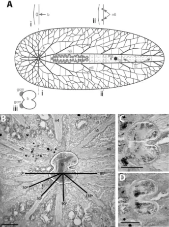
Six pairs of nerve cords covered by a sheath were found to branch out from the brain capsule (Figure 1A,B). The brain capsule did not extend along the branches; rather it appeared that the nerve cords perforated the capsule. The first four pairs innervated the region of the animal anterior to the brain in a well-defined radial pattern with respect to the longitudinal axis of the worm: n1 at 0°, n2 at 30°, n3 at 60°, and n4 at 90°; and the last two pairs (n5, n6) innervated the posterior region of the body in angles of approximately 135° and 180°, respectively (Figure 1B). Some nerve cords (n1/n2 and n5/n6) shared roots, whereas others (n3 and n4) did not share the same root but attached very close to each Figure 1- Polyclad nervous system organization: Category I.A. Sche-matic representation includes cross sections at the level of the brain (i) and at the level of the n6 nerve pair (ii); detail of the brain is shown below (iii)
B.Longitudinal section through the anterior region ofStyloplanocera fasciatashowing the distribution of the ventral nerve cords.Scale bar 250
mm.C.Section through the ventral region of theS. fasciatabrain, showing
the external globuli cell masses. Scale bar 250mm.D.Section through the
dorsal region of theS. fasciatabrain, showing the internal globuli cell masses. Scale bar 250mm. b brain, fg female gonopore, gcm globuli cell
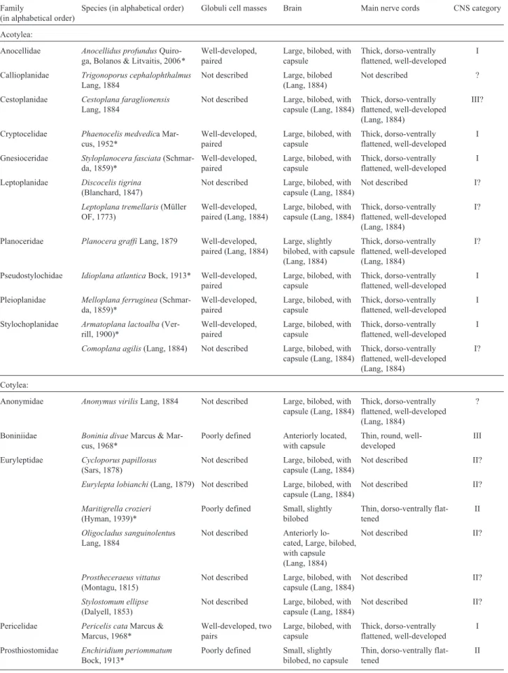
Table 1- Comparison of the nervous system in polyclads
Family
(in alphabetical order)
Species (in alphabetical order) Globuli cell masses Brain Main nerve cords CNS category
Acotylea:
Anocellidae Anocellidus profundus Quiro-ga, Bolanos & Litvaitis, 2006*
Well-developed, paired
Large, bilobed, with capsule
Thick, dorso-ventrally flattened, well-developed
I
Callioplanidae Trigonoporus cephalophthalmus
Lang, 1884
Not described Large, bilobed (Lang, 1884)
Not described ?
Cestoplanidae Cestoplana faraglionensis
Lang, 1884
Not described Large, bilobed, with capsule (Lang, 1884)
Thick, dorso-ventrally flattened, well-developed (Lang, 1884)
III?
Cryptocelidae Phaenocelis medvedica Mar-cus, 1952*
Well-developed, paired
Large, bilobed, with capsule
Thick, dorso-ventrally flattened, well-developed
I
Gnesioceridae Styloplanocera fasciata (Schmar-da, 1859)*
Well-developed, paired
Large, bilobed, with capsule
Thick, dorso-ventrally flattened, well-developed
I
Leptoplanidae Discocelis tigrina
(Blanchard, 1847)
Not described Large, bilobed, with capsule (Lang, 1884)
Not described I?
Leptoplana tremellaris(Müller OF, 1773)
Well-developed, paired (Lang, 1884)
Large, bilobed, with capsule (Lang, 1884)
Thick, dorso-ventrally flattened, well-developed (Lang, 1884)
I?
Planoceridae Planocera graffiLang, 1879 Well-developed, paired (Lang, 1884)
Large, slightly bilobed, with capsule (Lang, 1884)
Thick, dorso-ventrally flattened, well-developed (Lang, 1884)
I?
Pseudostylochidae Idioplana atlanticaBock, 1913* Well-developed, paired
Large, bilobed, with capsule
Thick, dorso-ventrally flattened, well-developed
I
Pleioplanidae Melloplana ferruginea (Schmar-da, 1859)*
Well-developed, paired
Large, bilobed, with capsule
Thick, dorso-ventrally flattened, well-developed
I
Stylochoplanidae Armatoplana lactoalba (Ver-rill, 1900)*
Well-developed, paired
Large, bilobed, with capsule
Thick, dorso-ventrally flattened, well-developed
I
Comoplana agilis(Lang, 1884) Not described Large, bilobed, with capsule (Lang, 1884)
Thick, dorso-ventrally flattened, well-developed (Lang, 1884)
I?
Cotylea:
Anonymidae Anonymus virilisLang, 1884 Not described Large, bilobed, with capsule (Lang, 1884)
Thick, dorso-ventrally flattened, well-developed (Lang, 1884)
?
Boniniidae Boninia divaeMarcus & Mar-cus, 1968*
Poorly defined Anteriorly located, with capsule
Thin, round, well-developed
III
Euryleptidae Cycloporus papillosus
(Sars, 1878)
Not described Large, bilobed, with capsule (Lang, 1884)
Not described II?
Eurylepta lobianchi(Lang, 1879) Not described Large, bilobed, with capsule (Lang, 1884)
Not described II?
Maritigrella crozieri
(Hyman, 1939)*
Poorly defined Small, slightly bilobed
Thin, dorso-ventrally flat-tened
II
Oligocladus sanguinolentus Lang, 1884
Not described Anteriorly lo-cated, Large, bilobed, with capsule (Lang, 1884)
Not described II?
Prostheceraeus vittatus
(Montagu, 1815)
Not described Large, bilobed, with capsule (Lang, 1884)
Not described II?
Stylostomum ellipse
(Dalyell, 1853)
Not described Large, bilobed, with capsule (Lang, 1884)
Not described II?
Pericelidae Pericelis cataMarcus & Marcus, 1968*
Well-developed, two pairs
Large, bilobed, with capsule
Thick, dorso-ventrally flattened, well-developed
I
Prosthiostomidae Enchiridium periommatum
Bock, 1913*
Poorly defined Small, slightly bilobed, no capsule
Thin, dorso-ventrally flat-tened
other in the brain (Figure 1B). Transverse commissures formed connections between the radial nerve cords (Figu-re 1A). Mo(Figu-re distant from the brain, the nerve cords of the first four pairs subdivided into dichotomous branches to-ward the margin, where they became thinner and connected with other nerve fibers originating from the proximal nerve cords. Thus, these nerve fibers formed polygons, and di-vided into a complex marginal net (Figure 1A). Cord pairs n5 and n6 innervated the region of the worm posterior to the brain. Pair n5 divided into dichotomous branches inner-vating the region from the level of the brain to the middle of the pharynx, and pair n6 started dividing where the pharynx began, innervating the remainder of the animal (Figu-re 1A). The number of main cords leaving the brain was constant in all the species analyzed within this category, al-though the degree of branching was variable. All nerve cords were dorsoventrally flattened and in transverse sec-tions, appeared oval (Figure 1A). The nerve cords of the n6 pair were the thickest ones with a diameter of about 250mm, identifying them as the main nerve cords for spe-cies in this category. The diameters of the remaining nerve cords varied from 125mm to 225mm. Pair n6 extended the entire length of the worm, lying parallel on either side of the pharynx and becoming thinner posteriorly (Figure 1A). Along their lengths, the two nerve cords were connected by transverse commissures and eventually, they divided into very thin branches, forming different plexi, such as the pha-ryngeal and reproductive plexi (Figure 1A).
Anterior to the brain was a pair of globuli cell masses (Figure 1C); This characteristic feature was common to all species in the category, except forPericelis cata, in which four well-developed globuli cell masses were located (above the roots of n2, n3, and n4). Distinctive from other species in the category that showed globuli cell masses only external to the brain capsule,S. fasciataglobuli cell masses were located both external to the capsule on the ventral side right between the roots of nerve cord pairs n2 and n3 (Fig-ure 1C), and dorsally in the interior of the capsule (Figure 1D).
Category II central nervous system
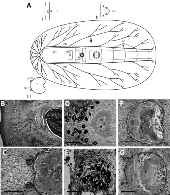
Species in this category (Table 1) showed: (a)slightly or not bilobed small brains(Figure 2), (b)thin main nerve cordsthat were dorsoventrally flattened and submerged in the parenchyma (Figure 2A, B), and (c) poorly defined globuli cell masses(Figure 2A-C, F, G) or thecomplete ab-sence of globuli cell masses. All species in this category be-longed to the suborder Cotylea (Table 1): Enchiridium
periommatum(Prosthiostomidae),Pseudoceros bolool, P.
bicolor (Pseudocerotidae), and Maritigrella crozieri
(Euryleptidae).In this category, the cerebral eyes were lo-cated in the close vicinity of and/or above the brain (Figure 2D, E).
Here we useEnchiridium periommatumas the repre-sentative species to describe the Category II NS. The brain consisted of a complex of neurites and neuronal bodies en-closed by a thin capsule. It was anteriorly located, at about 1/8 of the total length of the body. The cerebral eyes may be grouped into two clusters anterior to the brain as observed in this species (Figure 2D), but other species of this cate-gory may also present a single eye cluster directly above the brain (e.g. Pseudoceros bicolor, Figure 2E). The brain was only slightly bilobed with a shallow depression in the pos-terior (Figure 2A, B, D, F). However, in some species such
asPseudoceros bicolor(Figure 2E) andP. bolool(Figure
2G) it was not bilobed. In a mature, preserved specimen of
E. periommatum(19 mm x 9 mm) the brain was about 550
mm wide, about 525 mm long and about 300mm tall. All species in this category were characterized by having a small brain in relation to their body size when compared to species in Category I.
Six paired nerve cords radiated from the brain inE.
periommatumat different angles from those in category I
(Figure 2B). The main nerve cords apparently lacked the sheath observed in cords of Category I, or if such a sheath existed it was not well defined. Nerve cords (n1/n2 and n5/n6) also shared roots as inS. fasciata(Category I), and the arrangement and dichotomous branching of the main nerve cords were basically the same. However, the ramifi-cations had fewer transverse commissures connecting them (Figure 2A). The two main nerve cords (n6) had a diameter
Family
(in alphabetical order)
Species (in alphabetical order) Globuli cell masses Brain Main nerve cords CNS category
Prosthiostomum siphunculus
(Della Chiaje, 1822)
Not described Large, bilobed, with capsule (Lang, 1884)
Not described II?
Pseudocerotidae Pseudoceros bicolor
Verrill, 1902*
Absent Small, round, not bilobed
Thin, dorso-ventrally flat-tened
II
Pseudoceros boloolNewman & Cannon, 1994*
Absent Small, round, not bilobed
Thin, dorso-ventrally flat-tened
II
Thysanozoon brocchii
(Risso, 1818)
Poorly defined (Lang, 1884)
Small, round, not bilobed (Lang, 1884)
Thin, dorso-ventrally flat-tened (Lang, 1884)
II
*Examined in this study.
of about 90mm and ran parallel to the pharynx. These were connected by thin transverse commissures along the entire length of the body. The diameters of the remaining nerves cords varied from 35mm to 70mm.
The globuli cell masses were less defined than those in Category I. In most species, they consisted of few scat-tered cells above the roots of n1 and n2 cords (Figure 2C), and in Pseudocerotidae, globuli cell masses were com-pletely absent (Figure 2G). No interior globuli cells masses were observed in any of the specimens examined.
Category III central nervous system
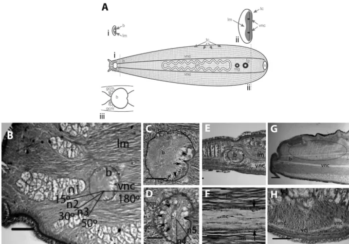
Species in this category (Table 1) showed: (a)a very anteriorly located brain(Figure 3A), (b)thick and round
main nerve cords (Figure 3A), and (c) poorly defined
globuli cell masses(Figure 3A, D). The sole species exam-ined in this category (Boninia divae) belongs to the Boniniidae (Table 1), a family currently classified in the Cotylea.In this species, the ventral longitudinal muscula-ture is extremely well-developed, and therefore the ventral nervous system of Boniniidae is completely submerged within the longitudinal muscles thus forming an intramus-Figure 2- Polyclad nervous system organization: Category II.A.Schematic representation includes cross sections at the level of the brain (i) and at the level of the n6 nerve pair (ii); detail of the brain is shown below (iii).B.Longitudinal section through the anterior region ofEnchirium periommatum
showing the distribution of the main ventral nerve cords. Scale bar 250mm.C.Anterior portion of theE. periommatumbrain, showing the poor
develop-ment of the globuli cell masses. Scale bar 100mm.D.Arrangement of the cerebral eyes inE. periommatum. Scale bar 100mm.E.Arrangement of the
cular plexus, in contrast to other rhabditophorans that gen-erally show a submuscular plexus. Both, brain and main nerve cords in this category are submerged within the well-developed longitudinal body wall muscles (Figure 3B, E, F).
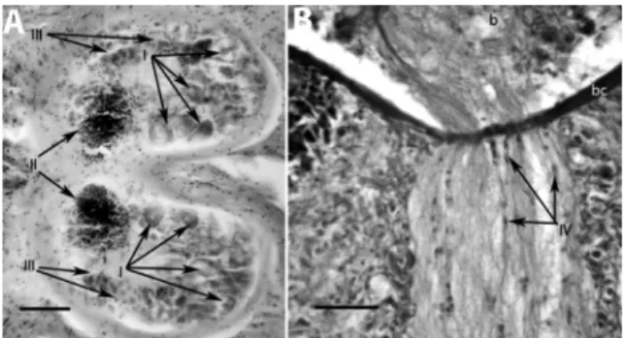
The well-defined brain capsule was about 175mm in diameter and was located very anteriorly at about 1/10 in relation to the total body length. The brain was bilaterally symmetrical and had a slight depression on the posterior area (Figure 3C, D). It contained Type I cells (see below) in the posterior and ventral parts of the brain (Figure 3C), and Type III cells (see below) in the anterior and dorsal sides (Figure 3D). Type II cells were also found exterior to the brain, some of them scattered anterior to the brain, forming very rudimentary globuli cell masses (Figure 3D).
The round main pair of nerve cords (n6) ran along the entire length of the body, parallel to each side of the phar-ynx (Figure 3A). In a mature individual (18 mm x 4 mm), these measured about 100mm in diameter. The main nerve cords connected ventrally to the brain (Figure 3B, E), and branched radially (Figure 3A). There were no clear perfora-tions of the brain capsule by these nerves cords. In addition to the main longitudinal cords, about 7 to 10 thinner cords ran parallel to them and all were connected by transverse commissures that crossed from one side of the body to the other (Figure 3A, G, H). This entire plexus formed a very consistent orthogonal pattern. At least three other pairs of nerve cords left the brain dorsally (not shown). These fibers subdivided into dichotomous branches, innervating the portion of the animal anterior to the brain, including the marginal tentacles. It is worthwhile to mention that a very Figure 3- Polyclad nervous system organization: Category III.A.Schematic representation includes cross sections at the level of the brain (i) and at the level of the n6 nerve pair (ii); detail of the brain is shown below (iii).B.Longitudinal section through the anterior region ofBoninia divae, showing the distribution of the ventral nervous branches and immersion of entire nervous system into the longitudinal musculature. Scale bar 150mm.C.Longitudinal
section through the ventral region of theB. divaebrain.Arrows indicate the Type I cells in the posterior portion of the brain. Scale bar 75mm.D.
Longitu-dinal section through the dorsal region of theB. divaebrain. Arrows indicate Type III cells in the anterior portion of the brain. Notice that the globuli cell masses are formed by Type II cells that are poorly defined. Scale bar 75mm.E.Sagittal section of theB. divaebrain, showing longitudinal musculature
surrounding the nervous system. Scale bar 150mm.F.Sagittal section of theB. divaebrain, showing a higher magnification through a ventral nerve cord
submerged in longitudinal musculature. Notice that the nerve cord is surrounded by a thin sheath (arrows). Scale bar 100mm.G.Anterior sagittal section showing the parallel branches and transverse commissures inB. divae, forming the orthogonal pattern (portion circled). Scale bar 250mm.H.Cross
sec-tion through a ventral commissure inB. divae. Scale bar 250mm. b brain, fg female gonopore, gcm globuli cell masses, mg male gonopore, n nerve cords,
diffuse, submuscular dorsal plexus was connected with the ventral plexus. A thin sheath was found covering all nerve cords in this category (Figure 3F).
Polyclad brain cell types
Within the encapsulated brains of all examined poly-clads, we identified four cell types (Types I-IV) based on their histology. The four cell types observed in the brain of Styloplanocera fasciataare shown in Figure 4:Type I cells were large ganglion cells with big nuclei located in the pos-terior part of the brain (Figure 4A), Type II cells were glob-ular cells which were characterized by a highly reduced cytoplasmic content and big nuclei rich in chromatin that occupied mostly the anterior rind of the brain and form the external globuli cell masses (Figure 4A), Type III cells were of medium size, and located mainly in the lateral parts along the periphery of the brain (Figure 4A), and Type IV cells were elongate and small, with sporadic occurrences along the nerve tracts, especially at bifurcations; a few were found in the posterior rind of the brain (Figure 4B).
Serotonin and FMRFamidein Boninia divae
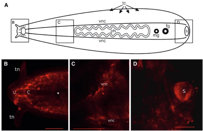
Serotonin (Figure 5) was expressed inBoninia divae in the apical area of the nerve cords (Figure 5B), which form a U-shaped structure (U) and a concentric C-shaped structure (C) anterior to the brain, along the main ventral nerve cords (Figure 5C), and in nerves associated with the sucker in the posterior of the animal (Figure 5D). Thinner longitudinal cords and the primary commissures that bran-ched off from the main longitudinal cords showed a faint expression of serotonin (Figure 5B, 5C), whereas the brain was characterized by a complete lack of serotonin expres-sion (Figure 5B).
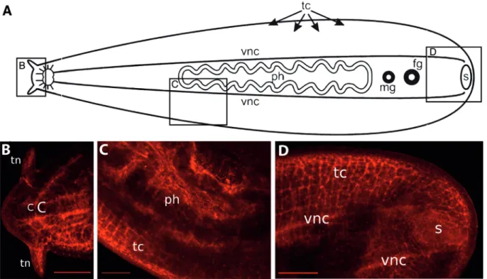
The neuropeptide FMRFamide expression is shown in Figure 6. Apical to the brain, FMRFamide was expressed in the transverse commissures that form a ladder-like net-work, in the concentric C-shaped structure (see above), and in nerve cords that extend into the tentacles (Figure 6B).
The brain also showed an absence of FMRFamide expres-sion in this species (Figure 6B). FMRFamide expresexpres-sion was observed in all longitudinal nerve cords and transverse commissures (Figure 6C), forming the typical orthogonal pattern that is completely submerged within the longitudi-nal muscles. FMRFamide expression was particularly strong in nerve cells of the pharyngeal plexus (Figure 6C) and in the ventral nerve cords (Figure 6D). Posteriorly, FMRFamide positive nerve cells were also found in the nerve plexus that surrounds the sucker, with a lesser expres-sion of in nerves directly associated with the sucker (Figure 6D).
Although some overlap of serotonin and FMRFamide expression was observed in the anterior of the animal, dis-tinct patterns of expression were observed in posterior structures. In the head, serotonin and FMRFamide expres-sion labeled the main and minor nerve cords, as well as the plexus innervating the tentacles. In addition, a bright and clear FMRFamide signal allowed observation of the main longitudinal ventral nerve cords, transverse commissures, and even thin nerves within the intramuscular plexus. In the posterior, seratonin primarily labeled the plexus of the sucker, whereas FMRFamide mostly labeled the pharyn-geal plexus.
Discussion
Evolution of the central nervous system in polyclads and the relevance of globuli cell masses
The CNS of all polyclad species examined in this study display the same overall configuration described in earlier studies (Hadenfeldt, 1929; Hyman, 1951; Bullock and Horridge, 1965; Minichev and Pugovkin, 1979). An anterior encapsulated brain, six pairs of ventral nervous branches, and the typical radial pattern forming a network seem to be constant features.
Minichev and Pugovkin (1979) contended that the nervous system in polyclads was not homologous to that of other flatworms. Contrary to their assertion, we identified a pair of distinctly thicker nerve cords (n6) that originated from an anteriorly located brain in all polyclad species ex-amined. The position, arrangement, and composition of these main nerve cords allow for homologizing of the polyclad CNS with the CNS of other flatworms. Spe-cifically, the polyclad CNS corresponds to the typical or-thogonal pattern common in other flatworms, in which two major longitudinal nerve cords are connected by transverse commissures at right angles in a ladder-like fashion (Rei-singer, 1925; Bullock and Horridge, 1965; Reuteret al., 1998). Kotikova (1986) already supported the homology of the polyclad CNS to that of other Platyhelminthes, and pro-posed that the dorsoventral flattening and spreading out of the polyclad body plan was driving the evolution of the pe-culiar polyclad orthogon.
Figure 4- Polyclad brain cell types. Longitudinal sections of the brain and nervous branches show distinguishable brain cells in Styloplanocera fasciata.A.Type I large ganglion cells with big nuclei; Type II globuli cells; Type III medium-sized cells. Scale bar 75mm.B.Type IV small cells of elongated shape located along the nerve tracts. Scale bar 25mm. b
In our comparative study, we have noticed that the shape and position of the pharynx, in association with spe-cies-specific behaviors, may also influence polyclad fea-tures, such as the organization of the orthogon, thickness of the nerve cords, the position and size of the brain, or the presence of globuli cell masses. In general, acotyleans show centrally located pharynges and hence, the position of the brain is shifted further posterior from the anterior body margin. Acotylean brains are generally larger, have better-developed external globuli cell masses, and the nerve cords are thicker, possibly indicating adaptive functions to a more complex neural integration, which may also be related to their generally more cryptic and benthic behaviors. In con-trast, cotyleans show more anteriorly located pharynges displacing the brain to a more rostral position, closer to the anterior body margin.
Well-developed external globuli cell masses are the diagnostic characteristic of all acotylean polyclads exam-ined in Category I. The cells are grouped in a cup-shaped fashion from which thin axons extend, forming a stalk that perforates the brain capsule. Similar structures have been found in nemerteans (Hanström, 1928), annelids including sipunculans (Golding, 1992; Åkesson, 1958), mollusks (Bullock and Horridge, 1965), and especially so in arthro-pods, where they are known as mushroom bodies
(Schürman, 1995; Strausfeldet al., 2006). The arthropod mushroom bodies are neuropiles of thin axons originating from clusters of small basophilic cells. They are part of the CNS and have been implicated in olfaction, learning, and memory (Strausfeldet al., 2006). The external globuli cell masses of all Category I species are morphologically simi-lar in composition and position to the mushroom bodies of arthropods and annelids, which in turn, share similarities with the vertebrate pallium (Tomeret al., 2010). However, their function in polyclads remains unresolved. If the exter-nal globuli cell masses of acotyleans prove to be homolo-gous to the mushroom bodies and the vertebrate pallium, then these structures may represent early sensory associa-tive centers in the evolution of invertebrate nervous sys-tems. However, further studies are needed to clarify the function of these structures and to establish homology.
include a centrally located ruffled pharynx, anteriorly di-rected uteri, and marginal eyes that surround the entire body. On the other hand, the presence of a ventral sucker, pseudotentacles, and uterine vesicles places the family into the Cotylea. A cladistic analysis of Cotylea resolved Pericelidae as a basal clade within the suborder and as sister group to another enigmatic polyclad family, the Boniniidae (Rawlinson and Litvaitis, 2008).
All other cotyleans examined in this study belong to our Category II, which is characterized by poorly devel-oped (e.g.,T. brocchii,see Lang, 1884) or absent globuli cell masses (e.g.,P. bicolor, P. bolool,this study). Further-more, globuli cell masses of this category never form mush-room-bodies. Finally, the position and size of our Type I brain cells appear to correspond well to the description of large ganglion cells in the posterior part of theT. brocchii brain (Lang, 1884). Although a functional correspondence remains to be established, we consider them homologous cells.
The phylogenetic position of Boniniidae is also con-troversial because it shares characteristics of cotyelans and acotyleans. Bock (1923) includes Boniniidae within Cotylea because of their marginal tentacles, the arrange-ments of the eyes, the sucker, and arrangement of uteri. Our study further supports Boniniidae as members of Cotylea because of their poorly defined globuli cell masses. How-ever, further and more complete molecular phylogenetic
studies with good taxon sampling of polyclad species are necessary to resolve this question.
Neurons and behaviourin Boninia divae
The particular fleshy consistency and elongated body shape of Boniniidae and their type of locomotion re-quire special modifications of their nervous systems. Spe-cies in this group are commonly found under smooth-surfaced rocks in the supralitoral zones. Their movement is best described as “leech-like.” Their ventral longitudi-nal musculature is extremely well developed, occupying almost half of the diameter of the worms. Although their CNS is still located ventrally, it is not submuscular as in other flatworms. Instead, the CNS of Boniniidae is com-pletely submerged in the longitudinal musculature, thus forming an intramuscular plexus. Thus, the unique charac-teristics of the boniniid CNS may represent an autapo-morphy of the family. However, the fact that Boninia divaeexhibits an orthogonal pattern demonstrates that this configuration may represent a plesiomorphy, or has evolved independently in different groups as previously suggested by Kotikova (1986, 1991).
Serotonin and FMRFamide expression along the main ventral nerve cords ofB. divaereveal their likely in-volvement in the regulation of motor neuron activities, as has been shown previously for other flatworms (Klaggeset al., 1996, Moneypenny et al., 2001, Gustafsson et al., Figure 6- FMRFamide expressing neurons inBoninia divae(category III).A.Diagram of the nervous system indicates the sites shown in panels B-D.B.
In the head, FMRFamide expression is observed in the anterior paired bilateral C-shaped nerve branches and neurons that innervate the tentacles. Notice that no expression is observed in the brain. Scale bar 100mm.C.FMRFamide expression in the pharyngeal plexus and in the submuscular plexus. Scale bar 200mmD.Mesh-like nerves surrounding the sucker also express FMRFamide. Scale bar 100mm. C C-shaped structure, vnc ventral nerve cords, ph
2002). Hence, it is possible that the close association be-tween motor neurons and musculature in this species al-lows for a faster locomotory response mediated by seroto-nin and FMRFamide. Serotoseroto-nin has also been reported to play important roles in muscular and sensory responses in polychaetes, and was found expressed in cells that inner-vate the somatic muscles (Golding, 1992).
Although a common and broad pattern of expression in the CNS of the worm is suggestive of relevant roles of both FMRFamide and serotonin for sensory and locomo-tory responses, only distinctive expression patterns allow for a predictive inference of these neuropeptides and the neurons expressing them. Because FMRFamide showed a relatively higher expression in nerve cells associated with the pharynx, we can predict that feeding or contractile pha-ryngeal action, sensory functions, absorption, secretion and/or digestion may be mediated by the FMRFaminergic system. Interestingly, FMRFamide has also been shown to regulate the motility of the digestive system in polychaetes (Krajniak and Greenberg, 1992) and the muscular contrac-tions of the pharynx in leeches (O’Garaet al., 1999). In contrast, because serotonin showed a relatively greater ex-pression in cells associated with the posterior sucker, we can predict that animal adhesion or posterior sucker action that operates the contraction and/or sensory response of this organ may be mediated preferentially by the serotonergic system. Expression of serotonin in the nerve plexus of the sucker has also been reported for cestodes (Webb and Mizukawa, 1985), and in a dense network of nerves at the tip of the tail of proseriates (Girstmairet al., 2014) and neodermatids (Biserova et al., 2000). Future studies are needed to test these hypotheses.
The brain of B. divae did not show any immuno-labelling of either FMRFamide or serotonin. A recent pub-lication describing the serotonergic system in Monocelis Eherenberg, 1831(Proseriata), which also has an encapsu-lated brain, only showed a faint immunosignal too in the brain (Girstmairet al., 2014). Electron microscopic studies in another polycladNotoplana acticolarevealed that most synaptic activity occurred in the rind of brain (Koopowitz, 1986), and therefore, the inner core of the brain may not be actively expressing neurotransmitters. In contrast, sero-tonergic immunoreactivity in the brain has been reported in flatworm species other than polyclads [e.g., Schmidtea
mediterranea(Cebrià, 2008),Fasciola hepaticaLinneaus,
1758 (Sukhdeo and Sukhdeo, 1988),Macrostomum juve-niles (Morriset al., 2007) andMacrostomumadults (Egger et al., 2007)]. Low immunoreactivity for serotonin in the brain ofB. divaemay be due to the presence of atypical cells, or may in fact, represents a biologically sound level of serotonin release for polyclads. Alternatively, it is possible that either suboptimal technical conditions or a lower affin-ity of the antibody during the ICC may have generated the observed differences in expression signal. Additional
neu-ral markers remain to be tested to further characterize polyclad brains.
Evolution of the central nervous system in Platyhelminthes
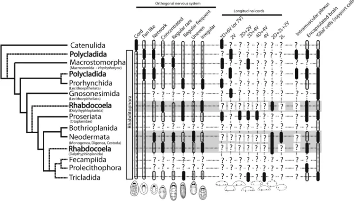
Trends that have been proposed for the evolution of the nervous system in flatworms include: (a) a transition from a diffuse nerve net of the bilaterian ancestor to the ventralized nervous system of protostomes (Lowe,et al., 2006); (b) reductions in the numbers of longitudinal cords (Reisinger, 1970, 1972); (c) a fusion of nerves of the sub-epidermal plexus to form longitudinal cords (Joffe and Reuter, 1993); (d) a conservation of the nervous circuits controlling the movement of the ciliated epidermis (Morris, 2007); (e) a transition from capsule-less brains in the ances-tor to encapsulated brains (Kotikova and Raikova, 2008); and (f) a convergent evolution of glial cells in different platyhelminth groups (Radojcic and Pentreath, 1979; Har-tenstein and Ehlers, 2000; Morriset al., 2004). To test these hypotheses, we used the most recent molecular phylogenies available for Platyhelminthes (Laumer and Giribet, 2014; Eggeret al., 2015; Laumeret al., 2015), and mapped rele-vant characters of flatworm nervous systems onto it. Char-acters for the phylogenetic reconstruction (Figure 7) were obtained from published literature and from our current findings on polyclad nervous systems. In spite of the exten-sive literature on the topic, many groups remain poorly studied (Figure 7), including the Gnosonesimida, Bothrio-planida, Fecampiida, and Prolecithophora. In the follow-ing, we discuss whether each of the trends finds support based on our current understanding of flatworm nervous system evolution.
(a) A transition from a diffuse nerve net of the
bilaterian ancestor to the ventralized nervous system of
protostomes:Current phylogenies place Catenulida as the
basal clade of Platyhelminthes. Although catenulids show a subepithelial, dispersed nerve network, a central nervous system develops by the presence of interconnected longitu-dinal cords (see cord-like orthogonal nervous system, Fig-ure 7), and some species such as Catenula Duges, 1832 even show a regular configuration of the peripheral nervous system (Moraczewskiet al., 1977). Studied species in all other platyhelminth groups show the well-defined central nervous system (i.e. orthogon) and a peripheral nervous system (Figure 7); thus, the prediction of an ancestor with a nerve net is not supported. However, proper outgroups re-main to be analyzed comparatively.
only include the brain plus one pair of main nerve cords in their definition. Our current descriptions of the polyclad nervous systems also showed a pair of main cords in all ex-amined species.
(c)A fusion of nerves of the subepidermal plexus to
form longitudinal cords:This trend may actually explain
the mechanism of the reduction of longitudinal cords dis-cussed in (b).
(d)A conservation of the nervous circuits controlling
the movement of the ciliated epidermis: The
well-con-served flattened body plan of flatworms may impose cer-tain developmental constraints onto the nervous system architecture and molecular circuitry that controls locomo-tion. To support this trend, the organization of the nervous system is generally found to be well-conserved, including the subepidermal plexus and the neurotransmitter expres-sion. However, two exceptions are observed: (1) in para-sitic worms that show the most variable organization of the nervous system of all groups (see shaded characters of the Neodermatida and the Rhabdocoela; Figure 7), most likely due to functional adaptations to a parasitic life style; and (2) in the Polycladida with inframuscular plexi that could ei-ther be a functional adaptation to quick escape response mechanisms and strong wave action in their habitat (rocky
intertidal environments), or simply an ancestral state sup-porting a more basal position of the Polycladida among the Platyhelminthes.
(e)A transition from neuropile brains in the ancestor
to encapsulated brains:There are two possible scenarios
for the evolution of encapsulated brains (Figure 7): (1) the encapsulated brains in flatworms evolved at least four times independently in Polycladida, Proseriata, Rhabdo-coela, and Prolecithophora, or (2) the encapsulated brain evolved once in the ancestor of the Rhabditophora, and was lost three times in Prorhynchida, Neodermata, and Tricla-dida. Although the rhabdocoels generally show a brain capsule, Strongylostoma simplexMeixner, 1915 is an ex-ception.
(f)A convergent evolution of glial cells in different
platyhelminth goups: Glial-like cells in flatworms have
evolved convergently at least five times to provide support, isolation, or metabolic functions to the nerve cells of the CNS.
molecular phylogenies, a deep evolutionary history of the phylum, and a serious lack of knowledge in many relevant groups are only some of the factors why this task has re-mained difficult. Consequently, this study provides not only original descriptive work on an understudied group of species, but also highlights some important gaps in our cur-rent understanding of the flatworn nervous system.
Acknowledgments
We thank Dr. Janet Voight from the Field Museum of Natural History, Chicago, Illinois, USA for specimens of Anocellidus profundus, and Dr. Bernhard Egger for the ex-perimental advice on the ICCs. A UNESCO-L’OREAL In-ternational Fellowships Programme for Young Women in Life Sciences was awarded to D.M.B., and a Sigma Xi Grants-in-Aid for Research was granted to E.C.B. This work was supported in part by the Facultad de Ciencias at Universidad de los Andes and Fundación Banco de la República de Colombia to D.M.B, E.C.B. and F.D.B, and an “Apoio aos Novos Docentes” Grant from Universidade de São Paulo to F.D.B. This work also was supported by NSF grant DEB-0412932 to M.K.L.
References
Åkesson B (1958) A study of the nervous system of the Sipun-culoideae, with some remarks on the development of the two speciesPhascolion strombiMontagu andGolfingia minuta Keferstein. Undersokningar over Oresund 38:1-249. Baguñà J and Riutort M (2004) Molecular phylogeny of the
Platyhelminthes. Can J Zool 82:168-193.
Bailly X, Reichert H and Hartenstein V (2013) The urbilaterian brain revisited: Novel insights into old questions from new flatworm clades. Dev Genes Evol 223:149-157.
Biserova NM, Dudicheva VA, Terenina NB, Reuter M, Halton DW, Maule AG and Gustafsson MK (2000) The nervous system of Amphilina foliacea (Platyhelminthes, Amphilinidea), an immunocytochemical, ultrastructural and spectrofluorometrical study. Parasitology 121:441-453. Bock S (1923)Boninia,a new polyclad genus from the Pacific.
Nov Act Reg Soc Uppsala Ser 46:1-32.
Böckerman I, Reuter M and Timoshkin O (1994) Ultrastructural study of the central nervous system of endemic Geocentrophora (Prorhynchida, Platyhelminthes) from Lake Baikal. Acta Zool 75:47-55.
Bullock TH and Horridge GA (1965) Structure and Function in the Nervous Systems of Invertebrates. Vol. 2. W.H. Free-man and Co, San Francisco, 1719 pp.
Carranza S, Baguñà J and Riutort M (1997) Are the Platyhel-minthes a monophyletic primitive group? An assessment us-ing 18S rDNA sequences. Mol Biol Evol 14:485-497. Cebrià F (2008) Organization of the nervous system in the model
planarian Schmidtea mediterranea: An immunocyto-chemical study. Neurosci Res 61:375-384.
Chien P and Koopowitz H (1972) The ultrastructure of neuro-muscular systems inNotoplana acticola, a free-living poly-clad flatworm. Z Zellforsch Mikroskop Anat 133:277-288.
Chien PK and Koopowitz H (1977) Ultrastructure of nerve plexus in flatworms. III. The infra-epithelial nervous system. Cell Tissue Res 176:335-347.
Day TA, Maule AG, Shaw C and Pax RA (1997) Structure-activity relationships of FMRFamide-related peptides con-tractingSchistosoma mansonimuscle. Peptides 18:917-921. Egger B, Gschwentner R and Rieger R (2007) Free-living flatworms under the knife: Past and present. Dev Genes Evol 217:89-104.
Egger B, Lapraz F, Tomiczek B, Müller S, Dessimoz C, Girstmair J, Skunca N, Rawlinson KA, Cameron CB, Beli Eet al. (2015) A transcriptomic-phylogenomic analysis of the evo-lutionary relationships of flatworms. Curr Biol 25:1-7. Ehlers U (1985) Das Phylogenetische System der Plathelminthes.
Gustav Fischer Verlag, Stuttgart, 317 pp.
Fernandes MC, Alvares EP, Gama P and Silveira M (2003) Sero-tonin in the nervous system of the head region of the land planarianBipalium kewense. Tissue Cell 35:479-486. Forest DL and Lindsay SM (2008) Observations of serotonin and
FMRFamide-like immunoreactivity in palp sensory struc-tures and the anterior nervous system of spionid poly-chaetes. J Morphol 269:544-551.
Girstmair J, Schnegg R, Telford MJ and Egger B (2014) Cellular dynamics during regeneration of the flatwormMonocelissp. (Proseriata, Platyhelminthes). Evo Devo 5:e37.
Golding DM (1992) Polychaeta: Nervous system. In: Harrison FW and Gardiner SL (eds) Microscopic Anatomy of Inver-tebrates. Vol. 7. Wiley-Liss, New York, pp 155-179. Gustafsson MKS, Halton DW, Kreshchenko ND, Movsessian SO,
Raikova OI, Reuter M and Terenina NB (2002) Neuro-peptides in flatworms. Peptides 23:2053-2061.
Hadenfeldt D (1929) Das Nervensystem von Stylochoplana maculata und Notoplana atomata. Z wiss Zool 133:586-638.
Halton DW and Gustafsson MKS (1996) Functional morphology of the platyhelminth nervous system. Parasitology 113:S47-S72.
Hanström B (1928) Vergleichende Anatomie des Nervensystems der wirbellosen Tiere, unter Berücksichtigung seiner Funktion. Springer Verlag, Berlin, 628 pp.
Hartenstein V and Ehlers U (2000) The embryonic development of the rhabdocoel flatwormMesostoma lingua(Abildgaard, 1789). Dev Genes Evol 210:399-415.
Hyman LH (1941) Terrestrial flatworms from Canal Zone, Pan-ama. Am Mus Novitat 1105:1-11.
Hyman LH (1951) The Invertebrates: Platyhelminthes and Rhyn-chocoela, the Acoelomate Bilateria. Vol. 2. McGraw-Hill, New York,. 550 pp.
Joffe BI and Reuter M (1993) The nervous system of Bothriomolus balticus (Proseriata): A contribution to the knowledge of the orthogon in the Plathelminthes. Zoomorphology 113:113-127.
Johnston RN, Shaw C, Halton DW, Verhaert P, Blair KL, Bren-nan GP, Price DA and Anderson PA (1996) Isolation, local-ization, and bioactivity of the FMRFamide-related neuro-peptides GYIRFamide and YIRFamide from the marine turbellarianBdelloura candida. J Neurochem 67:814-821. Karling TG (1967) Zur Frage von dem systematischen Wert der
Karling TG (1968) On the genus Gnosonesima Reisinger (Turbellaria). Sarsia 33:81-108.
Karling TG (1974) On the anatomy and affinities of the turbel-larian orders. In: Riser NW and Morse MP (eds) Biology of the Turbellaria. McGraw-Hill, New York, pp 1-16. Klagges BRE, Heimbeck G, Reifegerste R, Reisch D,
Godenschwege TA, Buchner S, Hofbauer A and Buchner E (1996) Invertebrate synapsins: A single gene codes for sev-eral isoforms in Drosophila. J Neurosci 16:3154-3165. Koopowitz H (1973) Primitive nervous systems. A sensory nerve
net in the polyclad flatwormNotoplana acticola.Biol Bull 145:352.
Koopowitz H (1974) Some aspects of the physiology and organi-zation of the nerve plexus in polyclad flatworms. In: Riser NW and Morse MP (eds) Biology of the Turbellaria. McGraw-Hill, New York, pp 198-212.
Koopowitz H and Chien P (1974) Ultrastructure of the nerve plexus in flatworms. I. Peripheral organization. Cell Tissue Res 155:337-351.
Koopowitz H and Chien P (1975) Ultrastructure of nerve plexus in flatworms. II. Sites of synaptic interactions. Cell Tissue Res 157:207-216.
Koopowitz H (1986) On the evolution of central nervous systems: Implications from polyclad turbellarian neurobiology. Hydrobiologia 132:79-87.
Kotikova EA (1986) Comparative characterization of the nervous system of the Turbellaria. Hydrobiologia 132:89-92. Kotikova EA (1991) The orthogon of the Plathelminthes and main
trends in evolution. Proc Zool Inst St Petersburg 241:88-111 [in Russian].
Kotikova EA and Raikova OI (2011) Architectonics of the central nervous system of Acoela, Platyhelminthes, and Rotifera. J Evol Biochem Physiol 44:95-108.
Krajniak KG and Greenberg MJ (1992) The localization of FMRFamide in the nervous and somatic tissues ofNereis virens and its effects upon the isolated esophagus. Comp Biochem Physiol C 101:93-100.
Kreshchenko N and Tolstenkov OO (2012) Some aspects of the immunolocalization of FMRFamide in the nervous system of turbellarians,Polycelis tenuisandGirardia tigrina.Acta Biol Hung 63(Suppl 2):83-87.
Ladurner P, Mair GR, Reiter D, Salvenmoser W and Rieger RM (1997) Serotonergic nervous system of two macrostomid species: Recent or ancient divergence? Invert Biol 116:178-191.
Lang A (1884) Die Polycladen (Seeplanarien) des Golfes von Neapel und der angrenzenden Meeresabschnitte: Eine Monographie. Atlas. Vol. 11. W. Engelmann Verlag, Leip-zig, 688 pp.
Laumer CE and Giribet G (2014) Inclusive taxon sampling sug-gests a single, stepwise origin of ectolecithality in Platy-helminthes. Biol J Linn Soc 111:570-588.
Laumer CE, Hejnol A and Giribet G (2015) Nuclear genomic sig-nals of the “microturbellarian” roots of platyhelminth evolu-tionary innovation. Elife 4:e05503.
Litvaitis MK and Rhode K (1999) A molecular test of platy-helminth phylogeny: Inferences from partial 28rDNA se-quences. Invert Biol 118:42-56.
Litvaitis MK, Bolanos DM and Quiroga SY (2010) When names are wrong and colours deceive: Unraveling thePseudoceros
bicolor species complex (Turbellaria, Polycldida). J Nat Hist 44:829-845.
Lowe CJ, Terasaki M, Wu M, Freeman RM, Runft L, Kwan K, Haigo S, Aronowicz J, Lander E, Gruber Cet al.(2006) Dorsoventral patterning in hemichordates: Insights into early chordate evolution. PLoS Biol 4:e291.
Mäntylä K, Halton DW, Reuter M, Maule AG, Lindroos P, Shaw C and Gustafsson MKS (1998a) The nervous system of Tricladida. IV. Neuroanatomy of Planaria torva (Paludi-cola, Planaridae): An immunocytochemical study. Hydro-biologia 383:167-173.
Mäntylä K, Reuter M, Halton DW, Maule AG, Brennan GP, Shaw C and Gustafsson MKS (1998b) The nervous system of Procerodes littoralis(Maricola, Tricladida). An ultrastruc-tural and immunoelectron microscopical study. Acta Zoo-logica 79:1-8.
Marks NJ, Johnston S, Maule AG, Halton DW, Shaw C, Geary TG, Moore S and Thompson DP (1996) Physiological ef-fects of platyhelminths RFamide peptides on muscle-strip preparations of Fasciola hepatica (Trematoda, Digenea). Parasitology 113:393-401.
Maule AG, Halton DW, Shaw C and Thim L (1993) GNFFRFamide: A novel FMRFamide-immunoreactive peptide isolated from the sheep tapeworm, Moniezia expansa. Biochem Biophys Res Comm 193:1054-1060. Minichev Y and Pugovkin AP (1979) Nervous system of the
polyclad flatwormNotoplana atomata(O.F. Müller). Cah Biol Mar 20:181-188.
Monneypenny CG, Maule AG, Shaw C, Day TA, Pax RA and Halton DW (1997) Physiological effects of platyhelminth FMRFamide-related peptides (FaRPs) on the motility of the monogenean Diclidophora merlangi. Parasitology 115:281-288.
Moneypenny CG, Kreshchenko N, Moffett CL, Halton DW, Day TA and Maule AG (2001) Physiological effects of FMRFa-mide-related peptides and classical transmitters on dispersed muscle fibres of the turbellarian,Procerodes littoralis. Para-sitology 122:447-455.
Moraczewski J, Czubaj A and Bakowska J (1977) Organization and ultrastructure of the nervous system in Catenulida (Turbellaria). Zoomorphologie 87:87-95.
Morris J, Cardona A, De Miguel-Bonet MDM and Hartenstein V (2007) Neurobiology of the basal platyhelminth Macrostomum lignano:Map and digital 3D model of the ju-venile brain neuropile. Dev Genes Evol 217:569-584. Morris J, Nallur R, Ladurner P, Egger B, Rieger R and Hartenstein
V (2004) The embryonic development of the flatworm Macrostomumsp. Dev Genes Evol 214:220-239.
Mousley A, Moffett CL, Duve H, Thorpe A, Halton DW, Geary TG, Thompson DP, Maule AG and Marks NJ (2005) Ex-pression and bioactivity of allatostatin-like neuropeptides in helminths. Int J Parasitol 35:1557-1567.
Newman LJ and Cannon LRG (1994) Pseudoceros and Pseudobioceros (Platyhelmithes, Polycladida, Pseudo-cerotidade) from eastern Australia and Papua New Guinea. Mem Qld Mus 37:205-266.
Presnell JK and Schreibman M (1997) Humason’s Animal Tissue Techniques. 5th edition The John Hopkins University Press, Baltimore, 572 pp.
Radojcic T and Pentreath VW (1979) Invertebrate glia. Prog Neurobiol 12:115-179.
Rawlinson KA and Litvaitis MK (2008) Cotylea (Polycladida): A cladistic analysis of morphology. Invertebr Biol 127:121-138.
Reisinger E (1925) Untersuchungen am Nervensystem der Bothrioplana semperiBraun. Z Morphol Ökol Tiere 5:119-149.
Reisinger E (1968) Xenoprorhynchus ein Modellfall für progres-siven Funktionswechsel. Z zoolog Syst Evolutionsforsch 6:1-55.
Reisinger E (1970) Zur Problematik der Evolution der Coelo-maten. J Zool Syst Evol Res 8:81-109.
Reisinger E (1972) Die Evolution des Orthogons der Spiralier und das Archicölomatenproblem. J Zool Syst Evol Res 10:1-43. Reuter M (1988) Development and organization of nervous
sys-tems visualized by immunocytochemistry in three flatworm species. Fortschr Zool 36:181-184.
Reuter M and Gustafsson MK (1995) The flatworm nervous sys-tem: Pattern and phylogeny. In: Breidbach O and Kutsch W (eds) The Nervous System of Invertebrates: An Evolution-ary and Comparative Approach. Birkhäuser, Basel, pp 25-59.
Reuter, M, Gustafsson MKS, Mäntylä K and Grimmelikhuijzen CJP (1996) The nervous system of Tricladida. III. Neuro-anatomy of Dendrocoelum lacteum and Polycelis tenuis (Plathelminthes, Paludicola): An immunocytochemical study. Zoomorphology 116:111-122.
Reuter M and Halton DW (2001) Comparative neurobiology of Platyhelminthes. In: Littlewood DTJ and Bray RA (eds) In-terrelationships of the Platyhelmithes. Taylor and Francis, London and New York, pp 239-249.
Reuter M, Mäntylä K and Gustafsson MKS (1998) Organization of the orthogon - Main and minor nerve cords. Hydro-biologia 383:175-182.
Reuter M, Maule AG, Halton DW, Gustafsson MKS and Shaw C (1995) The organization of the nervous system in Plathel-minthes. The neuropeptide F-immunoreactive pattern in Catenulida, Macrostomida, Proseriata. Zoomorphology 115:83-97.
Reuter M, Wikgren M and Lehtonen M (1986) Immunocyto-chemical demonstration of 5-HT-like and FMRF-amide-like substances in whole mounts of Microstomum lineare (Turbellaria). Cell Tissue Res 246:7-12.
Rieger RM, Tyler S, Smith JPS and Rieger GE (1991) Platy-helminthes: Turbellaria. In: Harrison FW and Bogitsh BJ
(eds) Microscopic Anatomy of Invertebrates. Platyhelmin-thes and Nemertinea. Wiley-Liss, NewYork, pp 7-140. Ruppert EE and Barnes RD (1994) Invertebrate Zoology.
Saun-ders College Publishing, New York, 1056 pp.
Schürmann FW (1995) Common and special features of the ner-vous system of Onychophora: A comparison with Arthropoda, Annelida and some other invertebrates. In: Breidbach O and Kutsch W (eds) The Nervous Systems of Invertebrates: An Evolutionary and Comparative Approach. Birkhäuser, Basel, pp 139-158.
Shishov BA (1991) Aminergic elements in the nervous system of helminthes. In: Sakharov DA and Winlow W (eds) Simpler Nervous Systems. Vol 13. Manchester University Press, Manchester, pp 113-137.
Sluys R (1989) A monograph of the marine triclads. A.A. Balke-ma, Rotterdam and Brookfield, 463 pp.
Steinböck O (1927) Monographie der Prorhynchidae (Turbellaria) Z Morphol Ökol Tiere 8:538-662.
Steinböck O and Reisinger E (1924) OnProrhynchus putealis Haswell, with a description of a new species of the genus. Quart J Micr Sci 68:443-451.
Strausfeld NJ, Strausfeld CM, Loesel R, Rowell D and Stowe S (2006) Arthropod phylogeny: Onychophoran brain organi-zation suggests an archaic relationship with a chelicerate stem lineage. Proc Biol Sci 273:1857-1866.
Sukhdeo SC and Sukhdeo MV (1988) Immunohistochemical and electrochemical detection of serotonin in the nervous system of Fasciola hepatica, a parasitic flatworm. Brain Res 463:57-62.
Timoshkin OA (1991) Turbellaria Lecithoepitheliata: Morphol-ogy, systematics, phylogeny. Hydrobiologia 227:323-332. Tomer R, Denes AS, Tessmar-Raible K and Arendt D (2010)
Pro-filing by image registration reveals common origin of annelid mushroom bodies and vertebrate pallium. Cell 142:800-809.
Villar D and Schaeffer DJ (1993) Morphogenetic action of neuro-transmitters on regenerating planarians - A review. Biomed Env Sci 6:327-347.
Von Graff L (1899) Monographie der Turbellarien II. Tricladida Terricola (Landplanarien). Wilhelm Engelmann Verlag, Leipzig 574 pp.
Webb RA and Mizukawa K (1985) Serotonin-like immunoreac-tivity in the cestodeHymenolepis diminuta.J Comp Neurol 234:431-440.
Wikgren MC and Reuter M (1985) Neuropeptides in a micro-turbellarian - Whole mount immunocytochemistry. Peptides 6:471-475.
Associate Editor: Igor Schneider