Anatomic variation of cranial
parasympathetic ganglia
Abstract: Having broad knowledge of anatomy is essential for prac-ticing dentistry. Certain anatomical structures call for detailed studies due to their anatomical and functional importance. Nevertheless, some structures are dificult to visualize and identify due to their small volume and complicated access. Such is the case of the parasympathetic ganglia located in the cranial part of the autonomic nervous system, which in-clude: the ciliary ganglion (located deeply in the orbit, laterally to the optic nerve), the pterygopalatine ganglion (located in the pterygopalatine fossa), the submandibular ganglion (located laterally to the hyoglossus muscle, below the lingual nerve), and the otic ganglion (located medially to the mandibular nerve, right beneath the oval foramen). The aim of this study was to present these structures in dissected anatomic specimens and perform a comparative analysis regarding location and morphology. The proximity of the ganglia and associated nerves were also analyzed, as well as the number and volume of ibers connected to them. Human heads were dissected by planes, partially removing the adjacent struc-tures to the point we could reach the parasympathetic ganglia. With this study, we concluded that there was no signiicant variation regarding the location of the studied ganglia. Morphologically, our observations con-cur with previous classical descriptions of the parasympathetic ganglia, but we observed variations regarding the proximity of the otic ganglion to the mandibular nerve. We also observed that there were variations regarding the number and volume of iber bundles connected to the sub-mandibular, otic, and pterygopalatine ganglia.
Descriptors: Anatomy; Ganglia, parasympathetic. Selma Siéssere(a)
Mathias Vitti(b)
Luiz Gustavo de Sousa(c) Marisa Semprini(b)
Mamie Mizusaki Iyomasa(d) Simone Cecílio Hallak Regalo(d)
(a) PhD in Anatomy, Assistant Professor; (b)Full
Professors; (c)BSc (Biology), Laboratory
Specialist in Anatomy; (d)Associate
Professors – Department of Morphology, Stomatology and Physiology, School of Dentistry of Ribeirão Preto, University of São Paulo.
Corresponding author:
Selma Siéssere
Faculdade de Odontologia de Ribeirão Preto – USP
Depto. de Morfologia, Estomatologia e Fisiologia
Avenida do Café, s/n Ribeirão Preto - SP - Brazil CEP: 14040-904 E-mail: [email protected]
Introduction
A ganglion is a mass of nervous tissues found in some peripheral nerves. Ganglia are located on the roots of spinal nerves and on the sensitive roots of the trigeminal, facial, glossopharyngeal, vagus, and vestibulochoclear nerves. Ganglia also appear in as-sociation with the autonomic nervous system. Each ganglion is covered by a smooth and dense capsule of ibrous connective tissue, with cells similar to as-sociated lattened ibrocytes, which extends to the nerves’ perineurium, sending numerous extensions to the ganglion’s interior. Ganglia vary considerably in size, shape1 and location. Hence, broad knowledge
on these structures is essential in dentistry, due to their anatomical and functional importance. Thus, detailed studies are called for. However, some are dificult to visualize and identify due to their small volume and complicated access. Such is the case of the parasympathetic ganglia. Located in the cranial section of the autonomic nervous system, they are the ciliary, pterygopalatine, submandibular, and otic ganglia. These structures are commonly mentioned in literature, but not frequently observed in ana-tomical material. Hence, the aim of this work was to present the ciliary, pterygopalatine, submandibu-lar, and otic ganglia in dissected anatomic pieces and perform a comparative analysis regarding location and morphology. The proximity of ganglia and asso-ciated nerves were also analyzed, as well as the num-ber and volume of inum-ber bundles connected to them.
Material and Methods
Forty human adult heads (males and females, av-erage age 40 years) from the Anatomy Laboratory, School of Dentistry of Ribeirão Preto, University of São Paulo were ixed in formalin (10%) and later sectioned in a median plane with a manual saw. By planes, the dissection was performed using scalpels numbers 3 and 4, curved scissors (sharp and rhomb-point), iris nippers, retractors, osteotomes and loupe, partially removing the adjacent structures to the point we could reach the parasympathetic ganglia.
Results
After the dissection, we could see that in 28 hu-man heads the ciliary ganglion was located deeply
in the eye socket, in its posterior third, laterally to the optic nerve, and it was functionally attached to the oculomotor nerve. In the remaining 12 human heads, we observed that the ganglion occupies a pos-teroinferior, intermediate or anterosuperior position in respect to the common tendinous ring and optic nerve. Regarding morphology, the ciliary ganglion had a small, oval, and lattened shape, measuring approximately 1 x 1 mm.
The pterygopalatine ganglion was located in the pterygopalatine fossa and was functionally attached to the facial nerve. This ganglion had the largest size when compared to the other ganglia and it had a lattened form.
The submandibular ganglion was located ally and superiorly to the hyoglossus muscle,
later-1
5
2 6
3
7
ally to the submandibular gland, under the lingual nerve, and was functionally attached to the facial nerve by the chorda tympani nerve. It appeared small and fusiform.
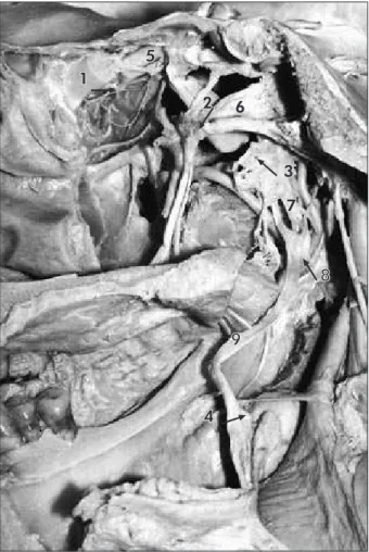
The otic ganglion was situated medially to the mandibular nerve, right under the oval foramen and was functionally attached to the glossopharyngeal nerve. The otic ganglion also had a lattened, small, and oval form (Figure 1).
The comparative study of the anatomical materi-al provided us with some disparities, such as the fol-lowing: for the submandibular ganglion, we noticed a variation in number and volume of iber bundles attached to it, as well as its association point to the lingual nerve. In addition, it presented an alteration
regarding the proximity to the lingual nerve, rang-ing from 2 mm to 6 mm (Figures 2 and 3).
The otic and pterygopalatine ganglia also showed a disparity regarding number and volume of iber bundles connected to them. As to the proximity of nerves, the otic ganglion also showed a variation concerning distance to the anterior section of the mandibular nerve, which ranged from 0 to 4 mm (Figures 4 and 5).
The pterygopalatine and ciliary ganglia did not show any variations in proximity to the maxillary and optic nerve, respectively.
Discussion
Our results are in agreement with those of other
1 5
2
3 4
1 5
2
3
4
Figure 3 - Medial view of a mandible. 1. Submandibular Ganglion; 2. Lingual Nerve; 3. Major Sublingual Duct; 4. Submandibular Gland; 5. Sublingual Gland.
Figure 2 - Medial view of a mandible. 1. Submandibular Ganglion; 2. Lingual Nerve; 3. Submandibular Gland; 4. Sublingual Gland; 5. Mylohyoid Nerve.
1
5 2
3
4
Figure 4 - Medial view of a human head (sagittal section). 1. Otic Ganglion; 2. Ophtalmic Nerve; 3. Maxillary Nerve; 4. and 5. Mandibular Nerve.
1 2
3
studies in the literature, regarding the fact that, in most of the studied human heads, it was observed that the ciliary ganglion is located close to the or-bital cavity’s apex, between the optic nerve and the lateral rectus muscle.1-5
Moreover, in terms of location, our study agrees with the results found by Tsybul’kin6 (2003), who
observed that the ganglion occupies a posteroin-ferior, intermediate or anterosuperior position in respect to the common tendinous ring (Zinn’s liga-ment) and optic nerve. This author also found that a posteroinferior position of the ganglion was typical to brachycephals, while the anterosuperior position was found in dolichocephals.
From a morphological point of view, the cili-ary ganglion is small and lat, about the size of a pinhead.1 This ganglion may also be oval shaped,2
quadrilateral shaped,7 of irregular form, egg-shaped,
or rectangular.4 In addition, measuring 1 mm by
2 mm2 or 3 mm by 3 mm,8 it can be as big as a corn
seed7 or be positioned with its largest diameter
par-allel to the optic nerve’s axis.4
The ciliary ganglion found in our anatomic spec-imens was also very small and with a morphology very similar to that described by Warwick, Wil-liams1 (1979) and by Dubrul2 (1991).
The pterygopalatine ganglion is important for intra-ocular pressure balance and cerebral vasodila-tation associated to vascular originated headaches.9
It is located deeply in the pterygopalatine fossa,1-3
close to the sphenopalatine foramen and in front of the pterygoid canal.1
Concerning morphology, there are several de-scriptions of the pterygopalatine ganglion. Accord-ing to some researchers, the pterygopalatine gan-glion is slightly lat,1,2 with a triangular and lat
form2 and is the biggest peripheral ganglion of the
cranial parasympathetic system. However, for oth-ers this ganglion is polymorphic, and can be rhom-boidal, pear-shaped, semi-lunar, triangular, or fusi-form, with volume similar to that of a lentil (5 mm to 7 mm length).7 In our study, the pterygopalatine
ganglion had a lat form and presented the largest size (3 to 5 mm); hence, our results concur with the description made by Warwick, Williams1 (1979).
According to the literature, the submandibular
ganglion is located in the upper part of the hyoglos-sus muscle.1 It is an autonomous nervous structure
with reduced dimensions and an aspect that could vary: triangular, egg-shaped, or plexiform. Its size can be compared to that of a lentil or a corn seed; therefore, its dissection is extremely dificult.7 Our
anatomic pieces showed that the submandibular ganglion is a small, fusiform structure (2 to 3 mm), which is in accordance with the description made by Warwick, Williams1 (1979). While investigating the
lingual nerve’s relationship to the submandibular ganglion in 32 adult bodies, it was observed that in 46.9% of the cases, the lingual nerve and the sub-mandibular ganglion were fused, and in 53.1% they were free.10 Our comparative analysis allowed us to
observe a variation in the lingual nerve’s association point to the submandibular ganglion, yet the fusion of these two anatomic structures was not observed.
The otic ganglion is located directly under the oval foramen.1-3,5 Regarding location, our results are
in accordance with what is stated in the literature. Morphologically, the otic ganglion is small, oval, and slightly lat1 or has the aspect of a lentil, and
its form can have slender variations.7 However, our
observations concur with the previous description made by Warwick, Williams1 (1979), i.e., the otic
ganglion seen in our anatomic specimens does not reach the size of a lentil.
fos-sae in two other cadavers. In the other eight fosfos-sae, no speciic anatomic structure could be discerned.11
Our observations revealed ganglionic forms bilater-ally in all the analyzed anatomic specimens.
Nothing was found in the literature in relation to the variation of number and volume of ibers con-nected to the submandibular, otic, and pterygopala-tine ganglia and the proximity of the otic ganglion to the mandibular nerve.
Conclusions
With this study, we concluded that there was no
signiicant variation regarding the location of the stud-ied ganglia. Morphologically, our observations concur with previous classical descriptions of the parasympa-thetic ganglia, but we observed variations regarding proximity of the otic ganglion to the mandibular nerve. We also observed that there were variations regarding the number and volume of iber bundles connected to the submandibular, otic, and pterygopalatine ganglia.
Acknowledgments
We would like to thank Luisa Caliri Juzzo for translating this paper into the English Language.
References
1. Warwick R, Williams PL. Gray anatomia. 35ª ed. Rio de Janeiro: Guanabara Koogan; 1979.
2. Dubrul EL. Anatomia oral. 8ª ed. São Paulo: Artes Médicas; 1991.
3. Machado ABM. Neuroanatomia funcional. 2ª ed. São Paulo: Atheneu; 1993.
4. Sinnreich Z, Nathan H. The ciliary ganglion in man (anatomi-cal observations). Anat Anz. 1981;150(3):287-97.
5. Tortora GJ, Grabowski SR. Princípios de anatomia e fisiologia. 9ª ed. Rio de Janeiro: Guanabara Koogan; 2002.
6. Tsybul’kin AG. Individual variability of external structure and topography of human ciliary ganglion. Morfologiia. 2003;124(6):34-7.
7. Figún ME, Garino RR. Anatomia funcional odontológica e aplicada. 3rd ed. Rio de Janeiro: Guanabara Koogan; 1994.
8. Izci Y, Gonul E. The microsurgical anatomy of the ciliary gan-glion and its clinical importance in orbital traumas: an ana-tomic study. Minim Invasive Neurosurg. 2006;49:156-60. 9. Oliveira SH, Freire CS, Costa WS, Mandarim-de-Lacerda
CA. Anatomic and quantitative study of the human pterygo-palatine ganglion: morphometry and stereology. Arq Neuro-psiquiatr. 1993;51(2):223-6.
10. Liao J, Lai H, Lu D. Applied anatomical study of the lingual nerve. Zhonghua Kou Qiang Yi Xue Za Zhi. 1996;31:101-3. 11. Roitman R, Talmi YP, Finkelstein Y, Sadov R, Zohar Y.