PRODUCTION OF REACTIVE OXYGEN (H2O2) AND NITROGEN (NO) INTERMEDIATES AND TNF-αααα IN MICE GENETICALLY SELECTED FOR HIGH (H) AND LOW (L) ANTIBODY RESPONSE AND EXPERIMENTALLY
INFECTED WITH LEPTOSPIRA SEROVAR POMONA
Haanwinckel, Maria Cristina Santos1,2*, Oliveira, Silvio Luis de1,2
1
Departamento de Microbiologia-Imunologia, Instituto de Biociências, Universidade Estadual Paulista, Botucatu, SP, Brasil;
2
Departamento de Doenças Tropicais e Diagnóstico por Imagem, Faculdade de Medicina, Universidade Estadual Paulista,
Botucatu, SP, Brasil.
Submitted: April 12, 2010; Returned to authors for corrections: May 24, 2010; Approved: January 13, 2011.
ABSTRACT
The aim of the present study was to evaluate the activity of macrophages, and the production of TNF- and
antibodies against experimental infection by Leptospira serovar Pomona in mice genetically selected for
High (H) or Low (L) humoral immune response. To evaluate macrophagic activity, peritoneal and splenic
lavages were performed for determination of oxygen (H2O2) and nitrogen (NO) intermediates. The
production of the tumor necrosis factor (TNF- ) was investigated through bioassays in serum and
homogenates of splenic and hepatic cells of control and infected animals, as was as specific antibodies
production. The immune response against serovar Pomonain those lines, was characterized by high antibody
production, especially in later periods of the infectious process, whereas values of bacterial recovery in
culture medium were lower. The production of reactives oxygen and nitrogen intermediate, also helped to
eliminate Leptospira Pomona in both lines; H2O2 production an important factor in HIV-A,as well as NO
production in LIV-A, especially in later post-inoculation periods. The same was detected for TNF-α. Results
suggest that such lines could be an important model to investigate the pathogenesis and the immune response
of animals against the several Leptospira serovars.
Keywords: Immunological aspects; Biozzi mice; Leptospira; Macrophages; Cytokines.
INTRODUCTION
Leptospirosis is a widespread zoonotic infection of results
ranging from subclinical infection, to a fatal presentation
named Weil's disease. Despite the overall impact of this
disease, the mechanisms of pathogenicity, host defense and
protective immunity of Leptospira still remain unclear,
especially for those related to the production of mediated
immune response for the infection control, which is not similar
in all hosts and depends on the infective serovar (8).
Protection against the several Leptospira serovars
involves different factors such as specific antibodies
Haanwinckel, M.C.S. et al. Production of reactive oxygen and nitrogen and TNF-α in mice
and the complement system. However, the phagocytic activity
of macrophages and granulocytes also seems to be efficient
against leptospirosis. Thus, phagocytic cells actively participate
in the inflammatory process, even in the absence of specific
antibodies (12, 16, 28).
Activated macrophages have microbicidal properties
through the action of lysosomal enzymes, release of cytokines
such as TNF-α and IFN- , and reactive products of nitrogen and oxygen (2).
In guinea pigs infected with Leptospira serovar
Icterohaemorrhagiae, there was a high production of the TNF-
relative to controls (20). In hamsters infected with Leptospira
serovar Pyrogenes, TNF- expression was identified in renal
tissues through real-time PCR from the third day post-infection
(18).
Initially from albino mice colonies outbred, Biozzi et al
(5) other lines were developed and they were genetic selected
for the study of the characterization of the mechanisms of
regulation that are involved in the immune response. After this
study, other studies have been done that generated the
development of five selections (I to V).
This model has been obtain based on the bidirectional
genetic selection of mice that had high response (H) and the
ones that had a low response (L) in terms of production of
antibodies against complex natural antigens for a period of 15
to 20 generations, until they have reached the highest
divergence in the immune humoral response. The selection has
modified the response not only of the selecting antigen but also
of others antigens not really related with this process (10). The
high and the low capacity of response are result of additional
effect of alleles localized in different independent loci
(polygenic control). Which have been accumulated
progressively in the groups H and L during the selection
process. These homozygotic lines represent extreme
phenotypes that can be found in natural heterogeneous
populations.
In the present study, genetically selected mice lines
(selection IV-A) were used for the quantitative production of
antibodies (6, 15). as well as changes in the metabolic activity
of macrophages. This two lines of mice, high line (HIV-A) and
low line (LIV-A) were genetically selected for their respective
high and low antibody formation after immunization with
sheep erythrocytes. The extensive quantitative difference
between them involves not only all classes of immunoglobulin
but also antibody synthesis in response to a wide range of
unrelated antigens, cytodynamics of their B cells response and
in the macrophage functions, namely those concerning the
metabolism and presentation of the antigen to lymphocytes (6).
Mice were experimentally infected with Leptospira
serovar Pomona in order to evaluate antibody production levels
and peritoneal and spleenic macrophages activity through
quantification of oxygen (H2O2) and nitrogen (NO) reactive
intermediates and TNF- production in serum and
homogenates of splenic and hepatic cells of control and
infected animals.
MATERIAL AND METHODS
Mice and Inoculum
In this study have been used thirty male mice of the IV-A
Selection for high humoral response (HIV-A) and other 30 mice
of the IV-A selection for low humoral response (LIV-A) with 4-6
weeks of age developed at the Immunology Lab of São Paulo
Biological Institute and they were maintain in the Department
of Microbiology and Immunology, Biosciences Institute, São
Paulo State University (UNESP).
Leptospira strains were obtained from the Bacterial
Zoonoses Lab, University of São Paulo (USP). Virulence of
Leptospira interrogans serovar Pomona strain Fromm was
maintained by iterative passages in Golden Syrian hamsters.
The mice were inoculated intraperitoneally with 1 mL
inoculum containing 2x10⁷ leptospires identified through
agglutinin-absorption tests (27) and quantified according to the
controls were also used. Before sacrifice, mice were
anesthetized by light ether inhalation and partially bled by
orbital plexus puncture to obtain serum for the in microscopic
agglutination test. The animals were sacrificed at days 4, 7, 14,
21, 28 and 35 post-inoculation under the supervision of the
Ethics Committee of Botucatu Medical College, São Paulo
State University, Brazil, process 383/2004.
Recovery of leptospires
To assess the degree of infection by leptospires, fragments
of kidneys and liver from the infected animals were cultured
and isolated through the by serial dilution technique, according
to Santa Rosa (27) and Passos et al (23).
Antibody titration
Anti-Leptospira interrogans serovar Pomona antibodies
were detected in sera through microscopic agglutination test on
slides according to the method described by Faine (11).
Pomona and 24 other serovars were used to exclude mice
previously infected or containing a different serovar.
Macrophage activity
After killing the animals, they were maintained in an
aseptic laminar flow hood and they were fixed in a lying back
position for dissection. The abdominal wall was exposed taking
the skin out from that region. After that a cold solution of 10ml
of PBS (Buffer solution phosphate pH 7,2) was inject in central
superior abdominal region. Subsequently, the abdominal cavity
was massaged and liquid was taken with needle from the
peritoneal cavity. This procedure was done three times. Then,
samples from spleen were taken and homogenised with PBS to
obtain suspensions. The abdominal suspension and spleen
suspension were kept in plastic tubes, maintain on ice and
centrifuge after being collect for 10 minutes at 1500 rpm.
After centrifugation, the cellular suspension was
maintained in complete medium for cell culture (MCCC).
From each peritoneal suspension 50 µl aliquots were made and
were supplemented with a solution that contained 0,45ml of
neutral red at 0,02% and incubate at 37ºC for 10 minutes for a
subsequent counting. The macrophages were identified by the
incorporation of the neutral red and counted Neubauer chamber
and the cellular concentration was adjusted to 2X10⁶
macrophages/ml of MCCC. Small volumes of 0,1ml of
suspension were distributed in micro plates (Corning plates) for
cell culture. After an incubation period of 2 hours at 37ºC with
5% of CO2, the non-adherents cells were taken using a wash
with RPMI 1640 medium.
Hydrogen peroxide production
After obtaining macrophages by peritoneal lavages and
splenic tissue suspensions, of hydrogen peroxide (H2O2)
production was quantified through the method described by
Pick & Keisari (24) and adapted by Pick & Mizel (25). H2O2
production was investigated without stimuli or with the
addition of interferon gamma (IFN-γ) and Phorbol-Myristate Acetate (PMA). Results are express as in nanomoles of H2O2
per 2x10⁵ cells, by comparing OD with standard curve of
known H2O2 concentration.
Nitrite production
NO2- production by the macrophages culture was assessed
through colorimetric method based on Griess Reaction (13).
NO production was analyzed by the addition or not of
IFN-γ. Results are express as in micromoles (umoles) of NO per 2x10⁵ cells, by comparing OD with standard curve of known
NO2_ concentration.
Tumour necrosis factor (TNF- ) bioassay
The murine tumor-line fibroblasts named L929, which is
sensitive to TNF- was used in the bioassay to detect this
cytokine in serum and homogenates of splenic and hepatic cells
from both lines through the procedure adapted from Di Giovine
et al (9) and Pourshafie et al (26). TNF- levels in the
Haanwinckel, M.C.S. et al. Production of reactive oxygen and nitrogen and TNF-α in mice
standard curve plotted with recombinant TNF- murine (R & D
Systems, Minneapolis, MN, USA) used at concentrations from
40 to 4000 U / mL.
Statistical Analysis
To compare the serological titers obtained from mice H
IV-A and LIV-A mice, parametric analysis of variance - ANOVA
was used for independent samples, besides non-parametric
Kruskal-Wallis test. Means of H2O2, NO and cytokine
productions were compared by using repeated measures
ANOVA and Student-Newman-Keuls t test for múltiple
comparisons (29). Significance level was 5% for all tests.
RESULTS
The results expressed in Figures 1A and 1B indicate that
the bacterial recovery from liver and kidneys occurred from the
4th day post-inoculation in both lines, peaking at the 7th day
post-inoculation in HIV-A and at the 7th, 14th and 21st days of
infection in LIV-A mice. After the 21st day, the values decreased.
In LIV-A line, the agent could be isolated for a longer period
compared to HIV-A.
Figure 1. Recovery of leptospires from kidneys (A) and liver (B) of High and Low mice, which were inoculated with 2x10⁷L.
interrogans serovar Pomona in specific periods of time. Results are expressed as percentage of recovery.
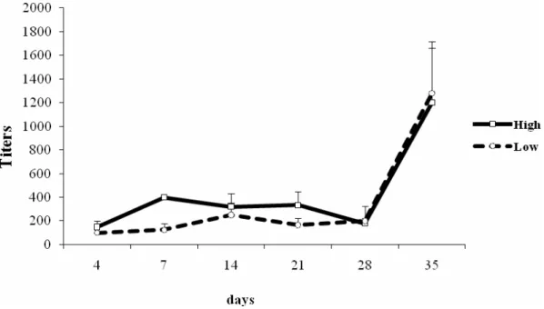
In the present study, antibody production in the lines was
associated with the leptospire recovery rates in renal and
hepatic tissues. The antibody titers produced by mice are
presented in Figure 2. In the negative control animals there was
no detection of any titration values.
The analysis of such results indicates that there was no
significant statistical difference (p< 0.05) between the
genetically selected lines HIV-A and LIV-A, although antibody
levels were higher in HIV-A than in LIV-A, especially between the
7th and 21st days post-inoculation.
The susceptibility and/or resistance were measured
according the rate recovery of leptospires in culture, the
presence of antibodies in a time graphical representation and
by the characteristics of the lesions in kidney tissues through
histopatological analysis (data not shown). Meanwhile,
although the lines have different levels of production of
antibodies and different rates of recovery of leptospires, mainly
in the initial period of post-inoculation, they maintained the
characteristics of resistance of the infectious agent has in the
murine model.
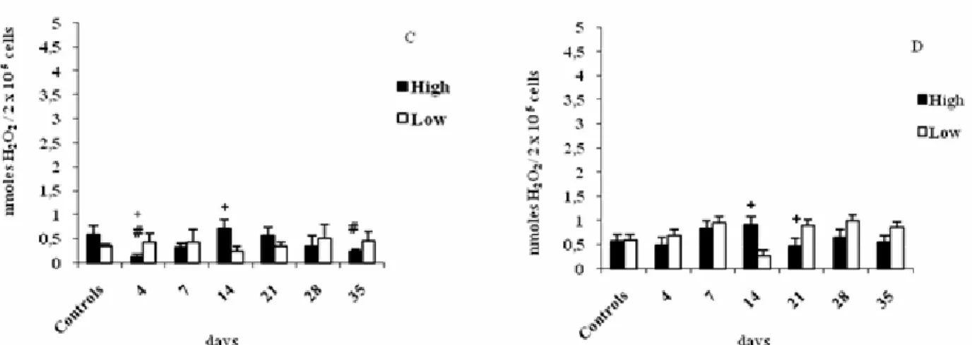
As regards H2O2 production by peritoneal macrophages
14th day in HIV-A compared to LIV-A lines (Figure 3A). When
stimulated with IFN- , macrophages of control HIV-A produced
higher H2O2 concentration than those of control LIV-A (Figure
3C). However, this apparent increase was not significant,
probably due to the great variability of individual data.
The treatment of peritoneal macrophages with PMA
significantly increased H2O2 production (Figure 3B).
Comparing the production of H2O2 between lines, significant
differences were observed at the 14th (H > L) and 21st (L > H) days of infection. The simultaneous stimulus with IFN-γ and PMA significantly increased H2O2 production at the 14
th
(H > L) and 21st (L > H) days of infection (Figure 3D).
Figure 2. Antibody titers detected by microscopic agglutination test against the antigen L. interrogans serovar Pomona in High
Haanwinckel, M.C.S. et al. Production of reactive oxygen and nitrogen and TNF-α in mice
Figure 3. Hydrogen peroxide production by peritoneal macrophages of High and Low mice from the IV-A selection infected with L.
interrogans serovar Pomona and not infected (controls). Macrophages were cultured in the absence of interferon-gamma (IFN-γ) without stimulus (A) or with PMA stimulus (B), or in the presence of IFN-γ without stimulus (C) or with PMA stimulus (D). Results represent the mean ± standard deviation of five animals per group. (#) indicates significant difference between infected and control mice, (+) indicates significant difference between lines (H x L). Student-Newman-Keuls test for multiple comparisons (p < 0.05).
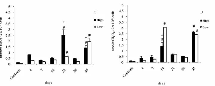
H2O2 production by splenic macrophages without stimuli
was higher at the 14th and 35th days in both HIV-A and LIV-A lines
(Figure 4A). However, the treatment with IFN-γ resulted in differences between lines: H > L at the 21st day and L > H at the 35th day (Figure 4C). In PMA-stimulated macrophages,
great alterations were observed at the 14th and 35th days
between the infected lines and their respective controls, as well
as between lines (Figure 4B). With the additional stimulus of
IFN-γ and PMA, there was a difference between lines: L>H at
the 14th day (Figure 4D).
As regards NO production by peritoneal macrophages,
differences between lines were only detected at the 21st day,
when LIV-A produced more NO than HIV-A mice (Figure 5A).
The treatment with IFN-γ increased NO production in both lines over the experiment (Figure 5B). NO production by
splenic macrophages stimulated or not with IFN- was not
significantly different between infected mice and their
Figure 4. Hydrogen peroxide production by splenic macrophages of High and Low mice from the IV-A selection infected with L.
interrogans serovar Pomona and not infected (controls). Macrophages were cultured in the absence of interferon-gamma (IFN-γ) without stimulus (A) or with PMA stimulus (B), or in the presence of IFN-γ without stimulus (C) or with PMA stimulus (D).
Results represent the mean ± standard deviation of five animals per group. (#) indicates significant difference between infected and control mice, (+) indicates significant difference between lines (H x L). Student-Newman-Keuls test for multiple comparisons
(p < 0.05).
Figure 5. Nitric oxide production by peritoneal macrophages of High and Low mice from the IV-A selection infected with L.
interrogans serovar Pomona and not infected (controls). Macrophages were cultured without stimulus (A) or with
interferon-gamma stimulus (B). Results represent the mean ± standard deviation of five animals per group. (#) indicates significant difference between infected and control mice, (+) indicates significant difference between liness (H x L). Student-Newman-Keuls test for
Haanwinckel, M.C.S. et al. Production of reactive oxygen and nitrogen and TNF-α in mice
TNF- production in serum and splenic cells of control
HIV-A andLIVA mice was not significantly different. However,
differences between lines were observed in the serum of LIV-A
line, which produced more TNF- than HIV-A at the 21 th
day
post-inoculation (Figure 6A). In splenic cells, significant
differences were observed at the 4th and 35th days of infection,
when HIV-A line produced more TNF- than LIV-A (Figure 6B).
TNF-α production by hepatic cells was higher than that observed in other analyzed tissues and fluids. In the liver, HIV-A
mice had a significant inhibition relative to controls at the 7th
and 14th days, recovering at the subsequent periods with a high
production at the 21st, 28th and 35th days (Figure 6C).
DISCUSSION
Mouse lines selected for the differentiation in the humoral
response may present varied degrees of antigenic response due
to genetic variations in immunoglobulin production and
macrophagic activity.
In the present study, the infectious agent was recovered
until the 21th day from the liver of LIV-A mice, whereas HIV-A
mice completely controlled the infectious process in the same
period. Such results may be related to the higher metabolism of
macrophages from low-responder relative to high-responder
animals, making difficult for macrophages from LIV-A mice to
present antigenic determinants to specific antibodies.
However, the formation of circulating antibodies probably
reduced drastically the agent possibility of diffusion in tissues,
contributing to the agent elimination after the 21th day in both
lines, which made difficult the isolation of leptospires in later
post-inoculation periods; this was also observed by Adler & Figure 6. TNF- levels detected in the serum (A),
spleen homogenate (B) and liver (C) of High and Low
mice inoculated with L. interrogans serovar Pomona
and not infected (controls). (#) indicates significant
difference between infected and control mice, (+)
indicates significant difference between lines (H x L).
Student-Newman-Keuls test for multiple comparisons
(p < 0.05).
A B
Faine (3, 4). The decreasing values of recovery from kidneys
and liver coincide with periods of increased detectable
immunoglobulin production in the microscopic agglutination
test, demonstrating a positive correlation between these
parameters, which corroborates other studies (22).
HIV-A mice are more resistant than LIV-A mice when the
humoral immune response represents the main mechanism of
protection against microorganisms. However, there was no
significant difference in the production of antibodies against L.
Pomona between HIV-A and LIV-A lines, although HIV-A animals
produced more antibodies between 7 and 21 days. These results
partially disagree with those obtained by Marinho et al (22),
who analyzed the production of antibodies against L.
Icterohaemorrhagiae. HIV-A line produced a statistically
significant response relative to LIV-A, maintaining the
multispecific effect normally observed in such lines.
The endogenous production of hydrogen peroxide (H2O2)
in peritoneal and splenic cells from HIV-A line may have helped
to control the infectious process, since a higher production of
such metabolite was observed at statistically significant
concentrations at the 14th day, which agrees with the results
obtained in other studies (21). Although LIV-A line presented a
basal H2O2 production in most of the evaluated periods, this
production was higher relative to controls at the 14th and 35th
days in the spleen and at the 28th and 35th days in the
peritoneum, when this line again completely controlled the
infectious process.
As regards NO production, there were differences between
lines considering infected animals without stimulus: LIV-A
produced more NO than HIV-A in the peritoneum at the 14 th
, 21st
and 28th days; in HIV-A line this difference was only observed at the
35th day (HIV-A > LIV-A).
Comparing such results with those obtained for bacterial
recovery, NO production contributed to control the infectious
process at later infection periods (from the 14th day), which
was similar to the results obtained by Marangoni et al (19).
Marinho (21) analyzed NO production in IV-A selection
lines against the infection triggered by Leptospira serovar
Icterohaemorrhagiae and observed that IFN-γ-treated cells had high NO levels. This effect was also observed in the present
study. The treatment with IFN-γ and/or PMA on H2O2
production and with IFN-γ on NO production indicated that in some evaluated compartments and periods L. Pomona was a
“messenger” for this cell to produce such metabolite.
Darrah et al (7) studied knockout mice to evaluate H2O2
and NO production and observed that the combination of both
radicals resulted in a synergistic effect, with rapid and efficient
pathogen killing by activated macrophages, especially under
IFN-γ stimulus. In the present study, a positive interaction between both metabolites seems to have occurred, since such
intermediaries were higher, especially in the infectious process
control periods.
NO production requires the presence of IFN-γ, which plays an important role as a macrophage activating factor and a
primary signal for the transcription of the enzyme Inducible
Nitric Oxide Synthase (INOS). In innate immunity, signals for
macrophage activation may originate from the pathogen and
natural killer cells (NK cells), which are a source of IFN-γ (1).
In addition, Marinho et al (22) analyzed TNF-α
production by HIV-A and LIV-A lines against Leptospira serovar
Icterohaemorrhagiae and observed a high production in LIV-A at
the beginning of the infection and this level remained stable
until the 14th day. Following this period, there was an inhibition
of TNF-α production, exactly at the moment leptospires were not recovered anymore.
Production of tumor necrosis factor (TNF-α) as a parameter for macrophagic activity was also evaluated in the
present experiment. TNF-α levels in the serum from infected LIV-A line were higher at the 7th, 21st and 35th days. However,
significant differences between lines were only observed at the
21st day. HIV-A splenic cells produced more TNF-α than those
of LIV-A at the 4 th
and 35th days post-inoculation. In hepatic
cells, TNF-α production was different between lines only at the 35th day (HIV-A> LIV-A).
Haanwinckel, M.C.S. et al. Production of reactive oxygen and nitrogen and TNF-α in mice
infectious process control in both lines, and the analysis of the
production of this cytokine in HIV-A hepatic cells evidenced its
importance in the infection control, which agrees with the data
obtained by Marangoni et al (20), who analyzed TNF-α
production by hepatic macrophages, correlating it to the
presence of lipopolysaccharides (LPS) from Leptospira serovar
Icterohaemorrhagiae.
The analyses of other cytokines can be determinant to
evaluate the profile of a broaden immune response of the
different serovars of leptospires in the lines genetically
selected. By Marinho et al. (22) the line HIV had the profile
Th2 with more production of antibodies, IL-4 and worst tissues
lesions, while the line LIV-A had the profile Th1 with higher
production of IFN- , higher macrophage activity and lesser
damaged tissues.
The differences in the recovery rates and in the cellular
and humoral immune response of Leptospira serovar Pomona
relative to other serovars investigated in previous studies may
be related to antibody production control mechanisms and
differentiated cellular response, but especially to the infectious
agent adaptability factors, since serovars had different
virulence in some hosts, i.e. they are species-adapted (14, 17).
In general, the study of the infection by Leptospira serovar
Pomona in lines indicated that macrophagic activity and TNF-
production play an important role in the infectious process
control, which varied according to the evaluated compartments
and periods. Compared to studies with other serovars, the
present results indicate partial differences in an immune trait
genetically selected by an external nonspecific agent.
ACKNOWLEDGMENTS
We thank the National Council for Scientific and
Technological Development (CNPq) for the financial support;
Prof. Dr. Silvio Arruda Vasconcellos and the biologist Zenaíde
Maria de Moraes from the Bacterial Zoonoses Lab, Department
of Veterinary Hygiene and Animal Health, College of
Veterinary Medicine and Animal Science, University of São
Paulo (USP), São Paulo City, São Paulo State, Brazil; Prof. Dr.
Jane Megid, Department of Veterinary Hygiene and Public
Health, College of Veterinary Medicine and Animal Science,
São Paulo State University (UNESP), Botucatu Municipality,
São Paulo State, Brazil.
REFERENCES
1. Abbas, A.K.; Lichtman, A.H.; Pober, J.S. (2002). Citocinas. In: Abbas, A.K., Lichtman, A.H., Pober, J.S. (eds). Imunologia celular e molecular. 4ª ed. Revinter, Rio de Janeiro, Brasil, p.235-269
2. Adams, H.R. (1996). Physiologic, pathophysiologic, and therapeutic implications for endogenous nitric oxide. J. Am. Vet. Med. Assoc. 209, 1297-1302.
3. Adler, B.; Faine, S. (1976). Susceptibility of mice treat with cyclophosphamide to lethal infection with Leptospira interrogans
serovar pomona. Infect. Immun.14, 703-708.
4. Adler, B.; Faine, S. (1978). Serological and protective-antibody response of rabbits to leptospiral antigens. J. Med. Microbiol. 11, 401-409. 5. Biozzi, G.; Stiffel, C.; Mouton, D.; Bouthillier, Y.; Decreusefond, C.
(1972). Cytodynamics of the immune response in the two lines of mice genetically selected for “high” and “low” antibody synthesis. J. Exp. Med. 135, 1071-1094.
6. Cabrera, W.H.K.; Ibañez, O.M.; Oliveira, S.L.; De Sant'anna, O.A.; Siqueira, M.; Mouton, D.; Biozzi, G. (1982). Evidence for distinct polygenic regulation of antibody responses to some unrelated antigens in lines of mice selected for high or low antibody responses to somatic antigens of Salmonella. Immunogenetics. 16, 583-592.
7. Darrah, P.A.; Hondalus, M.K.; Chen, Q.; Ischiropoulos, H.; Mosser, D.M. (2000). Cooperation between reactive oxygen and nitrogen intermediates in killing of Rodococcus equi by activated macrophages.
Infect. Immun. 68, 3587-3593.
8. Davis, J.M.; Haake, D.A.; Ramakrishnan, L. (2009) Leptospira interrogans stably infects zebrafish embryos, altering phagocyte behavior and homing to specific tissues. PLoS Negl Trop Dis. http://www.ncbi.nlm.nih.gov/pmc/articles/PMC2693671/?tool=pubmed 9. Di Giovine, F.S.; Nuki, G.; Duff, G.W. (1988). Tumour necrosis factor in
synovial exudates. Ann. Rheum. Dis. 47, 768–772.
11. Faine, S. (1982). Guidelines for the control of leptospirosis. World Health Organization, Geneva.
12. Faine, S. (ed) (1994). Leptospira and Leptospirosis, CRC-Press, Boca-Raton, Florida.
13. Green, L.C.; Tannenbaun, S.R. (1981). Nitrate synthesis in the germfree and conventional rat. Science. 212, 56-58.
14. Hathaway, S.C.; Blackmore, D.K.; Marshall, R.B. (1983). Leptospirosis and the maintenance host: a laboratory mouse model. Res. Vet. Sci. 34, 82-89.
15. Ibanez, O.M.; Mouton, D.; Oliveira, S.L.; Ribeiro Filho, O.G.; Piatti, R.M.; Sant´Anna, O.A.; Massa, S.; Biozzi, G.; Siqueira, M. (1988). Polygenic control of quantitative antibody responsiveness: restrictions of the multispecific effect related to the selection antigen. Immunogenetics.
28, 6-12.
16. Isogai, E.; Isogai, H.; Fujii, N.; Oguma, K. (1990). Macrophage activation by leptospiral lipopolysaccharide. Zentralbl. Bakteriol. 273, 200-208.
17. Klimpel, G.R.; Mathias, M.A.; Vinetz, J.M. (2003). Leptospira interrogans activation of human peripheral blood mononuclear cells: Preferential expansion of TCR yδ+ T cells vs TCRαβ+ T cells. J.
Immunol. 171, 1447-1455.
18. Lowanitchapat, A.; Payungporn, S.; Sereemaspun, A.; Ekpo, P.; Phulsuksombati, D.; Poovorawan, Y.; Chirathaworn, C. (2009). Expression of TNF- , TGF- , IP-10 and IL-10 mRNA in kidneys of hamsters infected with pathogenic Leptospira. Comp Immunol Microbiol Infect Dis. http://www.citeulike.org/article/5541032
19. Marangoni, A.; Accardo, S.; Aldini, R.; Guardigli, M.; Cavrini, F.; Sambri, V.; Montagnani, M.; Roda, A.; Cavenini, R. (2006). Production of reactive oxygen species and expression of inducible nitric oxide synthase in rat isolated Kupffer cells stimulated by Leptospira interrogans and Borrelia burgdorferi. World J. Gastroenterol. 12, 3077-3081.
20. Marangoni, A.; Aldini, R.; Sambri, V.; Giacani, L.; Di Leo, K.; Cevenini, R. (2004). Production de tumor factor by Treponema pallidum, Borrelia burgdorferi, s.l, and Leptospira interrogans is isolated rat Kupffer cells. FEMS Immunol. Med. Micriobiol. 40, 187-191.
21. Marinho, M. (2000). Resposta imune à infecção por Leptospira interrogans sorovar icterohaemorrhagiae em linhagens de camundongos geneticamente selecionados. Botucatu, São Paulo, Brasil, 102p. (D.Sc. Thesis. Faculdade de Medicina, UNESP).
22. Marinho, M.; Langoni, H.; Oliveira, S.L.; Carreira, R.; Perri, S.H.V.; Luvizoto, M.C. (2003). Resposta humoral, recuperação bacteriana e lesões histológicas em camundongos em geneticamente selecionados para bons e maus respondedores de anticorpos e balb/c, frente à infecção por Leptospira interrogans sorovar icterohaemorrhagiae. Pesq. Vet. Bras. 23, 5-12.
23. Passos, E.C.; Vasconcellos, A.S.; Ito, F.O.; Yasuda, P.H.; Nürmberger Jr, R. (1988). Isolamento de Leptospiras a partir do tecido renal de hamsters experimentalmente infectados com L. interrogans sorotipo pomona: Emprego das técnicas de Pipeta Pasteur e das diluições seriadas em meio de cultura de Fletcher tratado com 5-fluor-uracil ou sulfato de neomicina.
Rev. Fac. Med. Vet. Zoot. Univ. São Paulo. 25, 221-235.
24. Pick, E.; Keisari, Y. (1980). A simple colorimetric method for the measurement of hydrogen peroxide produced by cells in culture. J. Immunol. Methods. 38, 161-170.
25. Pick, E.; Mizel, D. (1981). Rapid microassays for the measurement of superoxide and hydrogen peroxide production by macrophages in culture using an automatic enzyme immunoassay reader. J. Immunol. Methods.
46, 211-226.
26. Pourshafie, M.; Ayub, Q.; Barrow, W.W.; (1993). Comparative effects of Mycobacterium avium glycopeptidolipid and lipopeptide fragment on the function and ultrastructure of mononuclear cells. Clin. Exp. Immunol. 93, 72–79.
27. Santa Rosa, C.A. (1970). Diagnóstico laboratorial das leptospiroses. Rev. Microbiol. 1, 97-109.
28. Werts, C.; Tapping, R.I.; Mathison, J.C.; Chuang, T.H.; Kravchenko, V.; Saint Girons, I.; Haake, D.A.; Godowski, P.J.; Hayashi, F.; Ozinsky, A.; Underhill, D.M.; Kirschning, C.J.; Wagner, H.; Aderem, A.; Tobias, P.S.; Ulevitch, R.J. (2001). Leptospiral lipopolysaccharide activates cells through a TLR2- dependent mechanism. Nat. Immunol. 2, 346-352. 29. Zar, J.H. (1996). Bioestatistical analysis. 3ª ed. Prentice Hall, Upper
Saddle River.