Desulfovibrio vulgaris defenses
against oxidative and nitrosative
stresses
Mafalda Cristina de Oliveira Figueiredo
Dissertation presented to obtain the Ph.D degree in Biochemistry
Instituto de Tecnologia Química e Biológica | Universidade Nova de Lisboa
Supervisor: Dr. Lígia M. Saraiva
Co-supervisor: Prof. Miguel Teixeira
From left to right: Carlos Romão (president of the jury), Fernando Antunes (3rd
opponent), Ana Melo (4th opponent), Lígia Saraiva (supervisor), Carlos Salgueiro
(2nd opponent), Mafalda Figueiredo, Alain Dolla (1st opponent) and Miguel Teixeira
(co-supervisor).
17th September 2013
Second edition, October 2013
Molecular Genetics of Microbial Resistance Laboratory
Instituto de Tecnologia Química e Biológica
Universidade Nova de Lisboa
2780-157
“Nothing in life is to be feared, it is only to be understood.
Now is the time to understand more, so that we may fear less.”
Acknowledgments
The present work would not have been possible without the help, the support and the friendship of several people whom I would like to formally express my sincere gratitude:
Dr. Lígia M. Saraiva
Firstly I would like to express my gratitude to my supervisor Dr. Lígia M.
Saraiva, without her ideas and persistence I would not have come this far. I thank
her for the constant support and encouragement when things did not go so well, for the trust she placed in me and in my work and for always being there when I
needed over these five years. I have to thank Dr. Lígia for the good advices and for the careful revision of this thesis. Thanks for everything!!!
Prof. Miguel Teixeira
To my co-supervisor Prof. Miguel Teixeira I would like to thankfor the very
interesting and helpful discussions and for all the help given whenever I needed.
Scientific Thesis Commission
To Prof. Cláudio M. Soares and Dr. Cecília M. Arraiano for being part of my
scientific thesis commission and for their suggestions and helpful advices.
Prof. David Turner
Prof. Judy D. Wall
To Prof. Judy D. Wall for providing the wild type and mutant strains of D.
vulgaris used in this work and for the collaboration on the work present in Part II -
Chapter 2.
João Carita
To João Carita for the collaboration on the work presented in Part II - Chapter 1, especially for performing the Desulfovibrio growths in the fermenter.
Prof. Simon Andrews
I have also to thank Prof. Simon Andrews for the excellent reception in his laboratory and also to the group members of the Bacterial Iron Homeostasis lab in particular to Helen Goodluck my companion during the month I was in the
University of Reading.
Molecular Genetics of Microbial Resistance Group
I would like to thank to all past and present members of Molecular Genetics
of Microbial Resistance group for making the lab such an enjoyable and pleasant
place to work.
Very special thanks to Susana that is my "third supervisor" who taught me everything in the lab, who always helped me and encouraged me, thank you also for being a great friend and confidant.
To Lígia by her enthusiasm when discussing ideas, for their laughter that
keeps the lab in a good mood and to be a good friend.
To Marta I would like to thank for always being available for my questions and issues and to always have a solution to solve the problems.
To Margarida for being a good friend who is always ready to help.
To Sara that is always a funny presence in the lab, for the conversations at
To Filipa for being a great friend and companion, for all conversations until late hours in the lab when I was doing the experiments with macrophages, and for always encouraging me to be better.
To Joana by her friendship and companionship, for supporting me whenever
it was necessary even though "the other side of the door" and often just talking by signs.
To Vera I would like to thank for their help in the early months in the lab, and to always be present whenever I needed.
To Cláudia for spend part of their time helping me when I started to give my
first steps in the lab, and for over the years to be a friend always ready to help. To André, for most of the time the only male presence in the lab, for always remember us that “The Summer is coming!”.
To the trainees who have passed by the lab, Fábio for all the work that we
have done together with Desulfovibrio, to Catarina, Luís, Cláudia and Marco, for all lunch talks and for contributing to the good environment.
Thank you all for your friendship and for the good moments shared over the last years.
Everyone in the 3rd floor of ITQB
I would like to thank also to everyone in the 3rd floor of ITQB, past and present people, for contributing to the helpful and friendly environment.
Aos meus amigos
Gostaria de agradecer também aos meus amigos de sempre e para sempre, o nosso “núcleo duro” vocês sabem quem são! Obrigado a todos pelo apoio e amizade ao longo destes anos. Quero agradecer especialmente ao João Daniel e
Viviane que mais que amigos neste momento se tornaram família e que sempre
estiveram lá quando foi preciso. Ao Daniel Coelho quero agradecer por sempre ter
demostrado interesse no meu trabalho, por ter feito um esforço para ler os meus
À minha Família
À minha família quero agradecer em primeiro lugar aos meus pais pelo vosso amor e apoio incondicional, por terem acreditado em mim e me darem força para ir sempre mais longe, é a vocês que devo aquilo que sou hoje.
À minha irmã Daniela, quero agradecer pela amizade, por estar sempre disponível para me ajudar e pelo apoio incondicional.
Aos meus avós, agradeço por me acompanharem e ajudarem ao longo de toda a minha vida e fico muito feliz de ver o vosso orgulho de terem uma netinha Doutora.
Aos meus tios, em especial à minha tiaMafalda, que ao longo destes 29 anos tem sido uma segunda mãe que me tem ajudado muito, sem ti teria sido muito mais difícil chegar onde estou hoje.
À minha prima Mara quero agradecer por todo o apoio, companheirismo e
amizade.
Francisco
Ao meu marido Francisco, quero agradecer por estar sempre ao meu lado,
por toda a paciência que tem comigo mesmo quando eu não tenho a mesma com
ele, por estar sempre disponível para me ouvir a falar de tudo o que se passou no meu dia, por ser o meu melhor amigo, e por me amar incondicionalmente e me fazer tão feliz.
Fundação para a Ciência e Tecnologia
Fundação para a Ciência e Tecnologia (FCT) for the financial support crucial for the accomplishment of this work and by awarding a PhD grant (SFRH/BD/44140/2008).
Thesis Outline
The work presented in this thesis was performed at the Molecular Genetics of
Microbial Resistance Laboratory from the Instituto de Tecnologia Química e
Biológica, Universidade Nova de Lisboa under the supervision of Dr. Lígia M.
Saraiva and co-supervision of Prof. Miguel Teixeira.
The main objective of this thesis was to elucidate the mechanisms of
resistance in Desulfovibrio vulgaris Hildenborough against oxidative and nitrosative
stresses.
This dissertation is divided in three main parts. Part I consists in three
introductory chapters: Chapter 1 describes the sulfate reducing bacteria group
with particular focus on Desulfovibrio genus; Chapter 2 addresses the iron
metabolism in Desulfovibrio; and Chapter 3 is an overview on the general
responses of Desulfovibrio to oxygen and nitric oxide stresses. Part II of this thesis
contains the experimental results achieved during this work, which is divided in
two chapters based on original publications. Part III presents a general discussion
Thesis Publications
This dissertation is based on the following original publications, listed by chronological order:
Figueiredo, MC, Lobo, SA, Carita, JN, Nobre, LS & Saraiva, LM (2012).
Bacterioferritin protects the anaerobe Desulfovibrio vulgaris Hildenborough against oxygen. Anaerobe, 18:454-458.
Abstract
The work presented in this dissertation aimed to unravel the defense
mechanisms of the anaerobic sulfate reducing bacterium Desulfovibrio (D.)
vulgaris Hildenborough against oxidative and nitrosative stresses.
Desulfovibrio spp. are usually found in anaerobic niches in soil, marine and
fresh waters and sediments, but also in zones periodically exposed to oxygen.
Ecologically, Desulfovibrio spp. play an important role in bioremediation of heavy
metals due to their capacity to precipitate toxic metal ions from environmental
contaminants. However, because of their ability to cause metal corrosion these
bacteria have a strong negative economic impact, constituting a major problem in
oil and gas industries. These microorganisms are also part of the normal microbial
flora present in the gut of animals and humans. Although recent reports revealed
that they can act as opportunistic pathogens, the function in the intestinal
ecosystem remains essentially unknown.
In spite of Desulfovibrio spp. being anaerobic microorganisms, their presence
in oxic-anoxic interfaces shows that pathways for defense against toxic reactive
oxygen species (ROS) must be active. Hence, in the first part of this work,
transcriptomic and physiological approaches were used to evaluate the effect of
oxygen on the expression of genes putatively involved in D. vulgaris oxygen
resistance. The results showed that the gene for the iron storage protein
bacterioferritin (bfr) exhibited high transcriptional induction upon exposure of
cells to high levels of oxygen. To clarify the contribution of bacterioferritin to
oxygen resistance, a bfr mutant strain was analyzed. The data revealed that the
absence of bfr significantly decreases the survival of D. vulgaris in oxygenated
ROS in relation to the wild type indicating that bacterioferritin contributes to
avoiding ROS formation.
To assess the regulation of D. vulgaris bacterioferritin, we examined the
transcription profile of the bfr gene in cells of two independent mutant strains
inactivated in two general regulators, namely the ferric uptake regulator (Fur) and
the peroxide regulator (PerR). While bfr gene transcription does not suffer
alteration in the ∆furmutant strain, it is repressed in the ∆perR mutant, showing that the expression of D. vulgaris bfr is under the control of PerR. Analysis of the
bfr putative promoter region allowed identification of a potential PerR binding site
located upstream of the start codon of bfr, with the sequence
TAAACGAATCTTTACACAC. Altogether, it was demonstrated that the iron storage
bacterioferritin provides protection for D. vulgaris against oxygen stress.
In recent years, Desulfovibrio spp. have been proposed to be associated with
gastrointestinal diseases such as inflammatory bowel disease, Crohn’s disease and ulcerative colitis. Due to its potential pathogenicity it is crucial to understand the
mechanisms whereby these microorganisms overcome the innate immune system
of the host. Cells of the innate immune system produce reactive oxygen species
and reactive nitrogen species (RNS) to fight pathogens and, in order to detoxify
these compounds, bacteria activate several protective systems. Therefore, in the
second part of this work, the response of D. vulgaris to nitric oxide was studied. D.
vulgaris was exposed to a nitric oxidedonor and the transcription of genes coding
for proteins potentially involved in NO protection, such as rubredoxin:oxygen
oxidoreductase proteins (roo1 and roo2) and hybrid cluster proteins (hcp1 and
hcp2) was analyzed. Under nitrosative stress conditions, the expression of hcp2
gene is highly induced while those of the other genes analyzed did not vary
significantly. Next, we tested the ability of D. vulgaris strains with the two roo and
hcp genes deleted to grow in the presence of a nitric oxide donor. The D. vulgaris
Moreover, the contribution of the Hcp and Roo proteins to the NO consumption of
D. vulgaris was evaluated by means of a NO-electrode in cell lysates of wild type
and mutant strains grown anaerobically and treated with a NO donor. Untreated
cells of roo and hcp mutants showed no major differences in the NO consumption
rates; however, upon nitrosative stress exposure roo1 and roo2 mutant strains
exhibited lower NO consumption in comparison with the wild type strain. A strain
of D. vulgaris with roo1 deleted had the lowest NO consumption ability, revealing
that, among the four proteins, Roo1 is the major contributor to the NO reduction
capability of the D. vulgaris cells.
In vivo studies performed with animal cells suggest that D. vulgaris is not
capable of intracellular replication in macrophages but survives in the extracellular
environment when co-cultured with macrophages. Importantly, the bacterium
proved to be able to activate one of the key weapons of the innate immune
system, namely the inducible nitric oxide synthase (iNOS) present in macrophages
that releases NO to fight pathogens. Indeed, exposure of D. vulgaris wild type to
macrophages significantly decreases the survival of the microorganism, which is
attenuated upon inhibition of iNOS.
To evaluate the role of Roo and Hcp in Desulfovibrio during infection of
macrophages, the viability of the mutant strains upon macrophage incubation was
analyzed. All mutant strains exhibited viability reduced by approximately 90%,
while that of the parental strain decreased by only 30%. This clearly shows that
Hcp and Roo proteins protect D. vulgaris during exposure to mammalian
macrophages.
In conclusion, this thesis contributed to better understanding the defense
mechanisms of D. vulgaris against oxygen and nitric oxide. In particular, the
relation between intracellular iron storage and oxidative stress response was
revealed through the role of D. vulgaris bacterioferritin in survival upon oxygen
system, with Roo and Hcp proteins contributing to the survival of the bacterium D.
Resumo
O trabalho apresentado nesta dissertação tem como objetivo elucidar os
mecanismos de defesa da bactéria anaeróbica redutora de sulfato Desulfovibrio
(D.) vulgaris contra os stresses oxidativo e nitrosativo.
Os membros do género Desulfovibrio são normalmente encontrados em
nichos anaeróbios do solo, nomeadamente ambientes marinhos e sedimentos,
mas também em zonas periodicamente expostas ao oxigénio. Ecologicamente, as
bactérias do género Desulfovibrio têm um papel importante, principalmente na
biorremediação de metais pesados visto terem a capacidade de separarem por
precipitação metais tóxicos de contaminantes ambientais. Contudo, devido
também à sua capacidade de causar corrosão de metais, estas bactérias têm um
forte impacto económico negativo, constituindo um sério problema para as
indústrias de petróleo e gás. Estes microrganismos fazem também parte da flora
microbiana normalmente presente no intestino de animais e humanos. Embora
estudos recentes tenham revelado que estas bactérias podem atuar como agentes
patogénicos oportunistas, a sua função no ecossistema intestinal permanece
essencialmente desconhecida.
Apesar de as espécies de Desulfovibrio serem microrganismos anaeróbios, a
sua presença em habitats expostos ao oxigénio demonstra que contêm
mecanismos de defesa contra as espécies reativas de oxigénio (ERO) tóxicas.
Assim, na primeira parte deste trabalho realizaram-se estudos transcriptómicos e
fisiológicos para avaliar o efeito do oxigénio na expressão de genes
hipoteticamente envolvidos na resistência ao oxigénio em D. vulgaris. Os
resultados mostraram que quando as células de D. vulgaris foram expostas a altas
concentrações de oxigénio o gene que codifica para a proteína armazenadora de
clarificar a contribuição da bacterioferritina para a resistência ao oxigénio, uma
estirpe mutada no gene bfr foi analisada. Os dados revelaram que a ausência do
gene bfr diminuiu significativamente a sobrevivência de D. vulgaris em ambientes
oxigenados. Além disso, a estirpe mutada no gene bfr apresentou um teor mais
elevado de ERO em relação à estirpe selvagem indicando que a bacterioferritina
contribui para evitar a acumulação de ERO.
Para avaliar a regulação da bacterioferritina de D. vulgaris, examinou-se o
perfil de transcrição do gene bfr em células de duas estirpes de D. vulgaris
mutadas independentemente em dois reguladores gerais, nomeadamente no
regulador de absorção de ferro (Fur) e no regulador de peróxido (PerR). Enquanto
a transcrição do gene bfr não sofre alteração na estirpe mutante ∆fur, a sua expressão é repressa no mutante ∆perR, demonstrando que a expressão da bfr de
D. vulgaris está sob o controlo do regulador PerR. Adicionalmente, a análise da
região promotora putativa do gene bfr permitiu a identificação de um potencial
local de ligação localizado a montante do codão de iniciação da bfr, com a
sequência TAAACGAATCTTTACACAC. Com estes estudos demonstramos que a
bacterioferritina protege D. vulgaris contra o stress oxidativo.
Nos últimos anos, algumas estirpes de Desulfovibrio têm sido propostas como
estando associadas a doenças gastrointestinais tais como doença inflamatória do
intestino, doença de Crohn e colite ulcerosa. Devido à sua potencial
patogenicidade é crucial compreender os mecanismos usados por estes
microrganismos para superar o sistema imune inato do hospedeiro. As células do
sistema imune inato produzem ERO e espécies reativas de nitrogénio (ERN) para
combater os patogénicos. Por forma a fazer a destoxificação destes compostos, as
bactérias contra-atacam ativando vários sistemas de proteção. Assim na segunda
parte deste trabalho, foi estudada a resposta de D. vulgaris ao óxido nítrico (NO).
Para isso, D. vulgaris foi exposto a um dador de óxido nítrico e a transcrição de
o NO, tais como as proteínas rubredoxina:oxigénio oxidoreductase (Roo1 e Roo2)
e “hybrid cluster protein” (Hcp1 e Hcp2), foi analisada. Sob condições de stress nitrosativo, a expressão do gene hcp2 é altamente induzida enquanto a expressão
dos outros genes analisados não variou significativamente. Em seguida, foi testada
a capacidade de estirpes de D. vulgaris mutadas nos genes roo e hcp para crescer
na presença de um dador de óxido nítrico. Verificou-se que a estirpe de D. vulgaris
mutada no gene hcp2 é a mais suscetível ao stress nitrosativo. A contribuição das
proteínas Roo e Hcp para o consumo de NO em D. vulgaris foi também avaliada,
por meio de um eléctrodo de NO, em lisados de células da estirpe selvagem e das
estirpes mutantes quando crescidas anaerobicamente e tratadas com um doador
de NO. As células não tratadas dos mutantes roo e hcp não apresentaram grandes
diferenças nas taxas de consumo de NO. No entanto, após a exposição ao stress
nitrosativo as estirpes mutantes roo1 e roo2 apresentaram menor consumo de NO
em comparação com a estirpe selvagem. A estirpe de D. vulgaris deletada no gene
roo1 exibiu a menor capacidade de consumo de NO revelando que, de entre as
quatro proteínas, a Roo1 é a principal proteína responsável pela capacidade de
redução de NO nas células de D. vulgaris.
Estudos in vivo realizados com células animais sugerem que D. vulgaris não é
capaz de replicação intracelular nos macrófagos, mas sobrevive no ambiente
extracelular quando co-cultivado com os macrófagos. Para além disso, estas
bactérias mostraram ser capazes de ativar uma das principais armas do sistema
imune inato, nomeadamente a proteína produtora de óxido nítrico (iNOS)
presente nos macrófagos, que liberta NO para combater organismos patogénicos.
De facto, a exposição da estirpe selvagem D. vulgaris aos macrófagos reduziu
significativamente a sobrevivência deste microrganismo, sendo atenuada com a
inibição da iNOS.
Para avaliar o papel de Roo e Hcp em Desulfovibrio durante a infeção dos
macrófagos foi analisada. Todas as estirpes mutantes exibiram a viabilidade
reduzida em aproximadamente 90%, enquanto a estirpe selvagem diminuiu
apenas 30%, mostrando claramente que as proteínas Roo e Hcp protegem D.
vulgaris durante a exposição a macrófagos de mamíferos.
Em conclusão, este trabalho contribuiu para uma melhor compreensão dos
mecanismos de defesa de D. vulgaris contra o stress oxidativo e nitrosativo. Em
particular, a relação entre a armazenagem de ferro intracelular e resposta ao
stress oxidativo foi revelada através do papel da bacterioferritina na sobrevivência
de D. vulgaris quando exposto ao oxigénio. Além disso, D. vulgaris mostrou ativar
o sistema imunitário inato, e que as proteínas Roo e Hcp contribuem para a
sobrevivência da bactéria D. vulgaris quando este enfrenta stresses prejudiciais
Abbreviations list
ALA δ-Aminoleaevulinic acid AMP Adenosine-5´-phosphate APS Adenosine-5´-phophosulfate ATCC American type culture collection ATP Adenosine triphosphate
Bp Base pair
cDNA Complementary DNA
CFU Colony Forming Units
CO Carbon monoxide
CO2 Carbon dioxide
CoA Coenzyme
DCFH-DA 2´,7´-dichlorofluorescein diacetate
DMEM Dulbecco´s Modified Eagle medium
DNA Deoxyribonucleic acid
e- Electron
eNOS Endothelial nitric oxide synthase
FDP Flavodiiron protein
Fe Iron
Fe-S Iron-sulfur cluster FI Fluorescence intensity FMN Flavin mononucleotide GSNO S-Nitrosoglutathione
H+ Proton
H2 Hydrogen
H2O Water
H2O2 Hydrogen peroxide H2S Hydrogen sulfide HMB Hydroxymethylbilane
HS- Sulfide
HSO3 Bisulfite INF-γ Interferon-γ
iNOS Inducible nitric oxide synthase
Km Kanamycin
LPS Lipopolysaccharides
M Molar
Mb Mega base pair
MOI Multiplicity of infection
mRNA messenger RNA
N2O3 Nitrogen trioxide
NADH β-Nicotinamide adenine dinucleotide, reduced form
NADPH β-Nicotinamide adenine dinucleotide phosphate, reduced form
ND Not determined
NH3 Ammonia
NH4 +
Ammonium ion
nm Nanometer
nNOS Neuronal nitric oxide synthase
NO Nitric oxide
•
NO2 Nitrogen dioxide
NO2 Nitrite
NO3
-Nitrate
NOS Nitric oxide synthase
O2 Dyoxygen
O2
•
-Superoxide anion OD Optical density
•
OH Hydroxyl radical
OH- Hydroxide ion
OHOO- Peroxynitrite
PBG Porphobilinogen
PBS Phosphate buffer
PCR Polymerase chain reaction
Phox NADPH oxidase
Pi Inorganic phosphate
PPi Pyrophosphate
RNA Ribonucleic acid
RNS Reactive nitrogen species
ROS Reactive oxygen species
RT-PCR Reverse transcriptase-polimerase chain reaction
S Supplemental
S2- Sulfide
S2O3
S3O6
Trithionate
SH Siroheme
SO4
2-Sulfate
spp. Species
SRB Sulfate reducing bacteria SRO Sulfate reducing organisms
TSAS Tryptic Soy Agar medium Supplemented
wt Wild type
Δ Deletion
Latin abbreviations
e.g. exempli gratia, for example
et al. et alia, and other people
Strains
B. brevis Bacillus brevis
B. melitensis Brucella melitensis
B. subtilis Bacillus subtilis
C. Campylobacter
D. Desulfovibrio
E. Escherichia
H. Helicobacter
N. Neisseria
P. Pseudomonas
S. aureus Staphylococcus aureus
Amino acids
A Ala Alanine M Met Methionine
C Cys Cysteine N Asn Asparagine
D Asp Aspartic acid P Pro Proline E Glu Glutamic acid Q Gln Glutamine F Phe Phenylalanine R Arg Arginine
G Gly Glycine S Ser Serine
H His Histidine T Thr Threonine
I Ile Isoleucine V Val Valine
K Lys Lysine W Trp Tryptophan
Table of Contents
Acknowledgments v
Thesis Outline ix
Thesis Publications xi
Abstract xiii
Resumo xvii
Abbreviations list xxi
Table of Contents xxv
Part I
–
Introduction
Chapter 1
Sulfate Reducing Bacteria
1.1 - General overview of Sulfate Reducing Bacteria 5
1.2 - Environmental impact of SRB 7
1.3 - Desulfovibrio genus 9
1.3.1 - Metabolism 10
Energy transduction 10
Energy transduction models 10
Electron carriers and membrane bound electron transport 12
Sulfate reduction 13
Nitrate and nitrite reduction 14
Heme biosynthesis 16
1.3.2 - Desulfovibrio in humans 17
Chapter 2
Desulfovibrio
and iron homeostasis
2.1 - Iron is an essential element 31
2.2 - Proteins involved in iron metabolism 32
2.2.1 - Iron acquisition systems 32
2.2.2 - Iron storage proteins 33
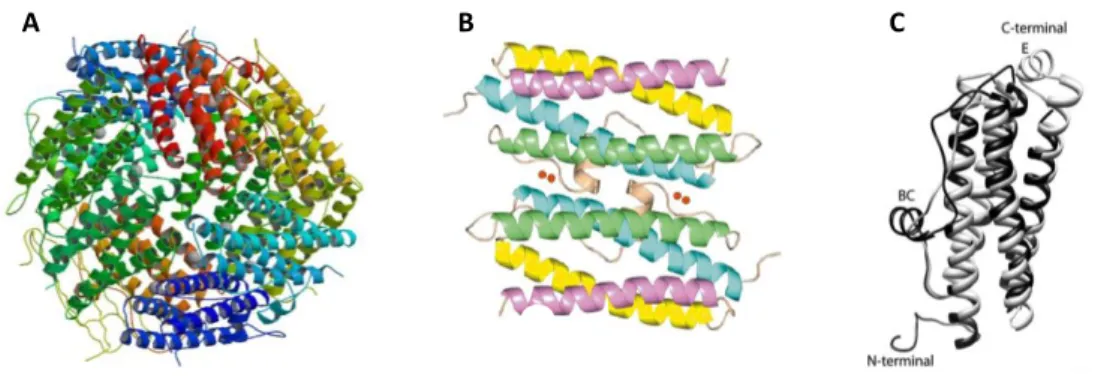
The ferritin superfamily 33
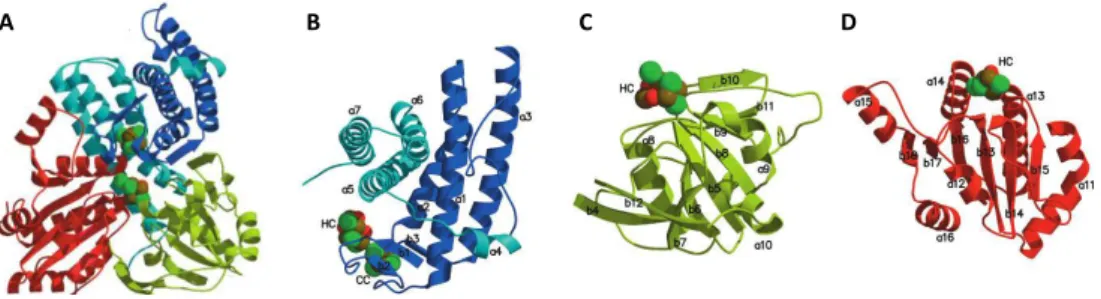
The Dps family 37
2.2.3 - Regulation of iron homeostasis 39
2.3 - References 42
Chapter 3
Desulfovibrio
responses to oxidative and nitrosative
stresses
3.1 - Oxidative and nitrosative stresses 49
3.1.1 - Oxidative stress 49
3.1.2 - Nitrosative stress 51
3.2 - Stress environmental challenges 53
3.2.1 - Desulfovibrio spp.in oxidative habitats 53
3.2.2 - Mammalian immune system response against invading bacteria 55
3.3 –Desulfovibrio systems putatively involved in oxygen
and nitric oxide detoxification 57
3.4 – Regulators of Desulfovibrio for oxidative and nitrosative
stress resistance 64
3.5 - D. vulgaris transcriptional response to stress conditions 65
Part II
–
Results
Chapter 1
Bacterioferritin protects the anaerobe
Desulfovibrio
vulgaris
Hildenborough against oxygen
1.1 - Introduction 81
1.2 - Materials and Methods 83
Bacterial strains and growth conditions 83
Viability assays and evaluation of ROS content 84
Real-time quantitative RT-PCR 84
1.3 - Results 86
Effect of oxygen on gene expression of iron storage
proteins and ROS protective enzymes 86
Bacterioferritin improves survival of D. vulgaris
in the presence of oxygen 89
Bacterioferritin contributes to lowering the formation of ROS 90
PerR regulates bfr 91
1.4 - Discussion 91
1.5 - Acknowledgments 94
1.6 - References 94
Chapter 2
Hybrid cluster proteins and flavodiiron proteins afford
protection to
Desulfovibrio vulgaris
upon macrophage
infection
2.2 - Materials and Methods 102
Bacterial strains and growth conditions 102
Quantitative real-time PCR analysis 102
NO consumption assays 104
Macrophage assays and determination of nitrite 104
2.3 - Results 106
Transcriptional response of hcp and roo genes to nitrosative stress 106
Sensitivity of D vulgaris wild type and mutant
strains to nitric oxide donors 107
NO consumption activity of D. vulgaris wild type and mutant strains 108
Infection of macrophages with D. vulgaris 109
Survival of D. vulgaris mutant strains upon contact with macrophages 111
2.4 - Discussion 112
2.5 - Acknowledgments 116
2.6 - References 116
2.7 - Supplementary data 120
Part III
–
General discussion
Chapter 1
General discussion
1.1 - General D. vulgaris response to oxidative stress 127
1.2 - Role of flavodiirion and hybrid cluster proteins
in D. vulgaris 133
Part I
Chapter 1
Sulfate Reducing Bacteria
1.1 - General overview of Sulfate Reducing Bacteria 5
1.2 - Environmental impact of SRB 7
1.3 - Desulfovibrio genus 9
1.3.1 - Metabolism 10
Energy transduction 10
Energy transduction models 10
Electron carriers and membrane bound electron transport 12
Sulfate reduction 13
Nitrate and nitrite reduction 14
Heme biosynthesis 16
1.3.2 - Desulfovibrio in humans 17
Pa
rt
I
Ch
apte
r
Chapter 1
Sulfate Reducing Bacteria
1.1 - General overview of Sulfate Reducing Bacteria
Sulfate reducing bacteria (SRB) are anaerobic prokaryotic microorganisms capable of dissimilatory sulfate reduction. This reduction is coupled with the oxidation of a wide range of electron donors, such as organic acids (e.g. formate, fumarate, or lactate), alcohols, fatty acids and molecular hydrogen. Although sulfate is the primary electron acceptor, SRB are metabolic versatile and therefore can use other terminal electron acceptors such as elemental sulfur, fumarate, nitrate, nitrite and transition metal ions (Widdel & Bak, 1992; Thauer et al., 2007; Gilmour et al., 2011).
This group of microorganisms is widespread in a multitude of environments such as soil, sediments, marine and fresh waters as well as in the gut of animals and humans (Beerens & Romond, 1977; Postgate, 1979; Thauer et al., 2007). These bacteria are major contributors to the global carbon and sulfur cycles, participating in the recycling of sulfur compounds by using simple organics as electron donors (Zhou et al., 2011). SRB are involved in biodegradation of environmental contaminants, having the capacity to reduce and precipitate toxic heavy metals such as uranium(VI), copper(II), chromium(VI) and manganese(II) (Hockin & Gadd, 2007; Muyzer & Stams, 2008). Nevertheless, SRB constitute a problem in oil and gas industries due to their metal corrosion ability, which causes serious deterioration of pipelines and industrial equipment (Hamilton, 1985; Beech & Sunner, 2007).
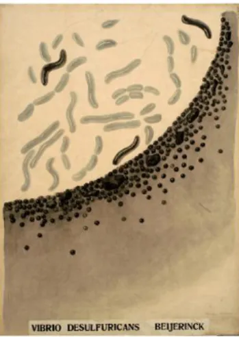
The first sulfate reducing organism, the Spirillum desulfuricans (later named Desulfovibrio desulfuricans) (Figure. 1.1), was isolated in 1985 by W. M. Beijerinck a Dutch microbiologist (Beijerinck, 1895). While he was studying the
in the contamination of sewage caused by large amounts of hydrogen sulfide produced by microorganisms, he detected isolated colonies with a surrounding black iron sulfide precipitate. Beijerinck observed that Spirillum desulfuricans grew only in the presence of aerobic bacteria, by consuming oxygen of the medium, which lead to the classification of this microorganism as strict anaerobe.
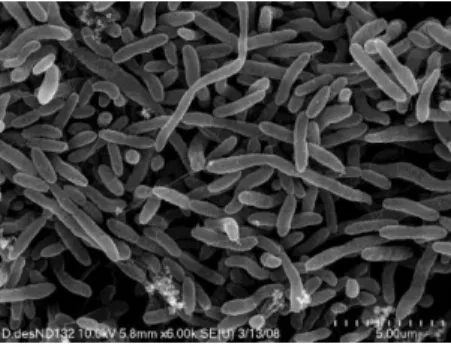
Figure 1.1 - Scanning electron microscopy image of D. desulfuricans. Adapted from (Gilmour et al., 2011).
comparative analysis of the sulfate reducing organisms (SRO) revealed that this group is formed by seven phylogenetic lineages, five within Bacteria (Deltaproteobacteria, Clostridia, Nitrospirae, Thermodesulfobacteria and Thermodesulfobiaceae) and two within Archaea (Euryarchaeota and Crenarchaeota) (Muyzer & Stams, 2008). The group that includes only bacteria is commonly named sulfate reducing bacteria (SRB). In spite of the 16S rRNA analysis being the most utilized technique in phylogenetic profiles of SRB, studies on lateral gene transfer events among the different species of essential genes have also been done. Enzymes of the sulfate respiration pathway like dissimilatory sulfide reductase (DsrAB) and dissimilatory adenosine-5’ -phosphosulfate (APS) reductase (AprBA) have been used as molecular markers providing a more precise evolutionary classification (Wagner et al., 2005; Meyer & Kuever, 2007).
1.2 - Environmental impact of SRB
The importance of SRB in the environment goes further beyond their role in the biogeochemical cycling of carbon and sulfur, mainly because the end product of sulfate reduction, hydrogen sulfide, can be harmful or beneficial.
and heterotrophic nitrate-reducing bacteria (hNRB), which remove sulfide by the production of nitrite and other reactive nitrogen species. Nitrite reduces the SRB activity and the corrosion rates, affecting especially the dissimilatory sulfite reductase, an enzyme that produces sulfide. With the use of this methodology, biocorrosion is significantly reduced when compared with the traditional biocide treatment (Bodtker et al., 2008; Voordouw, 2008).
Sulfate reduction can be applied beneficially to biotechnology, for instance in the removal of heavy metals. The ability of SRB to remediate heavy metals is dependent on the metal concentration in solution since the effect of these metals are toxic/inhibitory at higher concentrations and stimulatory at lower concentrations (Cabrera et al., 2006). Metal sulfates like cadmium, cobalt, copper, iron, nickel, uranium and zinc are highly soluble but the corresponding metal sulfides have low solubility. Hence, the sulfide produced during microbial sulfate reduction promotes metal precipitation allowing the recovery and reuse of the metals. The production of the insoluble mineral has been used as a mechanism for metal sequestration in metal-processing wastes and acid mine-drainage waters (Hockin & Gadd, 2007; Muyzer & Stams, 2008). This bioremediation process is performed in bioreactors that were designed for optimum hydrogen sulfide (H2S) production (Malik, 2004). Besides the chemical
indirect reduction of metals and metalloids due to the production of H2S, these
sulfate reducers can also reduce metals indirectly by an enzymatic way. Metalloenzymes, like hydrogenases and cytochromes, are involved in this reduction process. For example, in D. vulgaris, the tetraheme cytochrome c3
1.3 - Desulfovibrio genus
The most extensively studied genus among SRB is Desulfovibrio that
belongs to the class of δ-proteobacteria and is usually found in aquatic habitats, soil and sediments, oil and natural gas wells, sewages and in the digestive tract of animals and humans (Muyzer & Stams, 2008; Zhou et al., 2011). Cells of Desulfovibrio have a Gram-negative staining, are curved (vibrio) or rod-shaped (average width of 0.5 – 1.3 x 0.8 - 5 μm), often motile with a single polar flagellum (Figure 1.2). The DNA GC content is between 49 - 65% and the optimal growth temperature is from 30 to 38 ºC (Widdel & Bak, 1992).
Figure 1.2 – Scanning electron microscopy image of D. desulfuricans. Adapted from (Gilmour et al., 2011).
of sulfate reducing bacteria in a larger number of physiological and biochemical studies.
The work presented in this thesis was performed in D. vulgaris with the aim of understanding the behavior of sulfate reducers when exposed to oxidative and nitrosative stress conditions.
1.3.1 - Metabolism
Energy transduction
Energy transduction models
Desulfovibrio spp. are able to oxidize several substrates, such as, lactate, pyruvate, ethanol, molecular hydrogen, formate, malate, sugars, amino acids, aromatic hydrocarbons or alkanes (Rabus et al., 2006). The first mechanism proposed for energy conservation in Desulfovibrio spp. growing in lactate and sulfate is the so-called “hydrogen cycling” (Odom & Jr., 1981). Molecular hydrogen (H2) besides serving as sole electron donor with carbon dioxide (CO2)
and acetate as carbon sources (chemolithoheterotrophy) or only carbon dioxide (autotrophy), is also proposed to play a central role in the generation of a chemiosmotic gradient required for the reduction of sulfate (Odom & Jr., 1981; Liamleam & Annachhatre, 2007; Kanekar et al., 2012). In this model, the electrons produced during the oxidation of lactate to pyruvate and of pyruvate to acetyl-CoA are used by cytoplasmic hydrogenases to form H2, which diffuses
[NiFe] hydrogenases (Fauque et al., 1988; Heidelberg et al., 2004; Caffrey et al., 2007). The other two hydrogenases are included in cytoplasmic-facing membrane-bound associated complexes EchABCDEF and CooMKLXUH, and proposed to be involved in cytoplasmic proton reduction (Pereira et al., 2007). The principal drawback of this proposal is that the genes coding for these hydrogenases are not widespread in SRO (Pereira et al., 2011), and the production of hydrogen from lactate oxidation is an energetically unfavorable process that probably occurs only when the partial pressure of hydrogen is maintained at low levels (Traore et al., 1981; Odom & Peck, 1984). Hence, this mechanism is probably only one of the several possible pathways for energy conservation in Desulfovibrio.
Besides hydrogen cycling, other alternative mechanisms for energy conservation have been considered, such as formate and carbon monoxide (CO) cycling (Voordouw, 2002). The oxidation of formate to CO2 and H+ is catalyzed
by formate dehydrogenases (Fdh). In D. vulgaris there are three Fdh, being two of them periplasmic and the other a membrane-associated facing the periplasm (Sebban et al., 1995; Heidelberg et al., 2004; ElAntak et al., 2005). As well as hydrogen cycling, the formate cycling model proposes that formate is formed in the cytoplasm from lactate oxidation and then diffuses to the periplasm where is oxidized by formate dehydrogenases. The generated protons are used in adenosine-5’-triphosphate (ATP) synthesis and the electrons are donated to the periplasmic c-type cytochromes and membrane-bound complexes (Voordouw, 2002). The mechanism of CO cycling consists in cytoplasmic formation of CO from pyruvate generated in lactate oxidation, which is subsequent transformed in CO2 and H2 by a cytoplasmic CO dehydrogenase and a CO-dependent
CO cycling does not confer significant energetic advances, and probably, it does not serve as growth substrate (Rajeev et al., 2012).
Electron carriers and transmembrane electron transport
In terms of energy conservation, the components involved in the respiratory chain of SRB are not yet fully understood. Contrary to other anaerobic respiratory processes, in SRB the terminal reductases (APS reductase and DsrAB) are not directly involved in membrane charge translocation as they occur in the cytoplasm (Matias et al., 2005). Nevertheless, several membrane complexes had been proposed to contribute to the electron transfer during sulfate respiration (Pereira et al., 2006; Pereira, 2008). Among sulfate reducers two conserved membrane complexes have been identified: QmoABC (quinine-interacting membrane-bound oxidoreductase complex), which was first isolated from D. desulfuricans ATCC27774 (Pires et al., 2003), and the DsrMKJOP complex, first purified from Archaeoglobus fulgidus and named Hme complex
for “Hdr-like menaquinol-oxidizing enzyme” (Mander et al., 2002). Subsequently, in 2006, this complex was also isolated from D. desulfuricans ATCC27774 (Pires et al., 2006). QmoABC is proposed to transfer electrons from menaquinone pool to APS reductase (Pires et al., 2003), and DsrMKJOP complex, may be involved in electron transfer to the sulfite reductase (Pires et al., 2006).
Desulfovibrio spp. also contain a high number of periplasmic or membrane-bound c-type cytochromes that mediate the electron transfer. The c-type cytochromes include the monoheme cytochrome c553 and several multiheme
cytochromes, such as the tetraheme cytochrome c3 type I (TpIc3) and type II
(TpIIc3). Interestingly, the first multiheme cytochrome to be successfully
acts as electron acceptors for periplasmic hydrogenases and formate dehydrogenases. The periplasmic cytochromes c553 and c3 are proposed to
transfer electrons to the membrane-bound associated complexes, which contain a soluble cytochrome subunit of the c3 type family as electron acceptor.
The first transmembrane complex recognized in Desulfovibrio was the high molecular mass cytochrome c (Hmc) that contains 16 c-type hemes (Matias et al., 2005; Pereira, 2008). In D. desulfuricans the Hmc complex is not present, which instead contains a nine-heme cytochrome complex (9Hc) (Saraiva et al., 2001). Interestingly, the presence of both complexes in the same bacterium was not yet observed (Pereira et al., 2011). The third example is the transmembrane complex Tmc that was first isolated from D. vulgaris in 2006, this complex includes a tetraheme cytochrome c3 type subunit (TmcA or Type II cytochrome
c3), an integral membrane cytochrome b protein, and two cytoplasmic proteins
(Pereira et al., 2006). The expression of tmc genes is higher in cells growing in hydrogen/sulfate when compared with lactate/sulfate, suggesting a role of Tmc complex in hydrogen oxidation (Pereira, 2008).
Given the metabolic diversity of SRB, it is likely that, according to the electron donor used, several respiratory chains exist making difficult the development of a unique model of electron transport. The exception is the last set of reactions involved in sulfate activation and reduction which are common to all pathways (Rabus et al., 2006).
Sulfate reduction
eight electron process that occurs in the cytoplasm (Figure 1.3). After sulfate has been transported into the cell, it needs to be activated since this molecule is thermodynamically stable and by itself is not a suitable electron acceptor. Sulfate is first activated with ATP to yield adenosine phosphosulfate (APS), a reaction catalyzed by the ATP sulfurylase. The next steps of the pathway are the reduction of APS to bisulfite and adenosine-5´-phosphate (AMP) catalyzed by APS reductase, and the reduction of bisulfite to sulfide by the enzyme sulfite reductase, which in Desulfovibrio is known as desulfoviridin. Two different pathways for the reduction of bisulfite have been proposed: i) the sequential reduction in three two-electron reduction steps, with the formation of the intermediates trithionate and thiosulfate by trithionate and thiosulfate reductases; ii) a direct six-electron reduction without the formation of any intermediates (Figure 1.3) (Hansen, 1994; Rabus et al., 2006; Thauer et al., 2007).
Figure 1.3 – Pathway of dissimilatory sulfate reduction.
SO42- - sulfate; ATP - adenosine-5’-triphosphate;
APS - adenosine-5´-phosphosulfate; PPi - pyrophosphate; HSO3 - bisulfite; AMP -
adenosine-5´-phosphate; S3O6
- trithionate; S2O3
- thiosulfate; HS- - sulfide; e- - electron. Reaction catalyzed by (I) - ATP sulfurylase; (II) - APS reductase; (III) - sulfite reductase; (IV) - trithionate reductase; (V) - thiosulfate reductase and (VI) - sulfite reductase (desulfoviridin). Adapted from (Hansen, 1994).
Nitrate and nitrite reduction
Besides sulfate, Desulfovibrio spp. use several other electron acceptors, such as sulfite, thiosulfate, sulfur, nitrate and nitrite, elemental Fe3+, CO
2 and
SO42-+ ATP (I) APS + PPi 2e
-HSO3-+ AMP
S3O62 -2e
-2e
-S2O32
-2e- HS
fumarate. The dissimilatory reduction of nitrate and nitrite (also called ammonification) can function as the sole energy-conserving process in some SRB such as D. desulfuricans ATCC 27774 and D. gigas (Moura et al., 1997). This reaction occurs in a two step process (Figure 1.4), being the first the conversion of nitrate to nitrite, via two-electron reduction, catalyzed by the enzyme nitrate reductase. The crystal structure of the periplasmic nitrate reductase (Nap) from D. desulfuricans ATCC 27774 was the first to be solved for a dissimilatory nitrate reductase. The protein contains one molybdenum cofactor in the active site and a [4Fe-4S]+2/+1 cluster (Dias et al., 1999). Interestingly, nitrate reductase genes
are absent from D. vulgaris genome, and contrary to D. desulfuricans ATCC 27774, this strain is not able to grow on nitrate being a non-ammonifying microorganism (Pereira et al., 2000; Marietou et al., 2005). The second step consists in the six-electron reduction of nitrite to ammonia and is performed by nitrite reductase enzymes (NiR). Cytochrome c nitrite reductase (ccNiR) from D. desulfuricans ATCC 27774 is a membrane-bound protein composed by two subunits, the periplasmic pentaheme catalytic subunit NrfA and the transmembrane tetraheme subunit NrfH (Almeida et al., 2003). Although D. vulgaris Hildenborough is not capable of growing on nitrate, it contains a membrane protein complex identified as a cytochrome c nitrite reductase (NiR) formed by two cytochrome c subunits with nitrite and sulfite reductase activities, which reduce nitrite to ammonium in a reaction that does not sustain growth (Pereira et al., 2000).
Figure 1.4 – Pathway of dissimilatory nitrate reduction.
NO3- - nitrate; NO2- - nitrite; NH4+ - ammonium ion. Reaction catalyzed by (I) nitrate reductase
(Nap) and (II) cytochrome c nitrite reductase (NiR). Adapted from (Moura et al., 2007).
NO
3-(I) (II)
NH
4+
NO
2-Heme biosynthesis
The genus Desulfovibrio contains several single and multi-heme proteins. The most common heme containing proteins are the c-type cytochromes containing 1 (cytochrome c553), 4 (cytochrome c3), 9 (nine-heme cytochrome c),
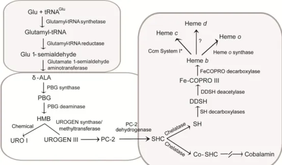
and 16 (dodecaheme cytochrome c) heme groups (Pereira & Xavier, 2005). In addition to heme c, other heme forms have been isolated from Desulfovibrio proteins, such as heme b, which is present in the membrane subunits of several respiratory complexes and enzymes, and heme d in the quinol oxygen reductases of the bd family (Lemos et al., 2001). Recently, it was shown the presence of an o-type heme associated with the protein heme-copper oxygen reductase (Lamrabet et al., 2011). This genus also contains unique heme derivatives such as Fe-coproporphyrin III isolated from D. desulfuricans ATCC 27774 bacterioferritin, (Romão et al., 2000a), and iron-uroporphyrin I present in D. gigas rubredoxin:oxygen oxidoreductase (Romão et al., 2000a). Siroheme also occurs in Desulfovibrio as a cofactor of the assimilatory and dissimilatory sulfite and nitrite reductases (Oliveira et al., 2008).
Figure 1.5 - Heme biosynthesis in Desulfovibrio.
δ-Aminolevulinic acid (δ-ALA), porphobilinogen (PBG), hydroxymethylbilane (HMB), uroporphyrinogen III (UROGEN III), uroporphyrin I (URO I), precorrin-2 (PC-2), sirohydrochlorin (SHC), cobaltsirohydrochlorin (Co-SHC), siroheme (SH), 12,18-didecarboxysiroheme (DDSH), and Fe-coproporphyrin III (Fe-COPRO III). Adapted from (Lobo et al., 2012).
1.3.2 - Desulfovibrio in humans
several isolates confirmed the presence of these bacteria in human feces (Beerens & Romond, 1977; Gibson et al., 1988; Gibson et al., 1993; Loubinoux et al., 2002b; Jia et al., 2012). Moreover, Desulfovibrio seem to be not only present in human feces but also in the gut. Measurements over 12 months revealed that mucosal Desulfovibrio numbers may vary significantly along time (Fite et al., 2004). In addition, fecal Desulfovibrios are also present in relatively high levels in children younger than six months reveling that SRB are acquired very early in life (Hopkins et al., 2005).
The implication of Desulfovibrio spp. in inflammatory bowel diseases has been suggested due to their metabolic end product hydrogen sulfide that at high concentrations promotes the damage of DNA and inhibits the butyrate oxidation pathway, which is an important energetic process for colonic epithelial cells (Pitcher & Cummings, 1996). Furthermore, patients presenting inflammatory bowel diseases showed significant high prevalence of Desulfovibrio strains (55%) in their feces when compared with healthy individuals (12%) (Loubinoux et al., 2002b). In spite of these results, a recent study does not show a clear correlation between Desulfovibrio numbers and inflammatory bowel diseases (Jia et al., 2012). This might be related with the differences in the methodologies used and the individual diversity in microbial flora within groups.
(Finegold, 2011b; Finegold, 2011a; Finegold et al., 2012). In spite of the proposals that Desulfovibrio spp. may be involved in disorders of the human gut tract, their role in the intestinal ecosystem remain unclear, as these microorganisms may be carried asymptomatic in the gastrointestinal tract or act as opportunistic pathogens. So far, five species of Desulfovibrio were isolated from human samples: D. piger (peritoneal fluid, abdominal collection and feces), D. desulfuricans (blood, peritoneal fluid and abdominal collection), D. fairfieldensis (blood, peritoneal fluid, abdominal collection, pelvic collection, colorectal collection, liver abscess, urine, and periodontal pockets), D. vulgaris (peritoneal fluid and abdominal collection) and, more recently, D. intestinalis from the vaginal flora (Johnson & Finegold, 1987; Tee et al., 1996; La Scola & Raoult, 1999; Loubinoux et al., 2000; Loubinoux et al., 2002a; Loubinoux et al., 2003; Goldstein et al., 2003; Pimentel & Chan, 2007; Urata et al., 2008; Ichiishi et al., 2010; Verstreken et al., 2012; Jia et al., 2012). Due to the slow growth of these microorganisms, they are difficult to identify and frequently overlooked in mixed cultures. Hence, it is possibly that their incidence in human remains underestimated (Goldstein et al., 2003; Verstreken et al., 2012).
Susceptibilities studies with commonly used antimicrobial agents have been done with clinical isolates from Desulfovibrio isolated from humans (Table 1.1) (Lozniewski et al., 2001; Warren et al., 2005; Nakao et al., 2009). In general, the results demonstrated that the Desulfovibrio isolates are susceptible to metronidazole, chloramphenicol, clindamycin and imipenem. In these studies Desulfovibrio spp. showed to be resistant to various antimicrobial agents
commonly used to treat mixed infections such as β-lactams (penicillin derivates)
and also β-lactams combined with β-lactamase inhibitors. Despite this
resistance to β-lactam antibiotics, only some of the analyzed isolates exhibit
be further investigated (Lozniewski et al., 2001; Warren et al., 2005; Nakao et al., 2009). So far, among all Desulfovibrio spp. D. fairfieldensis seems to be the species with higher pathogenic potential, possessing the higher antimicrobial resistance to the antibiotics tested. These differences in susceptibility pattern to antibiotics among Desulfovibrio species reveal that is important to identify the clinical isolates at the species level in order to perform the correct antibiotic administration (Loubinoux et al., 2000; Warren et al., 2005; Nakao et al., 2009).
Table 1.1 – Studies of antimicrobial susceptibilities of Desulfovibrio spp.
(Lozniewski et al., 2001)1 (Warren et al., 2005)2 (Nakao et al., 2009)3
Susceptible Metronidazole Chloramphenicol Clindamycin Imipenem Amoxicillin-clavulanate Ticarcillin-clavulanate Metronidazole Chloramphenicol Clindamycin Imipenem Metronidazole Chloramphenicol Clindamycin Sulbactam-ampicilin Meropenem
Resistant Penicillin G Piperacillin Piperacilin-tazobactam Cefoxitin Cefotetan Penincillin Piperacillin-tazobactam Cefoxitin Ceftriaxone Ertapenem Ticarcillin-clavulanic acid Piperacillin Piperacillin-tazobactam Cefoxitin Cefotaxime 1
16 strains of Desulfovibrio spp. isolated from patients hospitalized.
2
Human clinical isolates: D. fairfieldensis (n=10), D. piger (n = 2), D. desulfuricans (n = 3), D. vulgaris (n = 3).
3
23 Desulfovibrio isolates from humans: D. fairfieldensis (n = 8), D. desulfuricans Essex 6 (n = 7), D. desulfuricans MB (n = 6), D. piger (n = 2).
1.4 – References
Almeida, MG, Macieira, S, Goncalves, LL, Huber, R, Cunha, CA, Romão, MJ, Costa, C, Lampreia, J, Moura, JJ & Moura, I (2003). The isolation and characterization of cytochrome c nitrite reductase subunits (NrfA and NrfH) from Desulfovibrio desulfuricans ATCC 27774. Re-evaluation of the spectroscopic data and redox properties. Eur J Biochem, 270:3904-3915.
Badziong, W & Thauer, RK (1980). Vectorial electron transport in Desulfovibrio vulgaris (Marburg), growing on hydrogen plus sulfate as sole energy source. Arch Microbiol 125:167-174.
Bali, S, Lawrence, AD, Lobo, SA, Saraiva, LM, Golding, BT, Palmer, DJ, Howard, MJ, Ferguson, SJ & Warren, MJ (2011). Molecular hijacking of siroheme for the synthesis of heme and d1 heme.
Proc Natl Acad Sci U S A, 108:18260-18265.
Baron, EJ, Bennion, R, Thompson, J, Strong, C, Summanen, P, McTeague, M & Finegold, SM (1992). A microbiological comparison between acute and complicated appendicitis. Clin Infect Dis, 14:227-231.
Beech, IB & Sunner, JA (2007). Sulphate-reducing bacteria and their role in corrosion of ferrous materials In Sulphate-reducing Bacteria: Environmental and Engineered Systems, pp. 459-482. Edited by L. L. Barton & W. A. Hamilton. New York: Cambridge University Press.
Beerens, H & Romond, C (1977). Sulfate-reducing anaerobic bacteria in human feces. Am J Clin Nutr, 30:1770-1776.
Beijerinck, WM (1895). Ueber Spirillum desulfuricans als ursache von sulfatreduction. Zentralbl Bakteriol Parasitenkd, 1: 1-9, 49-59, 104-114.
Bodtker, G, Thorstenson, T, Lillebo, BL, Thorbjornsen, BE, Ulvoen, RH, Sunde, E & Torsvik, T (2008). The effect of long-term nitrate treatment on SRB activity, corrosion rate and bacterial community composition in offshore water injection systems. J Ind Microbiol Biotechnol, 35 :1625-1636.
Cabrera, G, Perez, R, Gomez, JM, Abalos, A & Cantero, D (2006). Toxic effects of dissolved heavy metals on Desulfovibrio vulgaris and Desulfovibrio sp. strains. J Hazard Mater, 135:40-46.
Caffrey, SM, Park, HS, Voordouw, JK, He, Z, Zhou, J & Voordouw, G (2007). Function of periplasmic hydrogenases in the sulfate-reducing bacterium Desulfovibrio vulgaris Hildenborough. J Bacteriol, 189:6159-6167.
Campbell, LL & Postgate, JR (1965). Classification of the spore-forming sulfate-reducing bacteria. Bacteriol Rev, 29:359-363.
da Costa, PN, Conte, C & Saraiva, LM (2000). Expression of a Desulfovibrio tetraheme cytochrome c in Escherichia coli. Biochem Biophys Res Commun, 268:688-691.
ElAntak, L, Dolla, A, Durand, MC, Bianco, P & Guerlesquin, F (2005). Role of the tetrahemic subunit in Desulfovibrio vulgaris Hildenborough formate dehydrogenase. Biochemistry, 44 :14828-14834.
Fauque, G, Peck, HD, Jr., Moura, JJ, Huynh, BH, Berlier, Y, DerVartanian, DV, Teixeira, M, Przybyla, AE, Lespinat, PA, Moura, I & et al. (1988). The three classes of hydrogenases from sulfate-reducing bacteria of the genus Desulfovibrio. FEMS Microbiol Rev, 4:299-344.
Finegold, SM (2011a). Desulfovibrio species are potentially important in regressive autism. Med Hypotheses, 77:270-274.
Finegold, SM (2011b). State of the art; microbiology in health and disease. Intestinal bacterial flora in autism. Anaerobe, 17:367-368.
Finegold, SM, Downes, J & Summanen, PH (2012). Microbiology of regressive autism. Anaerobe, 18:260-262.
Fite, A, Macfarlane, GT, Cummings, JH, Hopkins, MJ, Kong, SC, Furrie, E & Macfarlane, S (2004). Identification and quantitation of mucosal and faecal desulfovibrios using real time polymerase chain reaction. Gut, 53:523-529.
Gibson, GR, Macfarlane, GT & Cummings, JH (1988). Occurrence of sulphate-reducing bacteria in human faeces and the relationship of dissimilatory sulphate reduction to methanogenesis in the large gut. J Appl Bacteriol, 65:103-111.
Gibson, GR, Macfarlane, S & Macfarlane, GT (1993). Metabolic interactions involving sulphate-reducing and methanogenic bacteria in the human large intestine. FEMS Microbiol Ecol, 12 :117-125.
Gilmour, CC, Elias, DA, Kucken, AM, Brown, SD, Palumbo, AV, Schadt, CW & Wall, JD (2011). Sulfate-reducing bacterium Desulfovibrio desulfuricans ND132 as a model for understanding bacterial mercury methylation. Appl Environ Microbiol, 77:3938-3951.
Goldstein, EJ, Citron, DM, Peraino, VA & Cross, SA (2003). Desulfovibrio desulfuricans bacteremia and review of human Desulfovibrio infections. J Clin Microbiol, 41:2752-2754.
Hamilton, WA (1985). Sulphate-reducing bacteria and anaerobic corrosion. Annu Rev Microbiol, 39:195-217.
Hansen, TA (1994). Metabolism of sulfate-reducing prokaryotes. Antonie Van Leeuwenhoek, 66:165-185.
Hockin, SL & Gadd, GM (2007). Bioremediation of metals and metalloids by precipitation and cellular binding. In Sulphate-reducing Bacteria: Environmental and Engineered Systems, pp. 405-434. Edited by L. L. Barton & W. A. Hamilton. New York: Cambridge University Press.
Hopkins, MJ, Macfarlane, GT, Furrie, E, Fite, A & Macfarlane, S (2005). Characterisation of intestinal bacteria in infant stools using real-time PCR and northern hybridisation analyses. FEMS Microbiol Ecol, 54:77-85.
Ichiishi, S, Tanaka, K, Nakao, K, Izumi, K, Mikamo, H & Watanabe, K (2010). First isolation of Desulfovibrio from the human vaginal flora. Anaerobe, 16:229-233.
Jia, W, Whitehead, RN, Griffiths, L, Dawson, C, Bai, H, Waring, RH, Ramsden, DB, Hunter, JO, Cauchi, M, Bessant, C, Fowler, DP, Walton, C, Turner, C & Cole, JA (2012). Diversity and distribution of sulphate-reducing bacteria in human faeces from healthy subjects and patients with inflammatory bowel disease. FEMS Immunol Med Microbiol, 65:55-68.
Johnson, CC & Finegold, SM (1987). Uncommonly encountered, motile, anaerobic gram-negative bacilli associated with infection. Rev Infect Dis, 9:1150-1162.
Kanekar, PP, Kanekar, SP, Kelkar, AS & Dhakephalkar, PK (2012). Halophiles - Taxonomy. Diversity, Physiology and Applications. In Microorganisms in Environmental Management: Microbes and Environment, pp. 1-34. Edited by T. Satyanarayana, B. N. Johri & A. Prakash. New York: Springer.
Keller, KL, Wall, JD & Chhabra, S (2011). Methods for engineering sulfate reducing bacteria of the genus Desulfovibrio. Methods Enzymol, 497:503-517.
La Scola, B & Raoult, D (1999). Third human isolate of a Desulfovibrio sp. identical to the provisionally named Desulfovibrio fairfieldensis. J Clin Microbiol, 37:3076-3077.
Lamrabet, O, Pieulle, L, Aubert, C, Mouhamar, F, Stocker, P, Dolla, A & Brasseur, G (2011). Oxygen reduction in the strict anaerobe Desulfovibrio vulgaris Hildenborough: characterization of two membrane-bound oxygen reductases. Microbiology, 157:2720-2732.
Langendijk, PS, Hanssen, JT & Van der Hoeven, JS (2000). Sulfate-reducing bacteria in association with human periodontitis. J Clin Periodontol, 27:943-950.
Lemos, RS, Gomes, CM, Santana, M, LeGall, J, Xavier, AV & Teixeira, M (2001). The 'strict' anaerobe Desulfovibrio gigas contains a membrane-bound oxygen-reducing respiratory chain. FEBS Lett, 496:40-43.
Liamleam, W & Annachhatre, AP (2007). Electron donors for biological sulfate reduction. Biotechnol Adv, 25:452-463.
Lobo, SA, Brindley, A, Warren, MJ & Saraiva, LM (2009). Functional characterization of the early steps of tetrapyrrole biosynthesis and modification in Desulfovibrio vulgaris Hildenborough. Biochem J, 420:317-325.
Loubinoux, J, Mory, F, Pereira, IA & Le Faou, AE (2000). Bacteremia caused by a strain of Desulfovibrio related to the provisionally named Desulfovibrio fairfieldensis. J Clin Microbiol, 38:931-934.
Loubinoux, J, Bisson-Boutelliez, C, Miller, N & Le Faou, AE (2002a). Isolation of the provisionally named Desulfovibrio fairfieldensis from human periodontal pockets. Oral Microbiol Immunol, 17:321-323.
Loubinoux, J, Bronowicki, JP, Pereira, IA, Mougenel, JL & Faou, AE (2002b). Sulfate-reducing bacteria in human feces and their association with inflammatory bowel diseases. FEMS Microbiol Ecol, 40:107-112.
Loubinoux, J, Jaulhac, B, Piemont, Y, Monteil, H & Le Faou, AE (2003). Isolation of sulfate-reducing bacteria from human thoracoabdominal pus. J Clin Microbiol, 41:1304-1306.
Lozniewski, A, Maurer, P, Schuhmacher, H, Carlier, JP & Mory, F (1999). First isolation of Desulfovibrio species as part of a polymicrobial infection from a brain abscess. Eur J Clin Microbiol Infect Dis, 18:602-603.
Lozniewski, A, Labia, R, Haristoy, X & Mory, F (2001). Antimicrobial susceptibilities of clinical Desulfovibrio isolates. Antimicrob Agents Chemother, 45:2933-2935.
Macfarlane, GT, Cummings, JH & Macfarlane, S (2007). Sulphate-reducing bacteria and the human large intestine. In Sulphate-reducing Bacteria: Environmental and Engineered Systems, pp. 503-521. Edited by L. L. Barton & W. A. Hamilton. New York: Cambridge University Press.
Malik, A (2004). Metal bioremediation through growing cells. Environ Int, 30:261-278.
Mander, GJ, Duin, EC, Linder, D, Stetter, KO & Hedderich, R (2002). Purification and characterization of a membrane-bound enzyme complex from the sulfate-reducing archaeon Archaeoglobus fulgidus related to heterodisulfide reductase from methanogenic archaea. Eur J Biochem, 269:1895-1904.
Marietou, A, Richardson, D, Cole, J & Mohan, S (2005). Nitrate reduction by Desulfovibrio desulfuricans: a periplasmic nitrate reductase system that lacks NapB, but includes a unique tetraheme c-type cytochrome, NapM. FEMS Microbiol Lett, 248:217-225.
Matias, PM, Pereira, IA, Soares, CM & Carrondo, MA (2005). Sulphate respiration from hydrogen in Desulfovibrio bacteria: a structural biology overview. Prog Biophys Mol Biol, 89:292-329.
Meyer, B & Kuever, J (2007). Phylogeny of the alpha and beta subunits of the dissimilatory adenosine-5'-phosphosulfate (APS) reductase from sulfate-reducing prokaryotes-origin and evolution of the dissimilatory sulfate-reduction pathway. Microbiology, 153:2026-2044.
Michel, C, Giudici-Orticoni, M-T, Baymann, F & Bruschi, M (2003). Bioremediation of Chromate by Sulfate-Reducing Bacteria, Cytochromes c3 and hydrogenases. Water, Air, & Soil Pollution:
Focus, 3:161-169.