Arquivos Brasileiros de Cardiologia - Volume 83, Nº 2, Agosto 2004
141
Immunological factors may be involved in the etiopathogenesis of atherosclerosis. The role played by antibodies against phos-pholipids (PL) or against phospholipid cofactors in the atheros-clerotic process has not yet been elucidated 1.
Antiphospholipid antibodies, both anticardiolipin or aCL antibo-dies and lupus anticoagulant, are related to antiphospholipid syn-drome, which is characterized by arterial and venous thromboses and gestational morbidity, being currently considered the most
common cause of acquired hypercoagulability among young adults2.
Acute myocardial infarction occurs in 4 to 20% of the patients
with antiphospholipid syndrome 3. In a recent cohort of 1,000
patients with antiphospholipid syndrome, acute myocardial in-farction was observed in 2.8% of the cases 4.
The beta2-glycoprotein I (beta2-gpI) phospholipid cofactor is a natural anticoagulant 5. This cofactor was found in atherosclerotic plaque 6, and induction of atherosclerosis in receptor-LDL deficient
mice immunized with beta2-gpI has been reported 7.
Anti-beta2-gpI antibodies were found in the immunoassays of
patients with defined antiphospholipid syndrome 8, but also in
patients with thromboembolic pulmonary hypertension 9, cerebral
infarction 10, and coronary heart disease 11.
The frequency of anticardiolipin and anti-beta2-gpI antibodies, as well as their role in patients with acute myocardial infarction, has been a controversial issue. Our study provides a complete profile of anticardiolipin and anti-beta2-gpI antibodies in patients with acute coronary heart disease, analyzes their frequency in patients with acute myocardial infarction, and raises the possibility that anticardiolipin and anti-beta2-gpI antibodies act as independent risk factors for acute myocardial infarction.
Methods
This case-control study assessed the titers of anticardiolipin and anti-beta2-gpI antibodies in patients with acute myocardial infarction and in controls. Only incident cases were assessed.
The diagnosis of myocardial infarction was established by car-diologists according to previously reported algorithms, such as clinical history, serial electrocardiographic alterations, and labo-ratory tests confirming myocardial necrosis 12, and yet the cardio-logists continued to ignore the results of antibody titers.
The cases were patients older than 16 years with acute myocar-dial infarction, who were admitted to the hospital within the first 7 days of symptom onset. They were not selected by sex or race. The patient or his legal representative provided written informed consent. Race/ethnicity was determined by self-identification.
Original Article
Anti-Beta2-Glycoprotein I Antibodies as
Risk Factors for Acute Myocardial Infarction
Aline Ranzolin, Jussara Marilú Bohn, Gary L. Norman, Euler Manenti, Luis Carlos Bodanese,
Carlos Alberto von Mühlen, Henrique Luiz Staub
Porto Alegre, RS - Brazil
Hospital São Lucas da PUCRS
Mailing address: Henrique Luiz Staub - Reumatologia - Av. Ipiranga, 6690 - S/220 - Cep 90610-000 - Porto Alegre, RS - Brazil E-mail: henriquestaub@terra.com.br
Received: 5/30/03 Accepted: 1/6/04
English version by Stela Maris Costalonga
Objective
To determine whether high levels of antibodies against the phospholipid beta2-glycoprotein I (beta2-gpI) cofactor are asso-ciated with an increase in the risk of acute myocardial infarction.
Methods
The study comprised 82 patients with acute myocardial in-farction and 82 controls, who were assessed in regard to age, sex, race, hypertension, smoking, previous heart disease, history of diabetes mellitus, and hypercholesterolemia. The following antibodies were detected using immunoassay: anticardiolipin and anti-beta2-gpI IgA, IgG, and IgM. Adjusted odds ratios (OR) for risk factors were obtained through logistic regression.
Results
The mean ages of the cases and controls were, respectively, 57.7 and 51.1 years (P=0.003). Men (P=0.005) and the white race predominated in both groups (P=0.798). Of the risk factors, a history of diabetes (OR=5.3; 95% CI: 1.9 to 14.9; P=0.001) and previous heart disease (OR=4.7; 95% CI: 2.0 to 10.7; P<0.001) were the most consistent associations with myocardial infarction. The frequency of anticardiolipin IgG, IgM, and IgA antibodies did not differ between cases and controls (P=1.000). Anti-beta2-gpI IgA antibodies were more frequent in cases than in controls (P=0.054). The adjusted OR for anti-beta2-gpI IgA antibodies was 3.4 (95% CI: 1.3 to 9.1; P=0.015).
Conclusion
Anti-beta2-gpI IgA antibodies, but not anticardiolipin antibo-dies, seemed to behave as independent risk factors for myocardial infarction, which may represent a link between autoimmunity and atherosclerosis in patients with acute myocardial infarction.
Key words
Arquivos Brasileiros de Cardiologia - Volume 83, Nº 2, Agosto 2004
142
Anti-beta2-glycoprotein I antibodies as risk factors for myocardial infarction
The exclusion criteria were as follows: a) infective endocarditis; b) neoplasias (current or past); c) infection by the human
immu-nodeficiency virus or treponema pallidum; d) presence of known
hereditary causes of thrombosis, such as homocystinuria or mu-tation of factor V (Leiden); and e) previous diagnosis of antiphos-pholipid syndrome or another disease of the connective tissue.
The control group comprised patients without acute myocardial infarction admitted to orthopedic wards due to fractures or muscle-ligament disorders. The exclusion criteria were as follows: a) os-teonecrosis; b) infections, neoplasias, hereditary disorders, anti-phospholipid syndrome, or diseases of the connective tissue.
Historical, demographic, and clinical data were obtained through a review of medical records and interviews with patients and their families. The risk factors for myocardial infarction were as follows: 1) age, sex, race/ethnicity; 2) history of hypertension (diagnosis confirmed when the systolic or diastolic pressures were > 160 or 95 mmHg, respectively, or when the patient was using
antihypertensive medication) 12; 3) smoking, according to the
criteria of the British Council for Medical Research; 4) history of heart disease (atrial fibrillation or coronary heart disease, defined as previous myocardial infarction, angina, or revascularization pro-cedure); 5) history of diabetes mellitus, according to the medical history or the use of insulin or an oral antidiabetes drug; 6) hypercholesterolemia, based on total cholesterol > 200 mg/ dL, LDL-cholesterol > 130 mg/dL, or total cholesterol/HDL-cho-lesterol ratio > 5 13.
Blood samples were centrifuged and frozen within, at most, 2 hours after collection and stored at –70ºC until laboratory testing with ELISA (enzyme-linked immunoabsorbent assays).
ELISA IgG, IgM, and IgA anticardiolipin antibodies (INOVA Quan-talite cardiolipin kits, INOVA Diagnostics, Inc., San Diego, USA) were detected according to a previous report. The results for the IgG and IgM isotypes were reported in IgG phospholipid units (GPL) and IgM phospholipid units (MPL), in which 1 unit equals 1 mg/mL of IgG or IgM. Only samples with moderate to high IgG or IgM anticardiolipin antibody levels (above 20 GPL or 20 MPL) were considered positive in our study. Titers of IgA anticardiolipin antibodies were considered positive when above 15 units 14.
IgA, IgG, and IgM anti-beta2-gpI antibodies were measured according to the technique suggested in a previous report (INOVA Quantalite beta2-gpI kits, INOVA Diagnostics, Inc., San Diego, USA). Briefly, 50 µL of purified human beta2-gpI (at the
concen-tration of 10 µg/mL) was coupled to the orifices of polystyrene
plaques. Prediluted controls and diluted serum of patients (1/100) were added to certain orifices, allowing any anti-beta2-gpI antibody present to bind to the immobilized antigen. The samples not bound to the antigen were washed out. Human anti-IgG, anti-IgM, or
anti-IgA antibodies (100 µL) bound to peroxidase were added to
the orifices. A second incubation allowed antihuman antibodies to bind to any antibody of a patient, which had adhered to the plaque. After washing the unbound antihuman antibodies, the remaining enzymatic activity was measured by the addition of a chromogenic substrate. The assay was assessed with spectro-phototic measures. The intensity of the color developed by the sample of the patient was compared with that of the controls. The titers were considered positive when above 20 units for IgG, IgM, or IgA anti-beta2-gp antibodies 15.
Odds ratios with 95% confidence interval (95%CI) were
cal-culated through logistic regression adjusted for age, sex, race, history of hypertension, smoking, previous heart disease, history of diabetes, and hypercholesterolemia. All first-degree interactions between known risk factors for acute myocardial infarction and
antibody levels were examined. The Hopkins scale for OR 16 was
used as follows: OR between 1 and 1.5 was considered trivial; between 1.5 and 3.5 was considered low; between 3.5 and 9.0 was considered moderate; between 9.0 and 32 was considered strong; and above 32 was considered very strong. The Wald test 17 was used for assessing the significance of OR adjusted for logistic regression. The Fisher exact and chi-square tests were used for comparing categorical variables, and the Student t test was used for comparing continuous variables. The significance level of 5% (P < 0.05) was adopted. All analyses were obtained by using SPSS for Windows, version 8.0, Chicago, IL.
Results
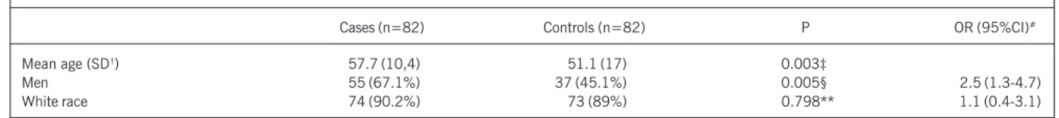
Our study comprised 82 patients with acute myocardial infarc-tion and 82 controls. The clinical and demographic characteristics of the cases and controls are shown in table I. Most patients with acute myocardial infarction were men and old (P=0.003) (P=0.005), which determined a low risk (OR 2.5; 95% CI: 1.3 to 4.7). The white race predominated among the cases and controls. The information on the risk factors for cases and controls are shown in table II, and the known risk factors for acute myocardial infarction were more frequent in cases than in controls. A history of diabetes (OR 5.3; 95%CI: 1.9 to 14.9; P=0.001) and previous heart disease (OR 4.7; 95%CI: 2.0 to 10.7; P<0.001) were the 2 most consistent associations with acute myocardial infarction. Table III categorizes the cases and controls according to the levels of anticardiolipin and anti-beta2-gpI antibodies. The frequency of anti-beta2-gpI IgA, but not of other antibodies, was greater among cases than among controls (P = 0.054).
The adjusted OR for risk factors (age, sex, race, history of hypertension, smoking, previous heart disease, history of DM, and hypercholesterolemia)are shown in table IV 18,19.
The positive test for the anti-beta2-gpI IgG antibody provided an OR of 0.1 (95%CI zero to 1.0); the adjusted P value in the Wald test was borderline for a protective role for this antibody (P=0.055). The occurrence of anti-beta2-gpI IgA antibody deter-mined a moderate risk for acute myocardial infarction (adjusted OR 3.4; 95%CI: 1.3 to 9.1; P=0.015).
Discussion
This case-control study of incident cases included a complete profile of anticardiolipin and anti-beta2-gpI antibodies in patients randomly chosen among adults with acute myocardial infarction. The mean age of the cases differed significantly from that of controls, and men predominated. It is worth noting that age and sex, as well as other risk factors, were adjusted for logistic re-gression. Of the known risk factors, a history of diabetes and previous heart disease were the most consistent associations with acute myocardial infarction.
Arquivos Brasileiros de Cardiologia - Volume 83, Nº 2, Agosto 2004
143
Anti-beta2-glycoprotein I antibodies as risk factors for myocardial infarctionTable - Demographic and clinical characteristics of patients with acute myocardial infarction and controls
Cases (n=82) Controls (n=82) P OR (95%CI)#
Mean age (SD†) 57.7 (10,4) 51.1 (17) 0.003‡
Men 55 (67.1%) 37 (45.1%) 0.005§ 2.5 (1.3-4.7)
White race 74 (90.2%) 73 (89%) 0.798** 1.1 (0.4-3.1)
* Odds ratio with 95% confidence interval; † SD - standard deviation; ‡ Student t test; § chi-square test.
Table II - Profile of the risk factors in cases and controls
Cases Controls P* OR (95%CI)†
(n=82) (n=82)
Risk factors
History of hypertension 46 (56.1%) 22 (26.8%) <0.001 3.5 (1.8-6.7)
Smoking 46 (56.1%) 28 (34.1%) 0.005 2.5 (1.3-4.6)
Previous heart disease 30 (36.6%) 9 (11%) <0.001 4.7 (2.0-10.7)
History of diabetes mellitus 21 (25.6%) 5 (6.1%) 0.001 5.3 (1.9-14.9)
Hypercholesterolemia 34 (41.5%) 16 (19.5%) 0.002 2.9 (1.4-5.9)
* Chi-square; † odds ratio with 95% confidence interval.
Table IV - Odds ratio for anticardiolipin and anti-beta2-gpI antibodies adjusted for risk factors
OR# IC95%† P‡
aCL IgG§ noncalculated ---
---aCL IgM 2.0 0.2-21.7 0.570
aCL IgA§ noncalculated ---
---anti-beta2-gpI IgG 0.1 0-1.0 0.055
anti-beta2-gpI IgM 1.2 0.4-4.0 0.726
anti-beta2-gpI IgA 3.4 1.3-9.1 0.015
*OR - adjusted odds ratio for demographic data and risk factors;
†95%CI - 95% confidence interval; ‡Wald test 17; § noncalculated logistic
regression due to null frequency in cases or controls. The nonadjusted OR after Agresti correction 18 was 0.3 for aCL IgG test and 3.0 for aCL IgA.
hypothesis (P = 1.000). Our group has already reported a very low prevalence (1.2%) of anticardiolipin IgG in acute myocardial
infarction 19. However, the presence of anticardiolipin IgG has
been linked to risk, although low, of infarction according to a
previous report 20. Two previous cohorts have reported a
time-dependent association of anticardiolipin IgG antibodies with acute myocardial infarction 21,22.
Our data regarding the anticardiolipin IgM isotype were not compatible with the association with acute myocardial infarction. The prevalence of anticardiolipin IgM in our study in 1993 was null 19. A recent study 23 associating IgM anticardiolipin and stroke triggers the discussion of the role played by infections in the anti-phospholipid IgM response.
The frequency of anticardiolipin IgA in our cases of acute myo-cardial infarction was very low (1.2%). No control was positive. The nonadjusted OR of 3.0 may suggest an association with is-chemic outcome. However, the nonadjusted P value of 1.000 makes this hypothesis unlikely. A prospective association of the anticardiolipin IgA isotype with acute myocardial infarction has been previously reported 21. Therefore, the aCL IgA isotype, whose immunoassay has not yet been internationally standardized, should be studied in these patients.
The relation between beta2-gp I and atherosclerosis is intri-guing. Atheromas contain beta2-gpI 6. Our study raises the possi-bility that anti-beta2-gpI antibodies may be associated with the risk of acute myocardial infarction.
In our study, the frequency of anti-beta2-gpI IgG antibodies was lower in cases than in controls. The low adjusted OR (0.1) and the adjusted P value of 0.055 point towards the possibility of a protective role of that antibody.
Farsi et al 11 reported an association of anti-beta2-gpI IgG
antibodies with coronary atherosclerosis (particularly unstable
angina). However, data from the Honolulu Heart Program 20 point
towards an insignificant frequency of anti-beta2-gpI IgG antibodies as compared with that of the controls. Two other studies have also ruled out the possibility of anti-beta2-gpI IgG antibodies being linked to coronary heart disease 3,24. In addition, the presence of anti-beta2-gpI IgG, as well as of anticardiolipin antibodies, does not seem to be a risk for coronary restenosis after angioplasty 25. In our study, the anti-beta2-gpI IgM isotype showed no asso-ciation with acute myocardial infarction. Theoretically, an occa-sional anti-beta2-gpI IgM response observed in myocardial infarction could result from infection or previous tissue necrosis.
Significant titers of anti-beta2-gpI IgA antibodies were detected in patients with acute myocardial infarction as compared with those in controls. The OR and the adjusted P value indicate that a positive test for anti-beta2-gpI IgA behaves as an independent risk factor for acute myocardial infarction. Likewise, an association between this antibody and the risk of cerebral infarction has been recently reported by our group 26.
The association of anti-beta2-gpI IgA antibodies with acute Table III - Frequency of anticardiolipin and anti-beta2-gpI antibodies for
cases and controls
Cases (n=82) Controls (n=82) P
aCL IgG positive 0 1 (1.2%) 1.000*
aCL IgM positive 4 (4.9%) 3 (3.7%) 1.000*
aCL IgA positive 1 (1.2%) 0 1.000*
anti-beta2-gpI 2 (2.4%) 4 (4.9%) 0.682*
IgG positive
anti-beta2-gpI 12 (14.6%) 8 (9.8%) 0.340**
IgM positive
anti-beta2-gpI 22 (26.8%) 12 (14.6%) 0.054**
IgA positive
Arquivos Brasileiros de Cardiologia - Volume 83, Nº 2, Agosto 2004
144
Anti-beta2-glycoprotein I antibodies as risk factors for myocardial infarction
1. Wick G, Xu Q. Atherosclerosis: an autoimmune disease. Exp Gerontol 1999; 34: 559-66.
2. Wilson WA, Gharavi AE, Koike T et al. International consensus statement on pre-liminary classification criteria for definite antiphospholipid syndrome. Arthritis Rheumatism 1999; 42: 1309-11.
3. Vaarala O. Antiphospholipid antibodies and myocardial infarction. Lupus 1998; S2 132-4.
4. Cervera R, Piette JC, Font J et al. Antiphospholipid syndrome: clinical and immu-nologic manifestation and patterns of disease expression in a cohort of 1,000 pa-tients. Arthritis Rheum 2002; 46: 1019-27.
5. Kandiah DA & Krilis S. Beta2-gpI. Lupus 1994; 3: 207-12.
6. George J, Harats D, Gilburd B, Afek A, Levy Y, Schneiderman J, Barshack I, Kopolovik J & Shoenfeld Y. Immunolocalization of beta2-gpI (apolipoprotein H) to human atherosclerotic plaques: potential implications for lesion progression. Cir-culation 1999; 99: 2227-30.
7. George J, Afek A, Gilburd B et al. Induction of early atherosclerosis in LDL-recep-tor-deficient mice immunized with beta2-gpI. Circulation 1998; 98: 1108-15. 8. Arvieux J, Roussel & Colomb MG (1994). Anticorps antiphospholipids et
anti-beta2-gpI. Ann Biol Clin 52: 381-5.
9. Martinuzzo ME, Pombo G, Forastiero RR et al. Lupus anticoagulant, high levels of anticardiolipin and anti-beta2-gpI antibodies are associated with chronic thromboembolic pulmonary hypertension. J Rheumatol 1998; 25: 1313-9. 10. Chen WH, Liu JS. An unusual increase of blood anti-beta2-gpI antibody but not
an-tiphospholipid antibody in cerebral ischemia: a case report. Angiology 2001; 52: 149-54. 11. Farsi A, Domeneghetti MP, Fedi S et al. High prevalence of anti-beta2-gpI antibodies
in patients with ischemic heart disease. Autoimmunity 1999; 30: 93-8. 12. Seventh report of the Joint National Commitee on the Prevention, Detection,
Eva-luation, and Treatment of High Blood Pressure (JNC 7): resetting the hypertension sails. Hypertension 2003; 41: 1178-9.
13. Donahue RP, Abbott RD, Reed DM, Yano K. Physical activity and coronary heart disease in middle-aged and elderly men. The Honolulu Heart Program. Am J Public Health 1988; 78: 683-5.
14. Gharavi AE, Harris EN, Asherson RA & Hughes GRV (1987). Antiphospholipid antibodies: isotype distribution and phospholipid specificity. Ann Rheum Dis 1987; 46: 1-6. 15. Lewis S, Keil LB, Binder WL & DeBari VA (1998). Standardized measurement of
major immunoglobulin class (IgG, IgA, IgM) antibodies to beta2-gpI in patients with antiphospholipid syndrome. J Clin Lab Anal 1998; 12: 293-7.
References
16. Hopkins WG: A new view of statistics. On line. Available: http://www.sportsci.org/ resource/stats/index. Captured in April 11th, 2002.
17. Hosmer DW, Lemeshow S. Applied logistic regression. New York, NY: John Wiley & Sons Inc; 1989.
18. Agresti A. Categorical data analysis. New York, NY: Wiley;1990.
19. Calcagnotto ME, Vitola D, Rabin M et al. Anticardiolipin antibodies and myocar-dial infarction. Rev Bras Reumatol 1993; 33: 217-20.
20. Brey RL, Abbott RS, Curb D et al. Beta2-gpI dependent anticardiolipin antibodies and risk of ischemic stroke and myocardial infarction. The Honolulu Heart Pro-gram. Stroke 2001; 32: 1701-6.
21. Vaarala O, Manttari M, Manninem V et al. Anticardiolipin antibodies and risk of myocardial infarction in a prospective cohort of middle-aged men. Circulation 1995; 91: 23-7.
22. Wu R, Nityanand S., Berglund L, Lithell H, Holm G, Lefvert AK. Antibodies against cardiolipin and oxidatively modified LDL in 50-year-old men predict myocardial in-farction. Arterioscler Thromb Vasc Biol 1997; 17: 3159-63.
23. Tuhrim S, Rand JH, Wu X et al. Elevated anticardiolipin antibody titer is a stroke risk factor in a multiethnic population independent of isotype or degree of positi-vity. Stroke 1999; 30: 1561-5.
24. Limaye V, Beltrame J, Cook R, Gillis D, Pile K. Evaluation of antibodies to beta2-gpI in the causation of coronary atherosclerosis as part of the antiphospholipid syndrome. Aust NZ J Med 1999; 29: 789-93.
25. George J, Harats D, Bakshi E et al. Anti-oxidized low density lipoprotein antibody determination as a predictor of reestenosis following percutaneous transluminal coronary angioplasty. Immunol Lett 1999; 68: 263-6.
26. Staub HL, Norman GL, Crowther T et al. Antibodies to the atherosclerotic plaque components beta2-glycoprotein I and heat-shock proteins as risk factors for acute cerebral ischemia. Arq Neuropsiquiatr 2003; 61: 757-63
27. Tsutsumi A, Matsuura E, Ichikawa K et al. Antibodies to beta2-gpI and clinical manifestations in patients with systemic lupus erythematosus. Arthritis Rheum 1996; 39: 1466-74.
28. Piette JC. Towards improved criteria for the antiphospholipid syndrome. Lupus 1998; 7 Suppl 2: S149-57.
29. Vaarala O. Antibodies to prothrombin imply a risk of myocardial infarction in mid-dle-aged men. Thromb Haemost 1996; 75: 456-9.
30. Roldán V, Marin F, Pineda J et al. Anexin V levels in survivors of early myocardial infarction. Rev Esp Cardiol 2002; 55: 1230-4.
myocardial infarction is controversial. The great majority of our patients with anti-beta2-gpI IgA antibodies are aCL IgA-negative. As previously suggested, anti-beta2-gpI IgA and aCL IgA may com-prise AAF of different specificities 27.
Whether patients with acute myocardial infarction, who are anti-beta2-gpI IgA positive, but have a negative anticardiolipin IgA test, should be managed as having antiphospholipid syndrome is still controversial. The 1999 international consensus for the diag-nosis of antiphospholipid syndrome does not include anti-beta2-gpI antibodies 2. The incorporation of these antibodies into the criteria of antiphospholipid syndrome has been recently proposed 28.
It is worth noting that antibodies against the prothrombin phospholipid cofactor have also been implicated as risk factors for acute myocardial infarction in middle-aged men according to a report 29. Low anexin V levels, a phospholipid cofactor with anti-coagulant properties, have been recently reported in patients with a history of early acute myocardial infarction 30.
In conclusion, anti-beta2-gpI IgA antibodies seemed to behave as independent risk factors for acute myocardial infarction in our study. The need for testing anti-beta2-gpI antibodies, particularly IgA, in patients with coronary heart disease should be discussed and their predictive value assessed.
Although beta2-gpI is found in atherosclerotic plaque 6, a
pathogenic role for anti-beta2-gpI IgA antibodies in acute myocardial infarction has not yet been confirmed. Epiphenomenon or not, the occurrence of these antibodies in acute myocardial infarction may represent 1 of the links between autoimmunity and coronary atherosclerosis. The clinical implications of such findings may be clarified in the near future.