S
CIENTIFICA
RTICLESIncentive spirometry and breath stacking:
effects on the inspiratory capacity of individuals
submitted to abdominal surgery
Inspirometria de incentivo e
breath stacking
: repercussões sobre a capacidade
inspiratória em indivíduos submetidos à cirurgia abdominal
Dias CM1,4, Plácido TR2, Ferreira MFB2, Guimarães FS1,3, Menezes SLS1,3
Abstract
Background: Respiratory complications are the main causes of increased morbidity and mortality in individuals who undergo upper abdominal surgery. The effi cacy of physical therapy procedures needs clarifi cation, and it is necessary to know which therapeutic approaches are the best ones to implement. Objective: To compare the inspiratory volume during the breath stacking maneuver with the volume during incentive spirometry, in abdominal surgery patients. Methods: Twelve patients, on their fi rst postoperative day, were instructed to take a deep breath through the Voldyne™ incentive spirometer and to make successive inspiratory efforts using a facemask that had been adapted for performing the breath stacking maneuver. Each technique was performed fi ve times according to the randomization. Before the operation, the patients performed a spirometric test. They were also assessed and instructed about the procedures. A Wright™ ventilometer allowed inspiratory capacity to be recorded. Results: The inspiratory capacity during breath stacking was signifi cantly higher than during incentive spirometry, both before and after the operation. There was a signifi cant reduction in volumes after the surgical procedure, independent of the technique performed. Conclusions: The breath stacking technique was shown to be effective. This technique was better than incentive spirometry for generating and sustaining inspiratory volumes. Since no adverse effects have been described, this technique can probably be used safely and effectively, particularly in uncooperative patients.
Key words: pulmonary volumes and capacities; respiratory complications; physical therapy; breath stacking.
Resumo
Contextualização: As complicações respiratórias são as principais causas de aumento da morbidade e da mortalidade em indivíduos submetidos à cirurgia de andar superior do abdômen. A efi cácia dos procedimentos fi sioterapêuticos precisa ser melhor defi nida, assim como é necessário o conhecimento da melhor estratégia terapêutica a ser implementada. Objetivo: Comparar o volume inspiratório mobilizado durante a técnica de breath stacking, com o volume na inspirometria de incentivo em pacientes submetidos à cirurgia abdominal. Materiais e métodos: Doze pacientes, no primeiro dia de pós-operatório, foram orientados a inspirar profundamente por meio do inspirômetro de incentivo Voldyne® e a realizar esforços inspiratórios sucessivos pela máscara facial adaptada para realização da manobra de breath stacking. Cada técnica foi realizada cinco vezes de acordo com a randomização. No período pré-operatório, os pacientes realizaram prova espirométrica, foram avaliados e instruídos quanto à realização das técnicas. Um ventilômetro de Wright®
permitiu o registro da capacidade inspiratória. Resultados: A capacidade inspiratória foi signifi cativamente maior durante o breath stacking do que durante a inspirometria de incentivo, tanto no pré quanto no pós-operatório. Houve redução signifi cativa dos volumes após o procedimento cirúrgico, independentemente da técnica realizada. Conclusões: A técnica de breath stacking mostrou-se efi caz e superior à inspirometria de incentivo para a geração e sustentação de volumes inspiratórios. Por não haver descrição de efeitos adversos, essa técnica pode, provavelmente, ser utilizada de forma segura e efi caz, principalmente em pacientes pouco cooperativos.
Palavras-chaves: volumes e capacidades pulmonares; complicações respiratórias; fi sioterapia; breath-stacking.
Received: 23/11/2006 – Revised: 13/08/2007 – Accepted: 30/01/2008
1 Centro Universitário Augusto Motta (Unisuam) – Rio de Janeiro (RJ), Brazil 2 Instituto Nacional de Câncer – HC-II – Rio de Janeiro (RJ), Brazil 3 Universidade Federal do Rio de Janeiro (UFRJ) – Rio de Janeiro (RJ), Brazil 4 Hospital de Força Aérea do Galeão – Rio de Janeiro (RJ), Brazil
Correspondence to: Cristina Márcia Dias, Physical Therapist, Ph.D, Physical Therapy Course Centro Universitário Augusto Motta, Paris Avenue, 72 – Bonsucesso – Zip Code : 21041020 -Rio de Janeiro, RJ, Brazil, e-mail: [email protected]
95
Introduction
h e associations between thoracic and abdominal sur-geries and the high incidence of respiratory complications1,2
are already well documented in the literature and itsmain characteristics are: atelectasis3,pneumonia4, respiratory
dys-function2 and pleural ef usion5. h e immediate postoperative
period may evolve with hypoventilation, due to the residual ef ects of the anaesthetic, and deep breathing may be im-paired as a function of pain from surgical incision6. h e rate
of prevalence of respiratory complications in upper abdomen surgeries ranges from 17 to 88%1.
One of the basic mechanisms involved in respiratory dis-orders is the lack of adequate pulmonary insul ation that results from monotonous and superi cial respiratory patterns7,
prolonged restraint in bed8 and temporary diaphragmatic
disfunctions9. h e mucociliary clearance is also impaired in
the postoperative period contributing to the reduction of the ef ectiveness of cough and increasing of risks associated with the retention of sputum10. h ere is a reduction in functional
residual capacity (FRC), of inspiratory (IRV) and expiratory reserve volumes (ERV) and vital capacity (VC), also causing a reduction in expiratory l ow, probably due to the reduced dia-phragmatic activity11.
All these respiratory complications can be minimized or avoided by the use of a protocol of respiratory physiotherapy, since the pulmonary atelectasis is considered the major cause of complications. h is assertion is based on the observation that lung compliance and partial pressure of oxygen (PaO2) return to their normal values after deep lung insul ations12.
Several methods have been studied such as: intermittent positive pressure ventilation, exercises with deep breathing, incentive spirometry and conventional chest physiotherapy, nevertheless, a meta-analysis coni rmed that all studied pro-tocols and methods were equally ef ective in reducing the frequency of pulmonary complications after upper abdominal surgery13. However, the ei cacy of physiotherapy in the
post-operatory period of abdominal surgery remains controversial. While Pasquina and colleagues suggest that the routine use of respiratory physiotherapy is not justii ed, since few clinical tri-als show its ei cacy as a prophylactic feature14; Lawrence and
colleagues describe that in the upper abdominal postoperative period, any technique for lung expansibility is superior than non-prophylaxis15.
h e incentive spirometer is an equipment that encourages the patient, through a visual feedback, to maintain a maximum inspiration, in one attempt, as one of the most commonly used strategies in the postoperative16. In 1986, Marini and colleagues
described an alternative method to estimate VC in low coopera-tive individuals, called breath-stacking. h e method proved to
be ef ective for the proposed purpose and also made maximum lung expansion possible with minimum patient cooperation17.
h e present study aimed to compare the ef ects of the technique called “Breath-Stacking” with those observed during the “Incentive Spirometry” in patients in the upper abdomi-nal post-operative period, assessing the inspiratory capacity achieved by patients with each technique.
Methods
Subjects
Twelve patients were sequentially recruited and evaluated in the pre-operative period for upper abdomen surgery admit-ted at the National Institute of Cancer – HC II (Instituto Nacio-nal de Câncer – HC II), in the period of August to November 2006. h e Research Ethics Committee of the National Institute of Cancer - HC II, approved this project, with the record number 31/06 in CONEP and written informed consent was obtained from all participants.
h e assessment of eligibility for participation in the study fol-lowed well dei ned criteria: 1) Inclusion criteria: patients in the upper abdomen pre-operative period who agreed to participate in the study; 2) Exclusion criteria: cognitive impairments or lack of coordination to perform the incentive spirometry, intolerance to use the breath-stacking mask, post-operative complications that led to admission on the Intensive Treatment Center or extubation in a period exceeding 24 hours after surgery, level of consciousness in the post-operative period incompatible with the incentive spirometry realization.
Interventions description
∙ Incentive Spirometry: after placing a nasal clip, the subject was instructed to inhale deeply until total lung capacity through the mouthpiece of the Voldyne 5000® equipment (Sherwood Medical, St Loius, MO - USA) from the func-tional residual capacity.
∙ Breath-stacking: a siliconized mask connected to a one-way valve was adapted to the patient’s face. Once the mask was set to allow only the inspiration (the expiratory branch re-mained occluded), the individual carried through successive inspiratory ef orts for a period of 20 seconds. h en, the expi-ratory branch was released and patient expired freely17,18.
Experimental protocol
96
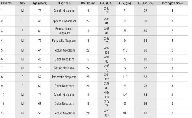
Table 1. Patients’ characteristics.
Patients Sex Age (years) Diagnosis BMI kg/m2 FVC (l, %) FEV
1 (%) FEV1/FVC (%) Torrington Scale
1 M 75 Gastric Neoplasm 18 2.45
73 71 72 4
2 F 45 Appendix Neoplasm 27 2.88
81 88 90 3
3 F 51 Retroperitoneal
Neoplasm 19
3.07
87 86 80 2
4 M 77 Pancreatic Neoplasm 18 2.42
70 64 68 4
5 M 41 Rectum Neoplasm 22 4.97
103 113 90 2
6 M 45 Colon Neoplasm 17 3.94
80 78 80 2
7 M 71 Gastric Neoplasm 24 2.98
73 84 87 3
8 F 57 Pancreatic Neoplasm 25 3.04
102 112 88 2
9 F 69 Colon Neoplasm 20 2.27
89 89 78 3
10 M 73 Gastric Neoplasm 19 4.09
110 122 84 3
11 M 68 Colon Neoplasm 18 3.79
78 95 96 3
12 M 56 Rectum Neoplasm 28 4.28
101 103 80 2
BMI= Body Mass Index; FVC= Forced Vital Capacity; FEV1= Forced Expiratory Volume, 1 second. use of each technique on the i rst postoperatory day after sur-gery. At the pre-operative evaluation, the patients had been trained for the accomplishment of the two techniques and, after learning, the registering of the mobilized volume was car-ried out. Additionally, the spirometric test was performed with the Pony Fx®,COSMED® equipment, USA, with the patient in the sitting position. h e Torrington range® was dei ned based on clinical and functional data.
On the i rst post-operatory day, the patient executed each of the techniques with a one hour interval, a period in which there were no modii cation or new medicines added. h e order of the techniques was randomly dei ned into two blocks of six individu-als, which means, for each block were made up of and ordered ran-domly into three envelopes corresponding to each treatment order (beginning by Voldyne® or Breath-stacking). h ese envelopes were externally numbered and after the recruitment and decision of the inclusion of the individual in the study, the envelope was opened to dei ne which technique would be i rst held. h e drawing up and the selection of the envelopes were conducted by a person who was not involved in the recruitment and selection of patients in the study. Five repetitions of each techniques were performed and a Wright® ventilometer (British Oxigen Company, London, Eng-land) was connected to the circuit of each equipment to measure the inspiratory capacity20. All the procedures were carried out
under the guidance and supervision of the same physiotherapist,
always in the morning, and the techniques were performed with the patient in the Fowler position of 45 degrees.
Statistical analyses
h e statistical analysis was done in the SigmaStat® pro-gram for Windows®(V 3.0). h e normality of the data (test of Kolmogorov-Smirnov with Lilliefors correction) and the equality of variance (Median Levene test) were tested. Due to the normality of the data, One-Way Repeated Measures ANOVA test was applied, followed by the Tukey test.
To correlate the volumes mobilized with the Torrington Scale, the Spearman Correlation was used. Values were ex-pressed by mean ± MSE and the selected level of signii cance was 5% (p< 0.05).
Results
97 h e analysis by the Wright® ventilometer showed signii
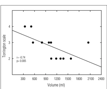
-cantly higher inspiratory volumes during the breath-stacking handling compared to the incentive spirometry, both in the pre-operative as in the post-operative periods (Figure 1). Comparisons of the values of each technique reported in the post-operative period with the values obtained before surgery showed signii cant reductions of the inspiratory volumes both in the breath-stacking treatment and in the incentive spirometry, and there was a more pronounced reduction in the latter condition, 76 ± 4 versus 61 ± 6, respectively. h e volumes deployed in the postoperative period during the incentive spirometry were signii cantly correlated with the Torrington Scale (Figure 2).
Discussion
h e present study showed that during the execution of the breath-stacking technique, largest mobilization of inspired volume occurred when compared to the incentive spirometry, both in the pre- as post-operative periods. h ere was a signii -cant reduction in volume after the surgical procedure, whatever the selected physiotherapeutic maneuver was performed. h e reduction of volumes in the post-operative period was more pronounced during the incentive spirometry when compared with the breath-stacking maneuver, with correlations between the volumes in incentive spirometry and the Torrington Scale. h ese correlations showed the importance of this scale as a predictor index of the risk of post-operative pulmonary com-plications, as well as, showing a higher dependency between
the volume in incentive spirometry and the risk of post-surgical pulmonary risks.
h e decrease in the inspired volume in the postoperative period observed in this study may be corroborated by previous i ndings that described impairments of the respiratory system functions during and after chirurgical procedures4, with
hy-poventilation, deep breathing impairments6, monotonous
re-spiratory patterns7 and decreases in coughing ef ectiveness10.
Prevention and reversion of atelectasis has shown to reduce pulmonary complications, and to this end, techniques and equipment are used to encourage patients to inspire deeply21,22.
h e i nal goal is the production of a large and sustained increase in the transpulmonary pressure, which will distend the lungs and re-expand the collapsed areas. h e ef ective treatment of the post-operative respiratory complications is still hard2, and
it is important to emphasize and establish the physiotherapeu-tic procedures for greater ef ectiveness.
h e i rst study that showed the benei ts of maximum in-spiration in the post-operative period was made by h oren in 1954. When analyzing 343 patients in the post-operative period after cholecystectomy, this study showed a 42% inci-dence of atelectasis in the group that was not submitted to the physical therapy procedures (including deep breathing) compared to 27% in the group that carried out the physical therapy treatments23.
h e ventilatory desynchronization causes dif erences in the spatial and temporal distributions of the inspired air in the lung regions with dif erent time constants18,24. Ward and colleagues
had shown that post-operative atelectasis were more ef ectively reversed when the deep inspiration was maintained for a three
Figure 1. The values are means ± SE of 12 patients. *Signifi cantly different from pre-operative Voldyne® values.
Volume (ml)
0 500 1000 1500 2000 2500 3000
PRE-OPERATIVE
POST-OPERATIVE
Voldyne Breath-stacking
*
# &
*
#Signifi cantly different from post-operative Voldyne® values.
&Signifi cantly different from pre-operative Breath-Stacking values. p< 0.05.
Figure 2. Spearman correlation.
• Post-operative Voldyne® values of each patient and Torrington Scale. Volume (ml)
0 300
Torring
ton scale
1 2 3 4 5
r= -0.74 p= 0.005
98
seconds pause post-inspiration, when compared to the deep breathing with multiple inspirations without sustenance25. h e
accomplishment of slow and deep inspirations, followed by a post-inspiratory pause, allows the air to distribute itself in a homogeneous form, with a necessary pause of, at least, i ve se-conds18, 24. h e primary therapeutic goal of the incentive
spirom-etry or any pulmonary insul ation technique is to increase the transpulmonary pressure and the functional residual capacity, reverting the alveolar collapse areas 26.
h e incentive spirometry is used clinically as an intrinsic part of the prophylactic and therapeutic routine in the respira-tory care in the post-operative period of abdominal, cardiac and thoracic surgeries. However, its ei cacy is still quite debated2.
h e success of incentive spirometry is quite variable, since pa-tients who are weak and with dyspnea are unable to perform enough of an inspiratory ef ort to achieve and sustain high inspiratory volumes and, even with very cooperative and moti-vated patients, the ability to perform the incentive spirometry is compromised by dyspnea, muscle weakness and pain18.
In the present study, there was greater inspiratory volume during the breath-stacking execution compared to incentive spirometry. Such i ndings were corroborated by the study of Baker and colleagues, conducted in 1990, which reported that the breath-stacking increases the amplitude and duration of thoracic expansion18. h e volume of multiple inspiratory ef orts
can be added through the use of a one-way valve that allows only the inspiration (the expiration is blocked), even in less co-operative patients17. During airway occlusion, the central drive
increases gradually and, and with the expiration blocked, the air inlet follows each inspiratory ef ort, consequently, increasing the thoracic volume18. h us, air can be involuntarily trapped18, not
requiring the patient’s cooperation, and favoring the distribution of air in areas with dif erent time constants24. h is increase of
the volume tends to diminish with successive breaths, a time that the complacency of the thoracic wall diminishes and the respiratory muscles are shortened and enter in a mechanical disadvantage18. Maximum inspirations cause increases of the
transpulmonary pressure and the post-inspiratory pause, with the maintenance of this raised pressure, contributes to the in-creases of the PaO2, presumably through the recruitment of the collapsed alveoli.
Many patients who breath spontaneously are able to generate sufficient pressures to achieve high pulmonary volumes17,27, however, the impairment of the respiratory
mechanics, dyspnea and pain compromise the maintenance
of the effort long enough to achieve the maximum volumes and sustained inspiration18. Breath-stacking makes the
pres-sure generated possible during successive inspiratory efforts to overcome the elastics (and non-resistive) forces. At the end of the stacking, small inspiratory volumes need smaller flows and cause less frictional pressure. The pressure peak is, then, available for the elastic work of the expansion of the thorax18.
To keep the lungs distended with the occlusion of the expi-ratory branch, this allows additional time so that the interde-pendent forces recruit volume, a process that is not complete in conventional spirometry18. Katz and colleagues showed that
a total recruitment volume attained from gradual increases of the PEEP, is only achieved after four-i ve breaths (20 to 25s) after application of the PEEP, or the duration of maneuver of breath-stacking28.
h is study shows as one limitation, the number of patients involved, as well as the specii city of the researched popula-tion, and does not allow the generalization of the results to other clinical situations. In this context, during the time that the analgesic medication dose used by each patient was not registered, the possibility that the drug action may have inl u-enced the volume mobilized by patients cannot be excluded. In the case that this ef ect may have been relevant, this does not invalidate the comparisons between the techniques, since the study was crossed and no medication was given during the interval between them.
The technique of breath-stacking could surpass the objectives of the incentive spirometry18 overall with
un-cooperative patients who demonstrated difficulties to generate high volumes and to sustain the inspired volume. Additionally, this technique could reveal to be efficient in patients with higher risks of pulmonary complications, since greater reduction of the volumes during the incen-tive spirometry were observed.
Conclusions
99
References
1. Overend TJ, Anderson CM, Lucy SD, Bhatia C, Jonsson BI, Timmermans C. The effect of incentive spirometry on postoperative pulmonary complications. Chest.2001;120:971-8.
2. Weindler J, Kiefer RT. The Effi cacy of postoperative incentive spirometry is infl uenced by the device-specifi c imposed work of breathing. Chest. 2001;119:1858-64.
3. Duggan M, Kavanagh BP. Pulmonary atelectasis: a pathogenic perioperative entity. Anesthesiology. 2005;102(4):838-54.
4. Pereira EDB, Fernandes ALG, da Silva Anção M, de Araúja Pereres CA, Atallah NA, Faresin SM. Prospective assessment of the risk of postoperative pulmonary complications in patients submitted to upper abdominal surgery. São Paulo Medical Journal. 1999;4:151-60.
5. Fischer BW, Majumdar SR, McAlistar FA. Predicting pulmonary complications after nonthoracic surgery: a systematic review of blinded studies.Am J Med. 2002;112:219-25.
6. Arozullah AM, Conde MV, Lawrence VA. Preoperative evaluation for postoperative pulmonary complications. Med Clinics North Am. 2003;87:153-73.
7. Bendixen HH, Smith GM, Mead J. Pattern of ventilation in young adults. J Appl Physiol. 1964;19:195-8.
8. Alexander JI, Spence AA, Parika RK, Stuart B. The role of airway closure in postoperative hypoxaemia. Br J Anaesth. 1973;45:34-40.
9. Ford GT, Rosenal TW, Clergue FC, Whitelaw WA. Respiratory physiology in upper abdominal surgery. Clin Chest Med. 1993;14:237-52.
10. Forbes AR, Horrigan RW. Mucociliary fl ow in the trachea during anesthesia with enfl urane, ether, nitrous oxide and morfi ne. Anesthesiology. 1977;46:319-21.
11. Ford GT, Whitelaw WA, Rosenal TW, Cruse PJ, Guenter CA. Diaphragm function after upper abdominal surgery in humans. Am Rev Respir Dis. 1983;127(4):431-6.
12. Bendixen HH, Hedley-Whyte J, Laver MB. Impaired oxygenation in surgical patients during general anesthesia with controlled ventilation. A concept of atelectasis. N Engl J Med. 1963;269:991-6.
13. Thomas JA, McIntosh JM. Are incentive spirometry intermittent positive pressure breathing, and deep breathing exercise effective in the prevention of postoperative pulmonary complications after upper abdominal surgery? A systematic overview and meta-analysis. Phys Ther. 1994;74:3-10.
14. Pasquina P, Tramèr MR, Granier JM, Walder B. Respiratory physiotherapy to prevent pulmonary complications after abdominal surgery. A systematic review. Chest. 2006;130:1887-99.
15. Lawrence VA, Cornell JE, Smetana GW. Strategies to reduce postoperative pulmonary complications after noncardiothoracic surgery: systematic review for the American College of Physicians. Ann Intern Med. 2006;144:596-608.
16. Wattie J. Incentive spirometry following coronary artery bypass surgery. Physiotherapy. 1998;84:508-14.
17. Marini JJ, Rodriguez RM, Lamb VJ. Involuntary breath-stacking. An alternative method for vital capacity estimation in poorly cooperative subjects. Am Rev Respir Dis. 1986;134:694-8.
18. Baker WL, Lamb VJ, Marini JJ. Breath-stacking increases the depth and duration of chest expansion by incentive spirometry. Am Rev Respir Dis. 1990;141:343-6.
19. Torrington KG, Henderson CJ. Perioperative respiratory therapy (PORT). A program of preoperative risk assessment and individualized postoperative care. Chest. 1988;93(5):946-51.
20. Caséca MB, Andrade LB, Britto MCA. Avaliação da função pulmonar em crianças e adolescentes no pré e pós-operatório de correção cirúrgica de valvulopatia reumática. J Pediatr. 2006;82:140-50.
21. Dohi A, Gold MI. Comparison of two methods of postoperative respiratory care. Chest. 1978;73:592-5.
22. Hedstrand U, Liw M, Rooth G, Ogren CH. Effect of respiratory physiotherapy on arterial oxygen tension. Acta Anaesthesiol Scand. 1978;22:349-52.
23. Thoren L. Posoperative pulmonary complications. Acta Chir Scand. 1954;107:194-205.
24. Postiaux G. Las técnicas inspiratórias lentas para la depuración de las vías respiratorias periféricas. In: Fisioterapia Respiratória en el Niño, Capítulo 6, McGraw-Hill Interamericana, 1999, 139-241.
25. Ward RJ, Danziger F, Bonica JJ, Allen GD, Bowes J. An evaluation of postoperative respiratory maneuvers. Surg Gynecol Obstet. 1966;123:51-4.
26. Douce FH. Incentive spirometry and others aids to lung infl ation. In: Barnes TA. Core Textbook of Respiratory Care Practice, Chapter 10, Mosby, 1994; 231-42.
27. Agostini E, Hyatt R. Static behavior of the respiratory system. In: Handbook of Physiology. Section 3. The respiratory system. Vol III: Mechanics of breathing. Bethesda, MD: American Physiology Society, 1986; 113-39.