Reliability of new software in measuring cervical
multifidus diameters and shoulder muscle strength in a
synchronized way; an ultrasonographic study
Leila Rahnama1, Asghar Rezasoltani2, Minoo Khalkhali-Zavieh3, Behnam Rahnama4, Farhang Noori-Kochi5
ABSTRACT | Objectives: This study was conducted with the purpose of evaluating the inter-session reliability of new
software to measure the diameters of the cervical multiidus muscle (CMM), both at rest and during isometric contractions
of the shoulder abductors in subjects with neck pain and in healthy individuals. Method: In the present study, the
reliability of measuring the diameters of the CMM with the Sonosynch software was evaluated by using 24 participants, including 12 subjects with chronic neck pain and 12 healthy individuals. The anterior-posterior diameter (APD) and the lateral diameter (LD) of the CMM were measured in a resting state and then repeated during isometric contraction of the shoulder abductors. Measurements were taken on separate occasions 3 to 7 days apart in order to determine inter-session reliability. Intraclass correlation coeficient (ICC), standard error of measurement (SEM), and smallest detectable difference (SDD) were used to evaluate the relative and absolute reliability, respectively. Results: The Sonosynch software has
shown to be highly reliable in measuring the diameters of the CMM both in healthy subjects and in those with neck pain. The ICCs 95% CI for APD ranged from 0.84 to 0.94 in subjects with neck pain and from 0.86 to 0.94 in healthy subjects. For LD, the ICC 95% CI ranged from 0.64 to 0.95 in subjects with neck pain and from 0.82 to 0.92 in healthy
subjects. Conclusions: Ultrasonographic measurement of the diameters of the CMM using Sonosynch has proved to
be reliable especially for APD in healthy subjects as well as subjects with neck pain.
Keywords: software; cervical multiidus muscle; neck pain; ultrasonography; reliability.
HOW TO CITE THIS ARTICLE
Rahnama L, Rezasoltani A, Khalkhali-Zavieh M, Rahnama B, Noori- Kochi F. Reliability of new software in measuring cervical multiidus diameters and shoulder muscle strength in a synchronized way; an ultrasonographic study. Braz J Phys Ther. 2015 July-Aug; 19(4):279-285. http://dx.doi.org/10.1590/bjpt-rbf.2014.0097
1 Department of Physical Therapy, University of Social Welfare and Rehabilitation Sciences, Tehran, Iran
2 Physiotherapy Research Center, Faculty of Rehabilitation Sciences, Shahid Beheshti University of Medical Sciences, Tehran, Iran 3 Department of Physical Therapy, Faculty of Rehabilitation Sciences, Shahid Beheshti University of Medical Sciences, Tehran, Iran 4 Department of Computer Science, Engineering, and IT, Shiraz University, Shiraz, Iran
5 Department of Radiology, Medical Imaging Research Center, Shiraz, Iran Received: Aug. 11, 2014 Revised: Nov. 06, 2014 Accepted: Feb. 26, 2015
Introduction
Real-time ultrasound (US) imaging is frequently
used to evaluate muscle activity1-3. It has the advantage
of being an accessible, inexpensive, yet reliable
and valid method of measuring muscle diameters both at rest and in contraction1,2. Therefore, it has
become a generally acceptable technique used to
assess muscle activity indirectly4-9. The reliability of
US measurements of muscle diameters has already
been established for deep neck muscles4,7,8,10, lumbar muscles11-13, and abdominal muscles5,14. During the
past decade, ultrasonography has been increasingly
employed to indirectly evaluate deep cervical muscle
activity as an alternative tool to the costly MRI15-18
when assessing the activation of these muscles.
Kristjansson7 reported the ultrasonography protocol
for detecting the size of the cervical multiidus muscle (CMM) as a reliable method; however, its reliability
for individuals with neck pain was reported only at an acceptable level7. Lin et al.8 evaluated the reliability
of deep dorsal neck muscle measurements at the
level of C4 both at rest and contracted. They reported
that ultrasonography was a highly reliable method of measuring the thickness of upper dorsal cervical muscles both at rest and when contracted8. Lee et al.10 assessed the reliability of ultrasonography of the
cervical multiidus muscle both at rest and contracted
and found it to be a reliable method to measure the
thickness of the cervical multiidus muscle in healthy
However, an important problem exists regarding
the practical use of ultrasonography. There is a need
to freeze an image and stop the procedure to allow
the measurement of the muscle diameters or amount of muscle force at a particular time. This limitation makes researchers unable to appraise the muscle diameter at different states of the contraction period
without interruptions to the procedure. The Sonosynch
software that was developed to overcome this limitation has the capability of simultaneously detecting and
recording US images and force data from the muscle for ofline measurements. Therefore, the aim of this
study was to assess the reliability of the measurement
of the CMM diameter as a sample muscle in a rested
state and during the isometric contraction of shoulder
abductors using the Sonosynch software.
Method
ParticipantsA total of 24 individuals, including 12 healthy males (mean age 27.45±4.37, mean BMI 23.28±1.67) and 12 males with chronic neck pain (mean age 28.90±5.53, mean BMI 23.44±1.59) voluntarily participated in this study. Any history of previous spinal surgery, congenital deformity, neck or back trauma, inlammatory
diseases like rheumatoid arthritis were considered
exclusion criteria. Participants with neck pain had to have experienced neck pain for at least 3 months in the last year. A full explanation of the impending
procedure was given to all participants before giving
their informed consent, followed by a practice of three random trials of the procedure in order to familiarize themselves with it. The Ethical Board of the Physical Therapy Research Center, Shahid Beheshti University of Medical Sciences, Tehran, Iran, approved the study procedure (approval number 1391-1-144-1058).
Procedure
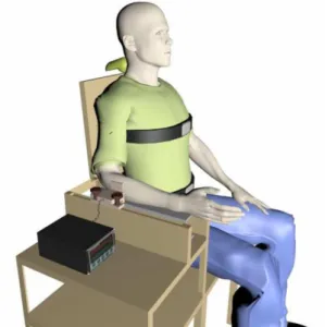
Recording isometric force
A ZEMIC load cell model H3-C3-100 Kg-3B
was placed on a U-shaped device located on the right armrest of a custom-made chair. This chair was designed to record the isometric forces of the shoulder muscles. The U-shaped device was placed on the right armrest of the chair to let the load cell
move. This design allowed the examiner to adjust the
load cell position to various anthropometric measures
or record different force directions. The expected direction for this study was abduction. Participants were instructed to sit on the chair, keeping their heads
neutral, put their right forearms on the armrest, and
gradually apply force against the load cell toward shoulder abduction (Figure 1). It has been reported that isometric contractions of shoulder muscles
cause CMM contraction, providing stability to the cervical spine. Therefore, participants were told to
contract their shoulder abductors so that changes in
the thickness of the CMM could be evaluated using the Sonosynch software19. Then, they were asked to
reach their maximal voluntary contraction (MVC) in 10 seconds. Three trials of MVC were done 60 - secs apart. The trial with the maximum amount of MVC
was chosen for data analysis and measurement of
CMM thickness16.
The trial was performed three times to ensure the
subject reached the maximum possible MVC rather
than calculating their average.
Ultrasound imaging
US imaging of the CMM was performed using an ultrasound device (Accuvix V20 prestige, Samsung Medison, Korea) with an 8 MHz, 4.5 cm linear array transducer. To measure CMM thickness, the spinous process of C4 was palpated. To conirm the spinal level,
ultrasonography guidance was used10. C4 was chosen as
it is claimed that CMM is easy to measure at this level7.
Further explanation is found in the Discussion section. Next, the examiner placed the transducer horizontally on the right side of the C4 spinous process. Then the
transducer was tilted slightly upward or downward to see the echogenic lamina and the interfacing fascia
clearly. At this level, the CMM was seen lateral to
the spinous process, rotator muscle and laminar junction, medial to the articular process and just
under the fascia of the semispinalis cervicis muscle10.
Images were taken irstly at rest and then during the
isometric contraction of the shoulder abductors until
participants reached their MVCs within the given 10 seconds. Anterior posterior diameter (APD) or the thickness of the CMM was measured as the longest
distance between the lamina and the interfacing fascia of semispinalis cervicis.
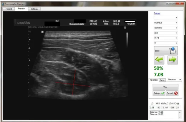
Software
The ofline measurement of the CMM thickness
at rest and at different states of isometric contraction of the shoulder abductor muscles was carried out by
the Sonosynch software. This software picked up and stored the US images in addition to the load cell data with a sampling rate of 20ps. This capability enables
us to appraise muscle diameters both at rest and at
different desired states of MVC. Sonosynch captures muscle forces from the state of rest to 100% MVC. Therefore, the examiner was able to measure the CMM thickness in every desirable amount of MVC. On average, 200 images corresponding to their force level can be stored in 10 seconds. Maximum force is considered as 100% MVC. Having all values with
a high sampling rate allows us to choose any value
between 0 to 100% MVC (Figure 2). In the present study, we assessed CMM thickness at rest (0%) and at 25%, 50%, 75%, and 100% MVC of isometric
contraction of shoulder abductors obtained from the
trials with higher MVCs19.
Reliability study
To evaluate the inter-session reliability for measuring
the thickness and lateral diameter of the CMM as well
as shoulder abductor strength captured and stored
by the software, the procedure was out by the same rater on two separate days, three to seven days apart.
The entire procedure was completed on both occasions and in both groups.
Statistical analysis
To estimate the relative reliability, a two-way mixed model of Intraclass correlation coeficient (ICC) with ICC3,1 was carried out. For the ICC, a
95% of conidence interval (CI) was reported in order to indicate the precision of estimates. To deine the absolute reliability, standard error of measurements (SEM) and the smallest detectable difference (SDD) were computed. SEM was measured as the square root of the mean square error term derived from analysis of
variance20 and SDD was deined as 95% CI of SEM,
calculated as 1.96 SEM4,21. The level of signiicance
deined as p<0.05.
Results
According to Munro’s classiication for reliability coeficients22, we found a high to very high level of
reliability with ICC ranging from 0.84 to 0.94, SEM ranging from 0.01 to 0.09, and SDD ranging from 0.03 to 0.25 for the APD and ICC ranging from 0.64 to 0.95, SEM ranging from 0.03 to 0.11, and SDD ranging
from 0.08 to 0.30 for the LD. The only exception
for the abovementioned results was regarding the
reliability of LD in 100% MVC of shoulder abductors which showed a moderate correlation, with ICC 0.64, SEM 0.14, and SDD 0.39 (Tables 1 and 2).
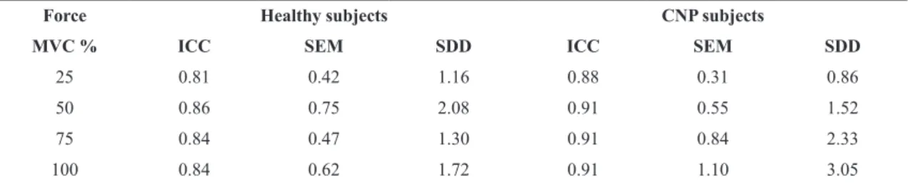
A very high reliability, with ICC ranging from 0.81 to 0.91, SEM ranging from 0.31 to 1.10, and SDD ranging from 0.86 to 3.05 (Table 3), was found
for the inter-session reliability of shoulder abductor
strength from 25% to 100% MVC.
Table 1. Inter-session reliability of the measurement of anterior-posterior dimension (APD) of the cervical multiidus muscle using the Sonosynch software.
APD Healthy subjects CNP subjects
MVC % ICC SEM SDD ICC SEM SDD
0 0.89 0.05 0.14 0.84 0.01 0.03
25 0.88 0.06 0.17 0.88 0.04 0.11
50 0.86 0.07 0.19 0.94 0.03 0.08
75 0.94 0.04 0.11 0.94 0.03 0.8
100 0.87 0.09 0.25 0.91 0.04 0.11
MVC: Maximal Voluntary Contraction; ICC: Intraclass Correlation Coeficient; SEM: Standard Error of Measurement; SDD: Smallest Detectable Difference; CNP: Chronic neck pain.
Table 2. Inter-session Reliability of the measurement of Lateral dimension (LD) of the cervical multiidus muscle using the Sonosynch
software.
LD Healthy subjects CNP subjects
ICC SEM SDD ICC SEM SDD
0 0.92 0.06 0.17 0.83 0.06 0.17
25 0.82 0.11 0.30 0.95 0.04 0.11
50 0.88 0.07 0.19 0.87 0.06 0.17
75 0.89 0.03 0.08 0.82 0.07 0.19
100 0.92 0.07 0.19 0.64 0.14 0.39
MVC: Maximal Voluntary Contraction; ICC: Intraclass Correlation Coeficient; SEM: Standard Error of Measurement; SDD: Smallest Detectable Difference; CNP: Chronic neck pain.
Table 3. Inter-session reliability of the measurement of shoulder muscle strength using the Sonosynch software.
Force Healthy subjects CNP subjects
MVC % ICC SEM SDD ICC SEM SDD
25 0.81 0.42 1.16 0.88 0.31 0.86
50 0.86 0.75 2.08 0.91 0.55 1.52
75 0.84 0.47 1.30 0.91 0.84 2.33
100 0.84 0.62 1.72 0.91 1.10 3.05
Discussion
The results of the present study showed that all
measurements of the muscle’s diameters and strength conducted by the Sonosynch software had high to very high inter-session reliability except for the lateral diameter of the cervical multiidus in 100% of MVC
of shoulder abductors which was shown to have moderate reliability.
We found a high inter-session reliability of APD and LD measurement of the CMM both at rest and contracted. However, Rankin et al.23 reported a very
high inter-session reliability (with ICC ranging from 0.98 to 0.99) for ultrasonographic measurement of deep dorsal neck muscles. In contrast, Kristjansson7
reported moderate to acceptable reliability when
measuring the size of CMM in healthy individuals.
This discrepancy between the results may be due to the fact that Rankin et al.23 reported the CSA
of CMM and semispinalis cervicis as one muscle. In the present study, we measured the APD and LD of the CMM separate from the semispinalis cervicis muscle. To the best of our knowledge, it is the irst
study to attempt to establish a new software that simultaneously records and measures muscle diameters
and strength conducted by a US device and load cell
respectively in both healthy subjects and patients with neck pain. This software enables researchers to
record US images while their subjects are doing the contractile task and to process them ofline. The high reliability of measuring the diameters of the CMM
and shoulder abductor strength shown in the present study encourages widespread usage of this software in studies aiming to evaluate activity of the muscles during functional daily tasks.
The only exception for the above-mentioned
results of the present study is a moderate inter-session
reliability of CMM lateral diameter at 100% of shoulder abductor MVC in patients with neck pain. Kristjansson also reported a good reliability of ultrasonographic measurement of CMM diameters
in healthy individuals but not in patients with neck pain7. The possible explanation for such a result may
possibly be due to the position that participants took
to produce the maximum abductor force in addition to the fact that recognizing interfacing muscle fascia in patients is more dificult than in healthy individuals10,24.
Lee et al.10 also argued that the anatomical structure
of the CMM causes the lateral boundaries to be less distinguishable in ultrasonography. In fact, the CMM comes from the spinous process of the
lower cervical vertebra and attaches to the articular
process of the upper process. Considering this fact,
the axial resolution of ultrasonography is better than its horizontal resolution, therefore, it is more precise to measure its APD relative to the LD16.
We also found a very low SEM and SDD for both APD and LD, which strengthens the ability to detect the diameters of CMM especially for APD with ultrasonography. This means that we require an average of 10% change in the APD to be detected by the Sonosynch software, which is in line with
previous studies4,25.
Regarding the inter-session reliability of MVCs, we found a very high ICC in both healthy individuals
and patients with neck pain. These results are higher
than those reported by Cadogan et al.26 and Celik at
al.27. This disagreement may be due to using different
devices for recording the muscle strength. However, our results support the indings of Adsuar et al.28 who
reported a moderate to very high relative reliability for the measurement of isotonic strength of shoulder muscles28.
We decided to evaluate the CMM thickness at the level of C4. The CMM is easily detected at this level7.
However, the CMM thickness has been measured in
other cervical levels as well10,29.
Limitations and future studies
There may be a few limitations to generalizing
the accomplishments of this research to all patients suffering from neck pain due to our focus on only
subjects with chronic neck pain. Therefore, future
study is recommended to evaluate the inter-session reliability of the software on subjects with other types
of neck pain, such as those with whiplash injury. In this research, we measured the CMM diameters as well as shoulder abductor muscle strength. However, future
reliability assessment of the software for measuring other muscle diameters and strengths is recommended
to expand the use of this software to other cases. In conclusion, the inter-session reliability of the Sonosynch software is high when tested by one examiner. This software provides the capability of capturing and saving the US images and load cell data in a synchronized way to allow ofline measurements
of muscle diameters during the muscle contraction period.
Acknowledgements
inclusion criteria among patients with neck pain.
We would also like to thank sincerely Roghayeh Dehghani for her continued technical support for the
software in the present study.
References
1. LeeJ-P, Tseng W-YI, ShauY-W, WangC-L, WangH-K,
WangS-F. Measurement of segmental cervical multifidus contraction by ultrasonography in asymptomatic adults.
Man Ther. 2007;12(3):286-94. http://dx.doi.org/10.1016/j.
math.2006.07.008. PMid:16987692.
2. Vasseljen O, Dahl HH, MorkPJ, Torp HG. Muscle activity onset in the lumbar multifidus muscle recorded simultaneously by ultrasound imaging and intramuscular electromyography.
Clin Biomech (Bristol, Avon). 2006;21(9):905-13. http://dx.doi.
org/10.1016/j.clinbiomech.2006.05.003. PMid:16822599.
3. PeolssonA, BrodinL-Å, PeolssonM. A tissue velocity ultrasound imaging investigation of the dorsal neck muscles during
resisted isometric extension.Man Ther. 2010;15(6):567-73.
http://dx.doi.org/10.1016/j.math.2010.06.007. PMid:20650674.
4. CagnieB, DereseE, VandammeL, VerstraeteK, Cambier
D, DanneelsL. Validity and reliability of ultrasonography for the longus colli in asymptomatic subjects. Man Ther.
2009;14(4):421-6. http://dx.doi.org/10.1016/j.math.2008.07.007.
PMid:18829376.
5. Hides JA, Miokovic T, BelavýDL, StantonWR, Richardson
CA. Ultrasound imaging assessment of abdominal muscle
function during drawing-in of the abdominal wall: an
intrarater reliability study. J Orthop Sports Phys Ther.
2007;37(8):480-6. http://dx.doi.org/10.2519/jospt.2007.2416.
PMid:17877284.
6. Hodges PW, PengelLH, Herbert RD, Gandevia SC.
Measurement of muscle contraction with ultrasound
imaging. Muscle Nerve. 2003;27(6):682-92. http://dx.doi.
org/10.1002/mus.10375. PMid:12766979.
7. KristjanssonE. Reliability of ultrasonography for the cervical multifidus muscle in asymptomatic and symptomatic subjects. Man Ther. 2004;9(2):83-8. http://dx.doi.org/10.1016/
S1356-689X(03)00059-6. PMid:15040967.
8. LinY-J, ChaiH-M, WangS-F. Reliability of thickness measurements of the dorsal muscles of the upper cervical
spine: an ultrasonographic study.J Orthop Sports Phys Ther.
2009;39(12):850-7. http://dx.doi.org/10.2519/jospt.2009.3005.
PMid:20026880.
9. PeolssonA, Löfstedt T, Trygg J, PeolssonM. Ultrasound imaging with speckle tracking of cervical muscle deformation
and deformation rate: isometric contraction of patients after
anterior cervical decompression and fusion for cervical disc disease and controls. Man Ther. 2012;17(6):519-25. http://
dx.doi.org/10.1016/j.math.2012.05.005. PMid:22703900.
10. LeeJP, Tseng WYI, Shau YW, Wang CL, Wang HK,
WangSF. Measurement of segmental cervical multifidus contraction by ultrasonography in asymptomatic adults.
Man Ther. 2007;12(3):286-94. http://dx.doi.org/10.1016/j.
math.2006.07.008. PMid:16987692.
11. KoppenhaverSL, Hebert JJ, FritzJM, ParentEC, Teyhen DS,
MagelJS. Reliability of rehabilitative ultrasound imaging of the transversus abdominis and lumbar multifidus muscles.
Arch Phys Med Rehabil. 2009;90(1):87-94. http://dx.doi.
org/10.1016/j.apmr.2008.06.022. PMid:19154834. 12. StokesM, Rankin G, NewhamDJ. Ultrasound imaging of
lumbar multifidus muscle: normal reference ranges for measurements and practical guidance on the technique. Man Ther. 2005;10(2):116-26. http://dx.doi.org/10.1016/j.
math.2004.08.013. PMid:15922232.
13. WallworkTL, Hides JA, StantonWR. Intrarater and interrater reliability of assessment of lumbar multifidus muscle thickness using rehabilitative ultrasound imaging. J
Orthop Sports Phys Ther. 2007;37(10):608-12. http://dx.doi.
org/10.2519/jospt.2007.2418. PMid:17970407.
14. Teyhen DS, MiltenbergerCE, DeitersHM, Del ToroYM,
PulliamJN, ChildsJD, et al. The use of ultrasound imaging of the abdominal drawing-in maneuver in subjects with low back pain. J Orthop Sports Phys Ther. 2005;35(6):346-55.
http://dx.doi.org/10.2519/jospt.2005.35.6.346. PMid:16001906.
15. Rezasoltani A, AhmadipoorA, Khademi-KalantariK,
JavanshirK. The sign of unilateral neck semispinalis capitis muscle atrophy in patients with chronic non-specific neck pain. J Back Musculoskelet Rehabil. 2012;25(1):67-72.
PMid:22398268.
16. RezasoltaniA, Ali-RezaA, KhosroKK, Abbass R. Preliminary
study of neck muscle size and strength measurements in
females with chronic non-specific neck pain and healthy control subjects. Man Ther. 2010;15(4):400-3. http://dx.doi.
org/10.1016/j.math.2010.02.010. PMid:20430684. 17. ElliottJ, Jull G, NoteboomJT, Darnell R, Galloway G,
Gibbon WW. Fatty infiltration in the cervical extensor
muscles in persistent whiplash-associated disorders: a
magnetic resonance imaging analysis. Spine (Phila Pa
1976). 2006;31(22):E847-55. http://dx.doi.org/10.1097/01.
brs.0000240841.07050.34. PMid:17047533.
18. ElliottJ, Jull G, NoteboomJT, Galloway G. MRI study of the
cross-sectional area for the cervical extensor musculature
in patients with persistent whiplash associated disorders
(WAD).Man Ther. 2008;13(3):258-65. http://dx.doi.
org/10.1016/j.math.2007.01.012. PMid:17383216. 19. Rahnama L, Rezasoltani A, Khalkhali ZaviehM, Noori
Kochi F, Akbarzadeh BaghbanA. The effects of isometric contraction of shoulder muscles on cervical multifidus muscle dimensions in healthy office workers. J Bodyw
Mov Ther. 2014;18(3):383-9. http://dx.doi.org/10.1016/j.
jbmt.2013.11.011. PMid:25042308.
20. WeirJP. Quantifying test-retest reliability using the intraclass
correlation coefficient and the SEM.J Strength Cond Res.
2005;19(1):231-40. PMid:15705040.
21. Angst F, AeschlimannA, Stucki G. Smallest detectable and minimal clinically important differences of rehabilitation
intervention with their implications for required sample sizes using WOMAC and SF-36 quality of life measurement
instruments in patients with osteoarthritis of the lower
extremities.Arthritis Rheum. 2001;45(4):384-91. http://
dx.doi.org/10.1002/1529-0131(200108)45:4<384::AID-ART352>3.0.CO;2-0. PMid:11501727.
22. DomholdtE.Rehabilitation research: principles and applications.
St. Louis: Elsevier Saunders; 2005.
23. Rankin G, StokesM, NewhamDJ. Size and shape of the
posterior neck muscles measured by ultrasound imaging:
Man Ther. 2005;10(2):108-15. http://dx.doi.org/10.1016/j.
math.2004.08.004. PMid:15922231.
24. JavanshirK, Mohseni-BandpeiMA, RezasoltaniA, Amiri
M, RahgozarM. Ultrasonography of longus colli muscle:
A reliability study on healthy subjects and patients with
chronic neck pain. J Bodyw Mov Ther. 2011;15(1):50-6.
http://dx.doi.org/10.1016/j.jbmt.2009.07.005. PMid:21147418.
25. CagnieB, O’LearyS, ElliottJ, Peeters I, Parlevliet T, Danneels
L. Pain-induced changes in the activity of the cervical
extensor muscles evaluated by muscle functional magnetic
resonance imaging. Clin J Pain. 2011;27(5):392-7. http://
dx.doi.org/10.1097/AJP.0b013e31820e11a2. PMid:21415716.
26. CadoganA, LaslettM, Hing W, McNairP, WilliamsM. Reliability of a new hand-held dynamometer in measuring shoulder range of motion and strength. Man Ther. 2011;16(1):97-101.
http://dx.doi.org/10.1016/j.math.2010.05.005. PMid:20621547. 27. CelikD, Dirican A, Baltaci G. Intrarater reliability of
assessing strength of the shoulder and scapular muscles. J
Sport Rehabil. 2012;Technical Notes 3:1-5. PMid:22495260.
28. AdsuarJC, Olivares PR, ParracaJA, Hernández-MocholíMA, Gusi N. Applicability and test-retest reliability of isokinetic shoulder abduction and adduction in women fibromyalgia patients. Arch Phys Med Rehabil. 2013;94(3):444-50. http://
dx.doi.org/10.1016/j.apmr.2012.08.198. PMid:22902889. 29. LeeJ-P, WangC-L, ShauY-W, WangS-F. Measurement
of cervical multifidus contraction pattern with ultrasound imaging. J Electromyogr Kinesiol. 2009;19(3):391-7. http://
dx.doi.org/10.1016/j.jelekin.2007.11.007. PMid:18207422.
Correspondence Leila Rahnama
University of Social Welfare and Rehabilitation Sciences Department of Physical Therapy