S
CIENTIFIC
A
RTICLES
Revista Brasileira de FisioterapiaValidity of computed photogrammetry for
detecting idiopathic scoliosis in adolescents
Validade da fotogrametria computadorizada na detecção de escoliose idiopática
adolescente
Döhnert MB, Tomasi E
Abstract
Introduction: Adolescent idiopathic scoliosis (AIS) is a three-dimensional abnormality of the spine, of unknown etiology. It starts at the beginning of puberty and its progression is associated with the growth spurt. Analysis of angular movement and body posture through the static imaging method known as photogrammetry could allow physical therapists to quantify and qualify their body posture/movement assessments. Objective: This study was carried out to evaluate the sensitivity of this instrument for detecting AIS in examinations in schools. Methods: This was a school-based cross-sectional study among fifth to eighth-grade elementary school students in public and private schools in Pelotas. Digital images were collected and radiographic examinations were performed in the anteroposterior and lateral planes. The sensitivity and specificity of the photogrammetry were investigated using three and two degrees of margin for the body surface asymmetry. Results: Two hundred twenty four students underwent the photogrammetry and standard radiological examinations at the schools. The prevalence of AIS was 4.5% (n=10), in eight girls and two boys with mean Cobb of 13.3º; mean vertebral rotation of 1.1 (Nash-Moe); dorsal kyphosis of 29.5º Cobb; iliolumbar angle of 3.6°; and Risser sign of 1.6. With three degrees margin, the sensitivity was 21.4% and the specificity was 90.7%. With two degrees margin, the sensitivity was 50% and the specificity was 61.2%. Conclusions: Based on these results, it was found that computerized photogrammetry could not be used as a screening method for detecting mild scoliosis in schools.
Key words: idiopathic scoliosis; photogrammetry; posture; physical therapy.
Resumo
Introdução: A escoliose idiopática adolescente (EIA) é uma alteração tridimensional da coluna vertebral. Sua etiologia é desconhecida e seu início ocorre no início da puberdade, tendo sua progressão associada ao estirão de crescimento. A análise angular de movimento e postura corporal através da imagem estática, conhecida como fotogrametria, permite ao fisioterapeuta quantificar e qualificar sua avaliação da postura/movimento corporal. Objetivo: Este estudo foi realizado para avaliar a sensibilidade deste instrumento na detecção da EIA no exame escolar. Métodos: Estudo transversal de base escolar sobre alunos de 5ª a 8ª série do ensino fundamental das redes pública e particular de Pelotas. Foram realizados coleta de imagem digital e exame radiográfico em postura antero-posterior e perfil. A sensibilidade e especificidade da fotogrametria foram verificadas utilizando três e dois graus de margem para desnivelamento da superfície corporal. Resultados: Duzentos e vinte e quatro alunos realizaram o exame de fotogrametria na escola e o exame radiológico padrão. A prevalência de EIA foi de 4,5% (n=10), sendo oito meninas e dois meninos, com média de 13,3º Cobb; média de 1,1 para rotação vertebral (Nash-Moe); 29,5º Cobb para cifose dorsal; 3,6º para ângulo íleo-lombar; e sinal de Risser em 1,6. Para trêsº, a sensibilidade foi de 21,4% e a especificidade de 90,7%. Utilizando dois graus, a sensibilidade foi de 50% e a especificidade de 61,2%.
Conclusões: Com base nestes resultados, verificou-se que a fotogrametria computadorizada não pode ser realizada como screening para detecção de escoliose de grau leve nas escolas.
Palavras-chave: escoliose idiopática; fotogrametria; postura; fisioterapia.
Received: 13/06/07 – Revised: 04/12/07 – Accepted: 06/05/08
MSc in Health and Behavior, Universidade Católica de Pelotas (UCPel), Pelotas (RS), Brazil
Correspondence to: Marcelo B. Döhnert, Rua Ercílio Farias Alves, 37, CEP 95560-000, Torres (RS), Brasil, e-mail: mdohnert@ig.com.br / mdohnert@pop.com.br
Introduction
Adolescent idiopathic scoliosis (AIS) is a three-dimensional abnormality of the spine, of unknown etiology. It starts at the beginning of puberty and its progression is associated with the growth spurt. he prevalence of scoliosis among adolescents ranges from 1 to 3% of the population1,2 and girls are more
afected than boys, in proportions of approximately four to one2. Many associated factors have been correlated with the
progression of the curvature3, such as the presence of double
curvatures, high magnitudes of curvature, early diagnosis, diag-nosis before menarche, low Risser sign and the female gender.
he vast majority of AIS cases are asymptomatic until high angulations are reached, normally more than 40º Cobb, and early detection of scoliosis gives rise to a threefold increase in the number of patients conservatively treated, thus reducing the percentage of patients who need surgery4.
he Cobb method continues to be the standard clinical me-asurement for evaluating the magnitude of scoliosis5. Other
non-radiological methods have been used in attempts to achieve early detection of AIS among school children. Amendt et al.6 used the
Scoliometer£, an instrument created in 1984 by Bunnell, to me-asure asymmetries or axial rotations of the trunk. Velezis, Sturm and Cobey2 used the Adams test ( forward bending test with
ante-rior spine lexion) to observe gibbosity on the back, resulting from vertebral torsion. hulbourne and Gillespie7 and Burwell et al.8
recorded the outline of the back by using a gibbogram1,2.
Turner-Smith Harris e homas9 proposed an integrated shape imaging
system (ISIS), while Stokes and Moreland10used a Raster
stereo-graph to study back shape abnormalities.
Over the last few years, many authors have questioned the existing models for scoliosis examinations among schoolchil-dren1-3. Although conventional radiography identiies spine
deformities, it is not recommended for basic examinations in schools. he risks involved in exposing the child to radiation and the high costs have justiied attempts to develop other methods for detecting and documenting scoliosis.
For some years now, physical therapists and other professio-nals within the ield of human movement studies have devoted themselves to kinematics: the angular analysis of movement and body posture through images11-14. When separately considered,
such images (photograms) can be analyzed with what is conven-tionally termed photogrammetry. Recent studies have tested the reliability of this examination technique applied to conventional postural evaluation and for measuring anterior trunk lexion, sho-wing good intra- and inter-examiner reliability15-19. Evaluation of
scoliosis through computed photogrammetry can make it possi-ble to analyze the shape of the child’s back, determine deformities and quantify in degrees the unevenness found and may be a useful tool for scoliosis examination14.
hus, the purpose of this study was to investigate the sensi-tivity of computed photogrammetry for examinations in scho-ols to detect early adolescent idiopathic scoliosis. It was also intended to determine the validity of angular kinematics as a diagnostic and analytical examination for scoliosis, compared to radiological examinations, and to establish cross-validation between the angular measurements by means of a surface pro-tocol, through angular kinematics, versus the Cobb angle from radiological images. he hypotheses were that, compared with conventional radiological examinations, angular kinematics would demonstrate high sensitivity and speciicity and that there would be positive and signiicant correlations between the surface angular kinematic analysis and the Cobb angle.
Materials and methods
Between March and July 2005, a school-based cross-sectional study was carried out among ifth to eighth grade elementary school students in the urban zone of Pelotas, Rio Grande do Sul. he sample size was calculated through the Epi-Info software, esti-mating a sensitivity of 90% with a margin of error of 3.5 percentage points, thus resulting in 279 students. For an estimated speciicity of 80%, with the same margin of error, 492 adolescents would be necessary. Adding 30% for losses and refusals, 650 students would be necessary. From a list containing all the schools in the urban zone of the municipality, 20 schools providing either a complete elementary level course or the inal four years were eligible. To en-sure representativeness among all students from the target popu-lation, the schools were stratiied by type: public (municipal and state) and private. Six hundred and ifty students were initially se-lected from eight schools. In each school, one class was randomly chosen from each of the last four years of elementary school and 20 students per class were consecutively selected according to the attendance list. One hundred and ifty-eight students did not participate in the study because of lack of authorization from their parents or other responsible adults. Another 178 students did not attend school on the appointed day to sit for the examination. Of the 314 students who underwent the photogrammetry at school, 224 also did the radiological examination and these, therefore, formed the inal sample.
After student selection, a written consent statement was sent to the parents or other adults responsible for the children, to obtain authorization for performing the photogrammetric examinations at school and the radiological examination in a specialized location.
he inclusion criteria consisted of prior parental authori-zation and that the child was included on the list of selected students. he exclusion criteria were lack of prior authoriza-tion, inadequate clothing for the examination to be performed and not undergoing both selected examinations (id est,
photo-grammetry and X-ray).
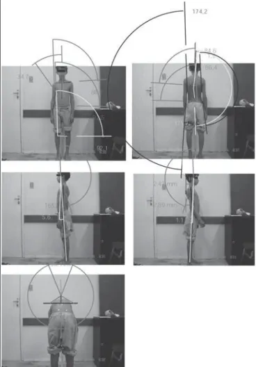
he digitized images were gathered at school, in a room set aside for this purpose. Crosses formed by adhesive tape on the loor marked the correct positions for the feet and alignments for the camera, at pre-established distances. here was ade-quate natural and artiicial light, and a large enough physical area for image gathering, perpendicular to the student under examination. he surface markers that were used were standar-dized: self-adhesive, white and spherical (13mm in diameter). hey were placed at anatomical points, from which the sym-metry of the body surface was delineated (Table 1). A digital camera (DSC-P73, Sony£) with a resolution of 2592x1944 pixels was used, and placed upon a photographic tripod at a height of 85cm from the loor, with levels for image alignment, auto-zoom and focal distances of 2.40m for the anterior-frontal, posterior-frontal and sagittal (right and left) planes, and 1.80m for the anterior lexion-posterior trunk position. According to a study found in the literature14, the degree of distortion in
the distance interval between 1.20 and 2.40m for angular me-asurements is the same. he image-gathering angle was 90º, at a height of 85cm from the loor, with a degree of distortion of approximately 1% for these parameters. Male students un-derwent the image gathering while wearing trunks, and the
female students wore bikinis and the subjects were barefoot. he digital images were stored on a CD for subsequent analyses and the symmetry and alignments measurements were carried out using the CorelDRAW 9.0 software.
In Figure 4, the symmetry and alignments measured in the diferent planes can be seen.
he biomechanical basis selected was that all the paired bone contralateral anatomical reference points were leveled, to form lines parallel with the loor, id est, with symmetry angles
of 0º between each other and both at 90º to the loor. Unpaired homolateral reference points also needed to be aligned with each other, forming perpendiculars with the loor, id est, with symmetry
angles of 0º between each other and both at 90º in relation to the longitudinal x-axis, parallel to the ground. Functional tolerance measurements of two and three degrees were used. A pathologi-cal condition was considered to be present when the angles were lower than 88 and 87º or greater than 92 and 93º, respectively, in relation to the 90º marks on the ordinate (y) and abscissa (x) axes.
he photographic interpretations of the hales triangles (∆hales) used the formula: ∆hales=halesRight - halesLeft. When the result of the equation was positive, there was a trunk/scapular waist inclination to the right; when negative, it was to the left. he same calculation was applied to deine the Head-Shoulder relationship (HSR) and the Head-Mal-leolus relationship (HMR), deining occurrences of rotation of the evaluated segments by using the following formulae, respectively: ∆HSR=HeadShoulderright - HeadShoulderleft and
∆HMR=HeadMalleolusright - HeadMalleolusleft. he
measure-ment references for ∆HSR, were the internal auditory canal and lateral shoulder acromion. For the ∆HMR measurements, the internal auditory canal and lateral ankle malleolus were used. For calculating the angle of gibbosity, the following formula was used: ∆Gibbosity= ∆Gibbosityright - ∆Gibbosityleft.
Pattern Anatomical points Alignment/Symmetry
Anterior frontal plane t"OUFSJPSCPSEFSPGBDSPNJPO 4IPVMEFSTBOE
t(MBCFMMBKVHVMBSWFJOJODJTJPO 0NQIBMJDBOE
t"OUFSPTVQFSJPSJMJBDTQJOFT 1FMWJTBOE
t"OUFSJPSUJCJBMUVCFSDMFT ,OFFTBOE
4BHJUUBM t&YUFSOBMBDPVTUJDNFBUVToMBUFSBMCPSEFSPGBDSPNJPO )FBETIPVMEFSSFMBUJPOTIJQ )43
t&YUFSOBMBDPVTUJDNFBUVToMBUFSBMNBMMFPMVTPGBOLMF )FBENBMMFPMVTSFMBUJPOTIJQ ).3 t $FSWJDBMMPSEPTJTBQFYoEPSTBMLZQIPTJTBQFYoFYUFSOBMWFSUFY $FSWJDBMUIPSBDJDMPSEPTJTQMBOF t -VNCBSMPSEPTJTBQFYoEPSTBMLZQIPTJTBQFYoFYUFSOBMWFSUFY -VNCBSUIPSBDJDMPSEPTJTQMBOF
1PTUFSJPSGSPOUBMQMBOF t1PTUFSJPSBDSPNJPO 4IPVMEFSTBOE
tthDFSWJDBMWFSUFCSBothUIPSBDJDWFSUFCSB 6QQFSTQJOF5$YBYJTBCTDJTTBBOE
tUIPSBDJDWFSUFCSBMVNCBSWFSUFCSB -PXFSTQJOF5-YBYJTBCTDJTTBBOE
t*OGFSPNFEJBMBOHMFPGTDBQVMB 4DBQVMBBOE
t1PTUFSPTVQFSJPSJMJBDTQJOFT 1FMWJTBOE
t"OHMFCFUXFFOYBYJTBOEMJOFUBOHFOUJBMUPHJCCVTZBYJT (JCCPTJUZBOHMFoAdams Table 1. "OBUPNJDBMQPJOUTBOEBMJHONFOUMFWFMTVTFEGPSQIPUPHSBNNFUSJDFYBNJOBUJPO
1SPQPTFECZUIFBVUIPSTGPSUIJTTUVEZ
To dei ne the physiological curvature measurements in the sagittal plane, the distances of the thoracic-cervical lordosis plane and the thoracic-lumbar lordosis plane were used, in which the thoracic-cervical lordosis plane was the distance in mm between the line tangential to the dorsal kyphosis apex and the line tangential to the cervical lordosis apex. h e thoracic-lumbar lordosis plane was the distance in mm between the line tangential to the dorsal kyphosis apex and the line tangential to the lumbar lordosis apex.
To determine the existence of any dorsal scoliosis, the upper spine alignment, anterior shoulder symmetry and pos-terior scapula symmetry were used. However, for scoliosis of lumbar form, the lower spine alignment and anterior and pos-terior pelvis symmetry were used. Dorsal-lumbar curvatures and double curvature were classii ed in consideration of all the above variables. h e determining factor for dei ning the side was the infra- or supra-symmetry of the anterior surface me-asurements. During the training, it was attempted to standar-dize the procedures for anatomical palpation when positioning the surface markers on overweight or obese children, so as not to distort the image gathering.
After the image gathering, all the students were referred to a radiology institute for standard radiological examination,
id est, total vertebral column in postero-anterior and lateral
planes (both in the orthostatic position). From these, the follo-wing variables were analyzed: localization (lateral curvature or not); type ( functional or structural); Cobb angle for lateral de-viations and dorsal kyphosis; initial specii c vertebral rotation angles (obtained by means of the Nash-Moe method); Risser sign (amount of calcii cation present in the iliac apophysis which measures the progressive ossii cation associated with the closing of bone growth) and the iliolumbar angle. Late-ral curvatures with Cobb angles greater than or equal to 11º were considered to be true scoliosis. Students with curvatures between 5 and 10º Cobb were classii ed as having a propen-sity for curve progression and development of scoliosis. h e radiological examinations were analyzed by two dif erent exa-miners and, if there was any discordance regarding any of the variables, a third examiner was asked to give the i nal opinion. h e sensitivity was dei ned as the percentage of patients with scoliosis who were also positive in the computed photogram-metry, while the specii city was the percentage of students who underwent radiography without showing scoliosis and who had a negative test results from the photogrammetry.
h e statistical analyses were predominantly descriptive, with observations of central trends and dispersion measure-ments for the quantitative variables. In the analyses of mea-surements according to gender, Student’s t-tests were used for comparisons between the means, and dif erences with p values less than 0.05 were taken to be signii cant.
Figure 4. #PEZ TZNNFUSZ BOE BMJHONFOUT NFBTVSFE CZ DPNQVUFS BJEFEQIPUPHSBNNFUSZ1FMPUBT34
Results
From the whole sample, just over half was male and the mean age was 12.3 years at the time of the examination at school (sd=1.6), without dif erences between the genders. Just under half of the students were from state schools, and the others were equally divided between municipal and private schools. Among the girls examined using photogrammetry, 85 (59.4%) had already had their i rst menstruation, at a mean age of 11.8 years (sd=1.2). None of these characteristics demonstra-ted any dif erences in relation to either the whole sample or the radiographed sample (Table 2).
n (total)
%
(total) n (Radiography) % (Radiography)
(FOEFS
.BMF 116
'FNBMF
"HF ZFBST
BOE
11
BOE
5ZQFPGTDIPPM
State
.VOJDJQBM 61
1SJWBUF 61
5PUBM
Table 2. %JTUSJCVUJPOPGTBNQMFPGTDIPPMDIJMESFOBDDPSEJOHUPHFOEFS BHFBOEUZQFPGTDIPPM
Idiopathic Scoliosis Functional Scoliosis Propensity to develop Without Scoliosis
n % n % n % n %
(FOEFS
.BMF 11
'FNBMF
"HF ZFBST
BOE 1
11
1
11
BOE 1
5ZQFPG4DIPPM
State 6
.VOJDJQBM
1SJWBUF
5PUBM
Table 3. 1SFWBMFODFPGTDPMJPTJTBNPOHTDIPPMDIJMESFOBDDPSEJOHUPSBEJPMPHJDBMFYBNJOBUJPOT
Sensitivity (%) Specificity (%) Positive predictive value (%)
Negative predictive value (%)
5IFQSFTFOUTUVEZ UP UP UP UP
#VSXFMM $PCCNJOJNVN UP UP UP o
)PXFMM$SBJHBOE%BXF $PCCJOJUJBM UP UP o o
"NFOEUFUBM B$PCC UP UP o UP
4BIMTUSBOE $PCCNJOJNVN UP UP o o
-BVMBOE4KCKFSHBOE)SMZDLF $PCCNJOJNVN o o UP UP
Table 4. 'JOBMSFTVMUTBOEDPNQBSJTPOTXJUIPUIFSTUVEJFTJOUIFMJUFSBUVSF
he prevalence of adolescent idiopathic scoliosis was 4.5% (10/224), and it was four times more common for girls than boys. Eight% (18/224) of the students showed scoliosis classi-ied as functional and 110 demonstrated curvatures with angu-lations ranging from 5 to 10º (Table 3).
The mean Cobb angle of the idiopathic scoliosis found by means of the radiological examinations were 13.3º Cobb, while for the functional scoliosis cases, 12.5º Cobb were found. The mean Cobb angle for dorsal kyphosis was 30.3º, and it was 29.5º among children with idiopathic scoliosis. The mean Risser sign was 1.6, without variations for the idiopathic scoliosis group (Table 4).
Among the curvatures, 77 were located in the dorsal region, 49 were in the lumbar region and 12 were double curvatures. Dorsal curvature predominated among the cases of functional scoliosis (13/18) and inclined scoliosis (62/110). For the idio-pathic scoliosis cases, the predominant location was lumbar and with double curvature (4/10). Analysis of the side of the convexity, the right side prevailed for the dorsal form curves (40/77) and the left side for the lumbar form curves (38/49).
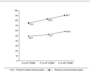
Figure 1. .FBO EJTUBODF NN PG UIF UIPSBDJDMVNCBS MPSEPTJT QMBOF BOE UIPSBDJDDFSWJDBM MPSEPTJT QMBOF JO SFMBUJPO UP $PCC BOHMF NFBTVSFNFOUT 100 90 80 70 60 50 40 30 20 10 0
8 to 20º COBB 21 to 40º COBB 41 to 58º COBB
74.3
82.5
89.7
49.0 51.7
58.3
Thoracic-lumbar lordosis plane Thoracic-cervical lordosis plane
The mean iliolumbar angle was 3.6º for all of the idio-pathic scoliosis cases, and 2.4 among those with functional scoliosis. Taking all the scoliosis cases with a single lumbar curvature, it was found through X-rays (n=9), that the mean iliolumbar angle was 4.1º, rising to 4.5º when analyzing only the idiopathic cases.
Observation of the sagittal physiological curvatures in the thoracic-cervical lordosis and thoracic-lumbar lordosis planes for all the children who underwent the radiological examination, it was found that the distances between these planes increased in the same proportions in which the Cobb angle increased for dor-sal kyphosis, as shown in Figure 1. Using the f test for linear trends, for diferences between the means of two variables according to the Cobb angle, it was observed that both showed statistical signi-icance (p=0.001 and p=0.015, respectively).
Using three degrees of asymmetry as the maximum measu-rement for body surface symmetry in photogrammetry, when cross-referenced with the radiological data, sensitivity of 21.4% was found for all scoliosis curvatures, 16.7% for functional sco-liosis and 30.0% for structural scosco-liosis. By using two degrees as the measurement reference, the sensitivity increased to 50.0%, 61.1% for functional scoliosis, while structural scoliosis remai-ned at 30.0% (Figure 2). he total speciicity was 88.8%, it was 90.7% for the group without scoliosis and 87.3% for those with a propensity towards it. By reducing the maximum symmetry measurement to two degrees of asymmetry, the total specii-city obtained was 61.2%, while for the group without scoliosis it was 68.6% and for those with a propensity, it was 55.5% (Figure 3). he positive predictive values were 16.0% (6/28) with three degrees as the asymmetry measurement and it rose to 50.0% using two degrees (14/28). he negative predictive values were 97.0% and 90.0%, respectively using three and two degrees as the body surface asymmetry measurements.
Discussion
Although the losses were notable both for the photo-grammetry and for the radiological examinations, they did not create differences in the characteristics of the group that followed through with the study. One of the limitations of this study was to have six different examiners for gathering the digitized images and placing the surface markers. Even with a significant period of training for these interviewers, inter-observer measurement errors may have occurred. It is understood that examinations should be carried out by the same interviewers but, for ethical reasons, female inter-viewers were chosen for the girls and male interinter-viewers for the boys. Another difficulty was the placement of surface markers on obese children. Angular measurement errors
due to difficulties in correctly marking the anatomical points because of the greater quantities of adipose tissue and skin were minimized by the standardization of point marking and image gathering carried out during the training and fiel-dwork, since there is no mention in the literature that there should be any exclusion of overweight or obese individuals from examinations in schools for detecting scoliosis.
he prevalence of adolescent idiopathic scoliosis, in this study, was within the rates found in the worldwide literature, which ranges from 2 to 4%. As reported in other studies, the girls showed a four times greater prevalence than did the boys. Among the students who demonstrated scoliosis, the
Figure 2. 4FOTJUJWJUZ PGDPNQVUFEQIPUPHSBNNFUSZXJUIMFWFMJOH PGUISFFBOEUXPEFHSFFT 100 90 80 70 60 50 40 30 20 10 0
Functional scoliose Structural scoliose All the curvatures 16.7
61.1
30.0 30.0
21.4
50.0 Three degress Two degress
Figure 3. 4QFDJmDJUZ PGDPNQVUFEQIPUPHSBNNFUSZXJUITZNNFUSZ PGUISFFBOEUXPEFHSFFT 100 90 80 70 60 50 40 30 20 10 0
segmental extension component (lordosis) was not observed, in comparison with the other children studied, because the mean Cobb angle for kyphosis was 30.3º for the group without scoliosis, and 29.5º for the group with idiopathic scoliosis. he scoliosis found among the students who were examined was all of a mild degree, with a mean Cobb angle of 13.3º and mean vertebral rotation of 1.1. he students examined were skeletally immature, with a mean Risser sign of 1.6. he predominant locations of the idiopathic scoliosis were double curvature and lumbar, followed by dorsal. In the functional scoliosis cases, the dorsal form prevailed followed by the lumbar form.
By analyzing the sensitivity, speciicity and positive and negative predictive value results, in comparison with other studies, variations were observed regarding the speciicity and sensitivity, according to the type of examination performed and the symmetry measurement criteria used. Burwell15
asses-sed 102 children with scoliosis with Cobb angles of at least 20º using the Scoliometer£ to measure trunk axial rotation, inding sensitivity ranging from 38 to 69% and speciicity ranging from 84 to 96%. Howell, Craig and Dawe16 assessed 54 children with
idiopathic scoliosis with an initial Cobb angle of 10º by means of photogrammetry and forward bending tests applied by phy-sical therapists and nurses, inding sensitivity levels of 29, 87 and 74% and speciicity of 81, 42 and 49%. Amendt et al.6 used
a Scoliometer£ on 65 patients with idiopathic scoliosis with 20 to 30º Cobb, and found sensitivity of 76 to 100% and speciicity of 54 to 90%. It is very likely that such results are due to the fact that in all of these studies, the evaluated subjects had a conirmed diagnosis of adolescent idiopathic scoliosis, id est, a
Cobb angle greater than 11º.
In this study, the sensitivity ranged from 21 to 50%, and the highest value occurred when using two degrees as the symmetry measurement (50%), while the specificity ranged from 61 to 89%, and the highest values occurred when using three degrees as the symmetry measurement (89%) (Table 4). It seems that the parameters used as significance me-asurements (two and three degrees) with photogrammetry in this study, together with a sample composed mostly of individuals without scoliosis, or with slight scoliosis, may have been overestimated.
he clinical use of an examination is not only determi-ned by its sensitivity and speciicity, but also by its predictive value. Although these two indicators are very important, an examination can also supply clinical evaluation and diagnostic information. Physical therapy professionals need to know the likelihood that computed photogrammetry might be positive or negative in the presence of scoliosis. By applying this princi-ple, a relatively low positive predictive value was found, id est,
the number of students with scoliosis divided by the total num-ber of students with positive photogrammetry with or without
scoliosis (true ones and false positives), with the symmetry me-asurement equal to two degrees (50%) and three degrees (16%). In the study carried out by Burwell15, the positive predictive
values ranged from 18 to 56%. In the study by Lauland, Søjbjerg and Hørlycke17who evaluated 195 children with scoliosis and
a minimum Cobb angle of 10º by means of Moiré topography and the forward bending test, the positive predictive value ran-ged from 18 to 29% (Moiré topography).
Inversely, the negative predictive value was observed to be extremely high, ranging from 90% to 97%, signifying that the proportion of students without scoliosis divided by the total number of students with negative photogrammetry (true ones and false positives). he studies by Lauland, Søjbjerg e Hør-lycke17 and Amendt et al.6 found negative predictive values
ran-ging from 97 to 100% and 40 to 100%, respectively. It seems that this high negative predictive value could have been overestima-ted because, in the present study, a higher level of signiicance was used (two and three degrees).
Computed photogrammetry in the present study, using three and two degrees for body surface symmetry, was not shown to be sensitive and speciic enough to be recommended alone as a school screening method for adolescent idiopathic scoliosis. It seems that using two degrees as an unevenness me-asurement level has been shown to be more reliable, given that it noticeably decreases the chance of not detecting a child with mild idiopathic scoliosis. In comparison with other studies, the mean Cobb angle found was one of the lowest (5.5º Cobb), which could in a way explain the tendency towards decreased sensitivity. It is believed that other studies will need to be car-ried out, with greater scoliosis curvatures and image gathering carried out by the same examiner, in order to verify these per-centages. Another factor is the number of subjects examined, since our study reported a higher number of subjects examined and, of these, only 4.5% presented idiopathic scoliosis, all in a mild form. Most of the studies used smaller samples and only subjects with scoliosis, with a minimum Cobb angle ranging from 5 to 30º Cobb, which seems to limit the predictive impli-cations of the examination to a general population. For detec-ting physiological curves in the sagittal plane, this examination was shown to be efective among this population.
he objective of examining schoolchildren was to achieve early identiication of scoliosis, id est, before curve progression
and before reaching skeletal maturity. Traditionally, classical school examination programs use the forward bending test. Computed photogrammetry allows quantiication of body surface symmetry that is not measured through the subjective clinical examination. hese data may contribute towards bet-ter monitoring of the progression, stabilization or reduction of scoliosis curvature over the course of therapy and bone growth, as well as helping to document the curvature.
,BSBDIBMJPT54PmBOPT+3PJEJT/4BQLBT(,PSSFT%/JLPMPQPVMPT, 5FOZFBSGPMMPXVQFWBMVBUJPOPGBTDIPPMTDSFFOJOHQSPHSBNGPSTDPMJPTJT *TUIFGPSXBSECFOEJOHUFTUBOBDDVSBUFEJBHOPTUJDDSJUFSJPOGPSUIFTDSFFOJOH PGTDPMJPTJT 4QJOF
7FMF[JT.4UVSN1'$PCFZ+4DPMJPTJTTDSFFOJOHSFWJTJUFEmOEJOHTGSPN UIF%JTUSJDUPG$PMVNCJB+1FEJBUS0SUIPQ
(SFJOFS,""EPMFTDFOUJEJPQBUIJDTDPMJPTJTSBEJPMPHJDEFDJTJPONBLJOH "N'BN1IZTJDJBO
5PSFMM(/PSEXBMM"/BDIFNTPO"5IFDIBOHJOHQBUUFSOPGTDPMJPTJTUSFBUNFOU
EVFUPFGGFDUJWFTDSFFOJOH.+#POF+PJOU4VSH"N
$IPDLBMJOHBN / %BOHFSmFME 1) (JBLBT ( $PDISBOF 5 %PSHBO +$ $PNQVUFSBTTJTUFE $PCC NFBTVSFNFOU PG TDPMJPTJT &VS 4QJOF +
"NFOEU -& "VTF&MMJBT ,- &ZCFST +- 8BETXPSUI $5 /JFMTFO %) 8FJOTUFJO4-7BMJEJUZBOESFMJBCJMJUZUFTUJOHPGUIF4DPMJPNFUFS1IZT5IFS
5IVMCPVSOF5(JMMFTQJF35IFSJCIVNQJOJEJPQBUIJDTDPMJPTJT.FBTVSFNFOU BOBMZTJT BOE SFTQPOTF UP USFBUNFOU + #POF +PJOU 4VSH #S
#VSXFMM 3( +BNFT /+ +PIOTPO ' 8FCC +, 8JMTPO :( 4UBOEBSEJTFE USVOLBTZNNFUSZTDPSFT"TUVEZPGCBDLDPOUPVSJOIFBMUIZTDIPPMDIJMESFO +#POF+PJOU4VSH#S
5VSOFS4NJUI " )BSSJT + 5IPNBT % *OUFSOBUJPOBM BTTFTTNFOU PG CBDL TIBQFBOEBOBMJTZTVTJOH*4*4*O4UPLFT"1FLFMTLZ+.PSFMBOE.FET 4VSGBDF5PQPHSBQIZBOE4QJOBM%FGPSNJUZ4UVUUHBSU(VTUBW'JTDIFS7FSMBH
4UPLFT *" .PSFMBOE .4 .FBTVSFNFOU PG UIF TIBQF PG UIF TVSGBDF PG UIF CBDL JO QBUJFOUT XJUI TDPMJPTJT 5IF TUBOEJOH BOE GPSXBSECFOEJOH QPTJUJPOT+#POF+PJOU4VSH"N
8PMUSJOH ) 0OF IVOESFE ZFBST PG QIPUPHSBNFUSZ JO CJPMPDPNPUJPO *O1SPDFFEJOHPGUIF4ZNQPTJVNPO#JPMPDPNPUJPO"$FOUVSZPG3FTFBSDI 6TJOH.PWJOH1JDUVSF'PSNJB*UBMZ
#FSNF / $BQP[[P " #JPNFDIBOJDT PG )VNBO .PWFNFOU "QQMJDBUJPO JO 3FIBCJMJUBUJPO4QPSUBOE&SHPOPNJDT8PSUIJOHUPO#FSUFD$PSQPSBUJPO
#PFOJDL 6 /BEFS . (BOHCJMEBOBMZTF 4UBOE EFS .FCUFDIOJL VOE #FEFVUVOH GàS EJF 0SUIPQÊEJF5FDIOJL *O*OUFSOBUJPOBMFT 4ZNQPTJVN (BOHCJMEBOBMZTF.FDLF%SVDLVOE7FSMBH#FSMJN
3JDJFSJ%7BMJEBÎÍPEFVNQSPUPDPMPEFGPUPHSBNFUSJBDPNQVUBEPSJ[BEBF RVBOUJmDBÎÍPBOHVMBSEPNPWJNFOUPUØSBDPBCEPNJOBMEVSBOUFBWFOUJMBÎÍP USBORàJMB EJTTFSUBÎÍPEFNFTUSBEPFN'JTJPUFSBQJB6CFSMÉOEJB$FOUSP 6OJWFSTJUÈSJPEP5SJÉOHVMP 6/*5
#VSXFMM 3( " .VMUJDFOUSF 4UVEZ PG #BDLTIBQF JO 4DIPPM $IJMESFO" 1SPHSFTT3FQPSU1PTJUJPOBM$IBOHFTJO#BDL$POUPVSJO3FMBUJPOUPB/FX 4DSFFOJOH 5FTU GPS 4DPMJPTJT *O 1SPDFFEJOHT PG UIF 4DPMJPTJT 3FTFBSDI 4PDJFUZ $PNCJOFE XJUI UIF #SJUJTI 4DPMJPTJT 4PDJFUZ 4PVUI )BNQUPO #FSNVEB4FQUFNCFSQ
)PXFMM+.$SBJH1.%BXF#(1SPCMFNTJOTDPMJPTJTTDSFFOJOH$BO+ 1VCMJD)FBMUI
-BVMVOE*4KCKFSH+)SMZDL&.PJSÏUPQPHSBQIZJOTDIPPMTDSFFOJOHGPS TUSVDUVSBMTDPMJTJT"DUB0SUIPQ4DBOE
*VOFT%)$BTUSP'"4BMHBEP)4.PVSB*$0MJWFJSB"4#FWJMBRVB(SPTTJ % $POmBCJMJEBEF *OUSB F *OUFSFYBNJOBEPSFT F SFQFUJCJMJEBEF EB BWBMJBÎÍP QPTUVSBMQFMBGPUPHSBNFUSJB3FW#SBT'JTJPUFS
4BUP507JFJSB&3(JM$PVSZ)+$"OÈMJTFEBDPOmBCJMJEBEFEFUÏDOJDBT GPUPNÏUSJDBT QBSB NFEJS B nFYÍP BOUFSJPS EP USPODP 3FW #SBT 'JTJPUFS
4BIMTUSBOE 5 5IF WBMVF PG .PJSÏ UPQPHSBQIZ JO UIF NBOBHFNFOU PG TDPMJPTJT4QJOF

