Clastogenic effects of different
Ureaplasma urealyticum
serovars
on human chromosomes
1Departamento de Análises Clínicas, Faculdade de Ciências Farmacêuticas,
Universidade de São Paulo, 05508-900 São Paulo, SP, Brasil
2Laboratório de Citogenética, Unidade de Aconselhamento Genético,
Departamento de Biologia, Instituto de Biociências, Universidade de São Paulo, 05508-900 São Paulo, SP, Brasil
R.A.F. Cunha1, C.P. Koiffman2, D.H. Souza2 and K. Takei1
Abstract
The possibility that Ureaplasma urealyticum might play an important role in human infertility was first raised more than 20 years ago, but this association remains speculative. Considering the hypothesis that the pathogenicity of Ureaplasma urealyticum may depend on its serotypes, the clastogenic effects of different strains of Ureaplasma urealyticum, at concentrations of 103 CCU (color changing units)/ml,
104 CCU/ml and 105 CCU/ml, were evaluated in vitro in short-term
cultures of human lymphocytes. Total or partial mitotic inhibition was produced by Ureaplasma urealyticum serotypes 2, 3 and 10 independ-ent of the concindepend-entration (103 CCU/ml, 104 CCU/ml or 105 CCU/ml) of
the microorganisms employed. In contrast, the clastogenic effects observed with serotypes 1, 7 and 12 varied according to the concentra-tion employed in the test. Mitotic alteraconcentra-tions were observed in Urea-plasma urealyticum serotypes 5, 6, 7, 8, 9, 11 and 12. Chromatid gaps (53.0%) and chromatid breaks (13.9%) were the most frequent types of alterations observed. The results of this in vitro assay demonstrated that the clastogenic effects varied with the Ureaplasma urealyticum
serotypes evaluated. Correspondence
R.A.F. Cunha
Departamento de Análises Clínicas e Toxicológicas
Faculdade de Ciências Farmacêuticas Universidade de São Paulo Av. Lineu Prestes, 580 05508-900 São Paulo, SP Brasil
Research supported by CNPq (No. 040-3996/90). Publication supported by FAPESP.
Received April 30, 1996 Accepted March 18, 1997
Key words •Mycoplasma
•Chromosome alterations
•Ureaplasma urealyticum
•Clastogenic effects
Introduction
Mycoplasmas are the smallest prokary-otes able to self-replicate and their main feature is the absence of a cell wall. These two facts account for some of the distinctive characteristics peculiar to the group. Myco-plasmas are responsible for many diseases affecting animals and plants, some of them fatal. However, the role of these microorgan-isms in human pathologies is only partially known. The search for evidence indicative
of any correlation between the presence of these microorganisms and human morbidity and mortality has been the subject of many studies.
of fetal membranes and low-weight new-borns observed among women with Urea-plasma-positive placentas is evidence of the vertical transmission of the microorganism. The rate of vertical transmission ranges from 18% to 55% among full-term infants and from 29% to 55% among preterm infants. The high colonization rates in pregnancy make it difficult to validate the concept of fetal/neonatal infection based only on the isolation of Ureaplasma urealyticum. How-ever, the possible consequences of this colo-nization cannot and must not be ignored in pregnant women with problems of reproduc-tive wastage (1,2).
For a long time Mycoplasmas sp have been indicated as the microorganisms re-sponsible for many changes in eukaryotic cells with varying deleterious effects (3,4). Different mycoplasma species can lead to different changes in a certain cell, while the same mycoplasma species can lead to differ-ent changes in cells of the same type belong-ing to different host species. Chromosomal aberrations, mitotic inhibition and stimula-tion and various other cytopathic effects have been observed (5-9).
The difficulties in establishing the role of
Ureaplasma urealyticum in diseases of the reproductive tract and the antigenic varia-tion among strains suggest that only some of the 14 serotypes are associated with disease (10-12).
The objective of the present study was to determine theclastogenic effects of differ-ent Ureaplasma urealyticum serotypes on human chromosomes in vitro. The under-standing of these interactions may be rel-evant for the study of some of the disease-inducing mechanisms of mycoplasmas.
Material and Methods
Lymphocytes were obtained from a single donor and 11 serotypes of Ureaplasma urealyticum (1, 2, 3, 5, 6, 7, 8, 9, 10, 11, 12) were obtained from the American Type
Cul-ture Collection. Four supernatants obtained by centrifugation of the culture broths of serotypes 2, 3, 7 and 10 at 14,200 g for 40 min were also assayed.
Strains were prepared by culturing in modified U10 medium (13) and incubating
under microaerophilic conditions for 18 to 24 h. A7 solid medium (14) was used to
observe the growth and viability of the mi-croorganisms. The inocula were microtitrated in such a way that 225 µl of U10 medium was
added to each microtiter plate well. Twenty-five µl of the culture medium for each sero-type was added to the first well after homog-enization. Successive dilutions were prepared from 25 µl of this first dilution. After incuba-tion, the highest dilution in which the microorganism grew (this growth was indi-cated by the change in color of the culture medium) was taken as the titer, and the results were compared with those obtained with a control (U10 medium without the
in-oculum).
Lymphocyte cultures were prepared by the method of Moorhead et al. (15) with some modifications. For each assay, 20 ml of aseptically collected venous blood was allowed to sediment by keeping the syringe in a vertical position for 2 to 3 h. After red cell sedimentation, the supernatant was trans-ferred to a sterile flask and homogenized and 0.5 ml of this material was added to each flask together with 4 ml of 199 medium supplemented with 10% fetal calf serum and Bacto phytohemagglutinin-P (Difco, cod. 3110) at a concentration of 125 µl/100 ml.
Test culture
Culture broth (0.5 ml) from each sero-type was added to the lymphocyte cultures at concentrations, expressed as CCU/ml (color changing units/ml), of 103 CCU/ml, 104 CCU/
ml and 105 CCU/ml, and the preparations
were then incubated at 36.5oC for 72 h. After
this period, 0.1 ml of 4 x 10⁻⁵ M colchicine
for 1 h. The lymphocyte viability test was carried out using 1% Trypan blue.
Control culture
The procedure for lymphocyte culture was the same as that used for the test culture, except that Ureaplasma urealyticum was not added.
Slide preparation
The contents of the flasks were centri-fuged at 300 g for 5 min, the supernatant was discarded, 4 ml of hypotonic solution (75 mM KCl) was added, and the flasks were incubated for 20 min. After incubation and centrifugation, a few drops of fixative (metha-nol/acetic acid, 3:1) were added to the sedi-ment. After homogenization, 4 ml of fixative was added and the preparation was centri-fuged again at 300 g for 5 min. The superna-tant was removed and the same procedure was repeated once more. Two or 3 drops of this material were then transferred to slides and stained with Giemsa in sodium phos-phate buffer, pH 6.8. After staining, the slides were submitted to microscopic analysis for the determination of mitotic index and for chromosome analysis.
Chromosome analysis
Metaphases were analyzed and classified according to the presence of structural aber-rations. Metaphases presenting chromosomes with structural alterations were drawn and then photographed. The mitotic index was determined by counting the metaphases ob-tained out of 1,000 stimulated cells. In this study we considered a mitotic index of less than 10 to indicate partial inhibition of mitosis. The following structural chromosome abnormalities were considered: chromatid gap (chtg), chromatid breaks (chtb), chro-mosome break (chrb), rearrangement (rea), ring chromosome (r), premature centromeric
disjunction (pcd) and centromeric decon-densation (cd) (16).
Data were analyzed statistically by the Pearson chi-square test to determine the ef-fect of inoculum concentration on the mi-totic process, and by the significance test for the difference between two population pro-portions, applied to determine the signifi-cance of the results obtained for the test cultures compared with the control cultures. The level of significance was set at α = 5% (17).
Results
The effects of 11 serotypes of Urea-plasma urealyticum on human chromosomes were studied, together with 4 supernatants obtained from cultures of serotypes 2, 3, 7 and 10. All tests were conducted with their respective controls and the slides were ana-lyzed in a blind test.
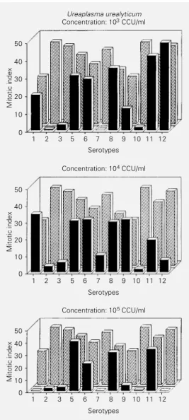
We studied the effects of the microorgan-ism on mitosis and mitotic index. Among the results observed, the main feature was total and partial mitotic inhibition (mitotic in-dexes equal to zero and below 10, respec-tively). Figures 1 and 2 show the values of the mitotic indexes obtained after stimula-tion with 11 serotypes at concentrastimula-tions of 103 CCU/ml, 104 CCU/ml and 105 CCU/ml
and for the supernatants from serotypes 2, 3, 7 and 10. All control cultures were also submitted to determination of mitotic index and karyotype analysis.
Mitotic indexes equal to zero or below 10 were observed for serotypes 1, 2, 3, 7, 9, 10 and 12. Similar results were obtained for supernatants 2, 3 and 7. Some of these sero-types presented these effects only at certain microorganism concentrations: serotype 1 (105 CCU/ml), serotype 7 (103 and 105 CCU/
ml), serotype 9 (105 CCU/ml), serotype 10
(105 CCU/ml) and serotype 12 (104 and 105
the concentration of 104 CCU/ml, serotype 8
at concentrations of 103, 104, 105 and >105
CCU/ml, serotype 9 at concentrations of 103,
104 and 105 CCU/ml, serotype 11 at
concen-trations of 103, 104 and 105 CCU/ml,
sero-type 12 at the concentration of 103 CCU/ml,
and the supernatants from serotypes 3 and 7. Serotypes 5 and 6 showed chromosome ab-errations at concentrations of 103 and 105
CCU/ml, but these results were not statisti-cally significant when compared to the re-spective controls. Serotype 8 at concentra-tions of 104, 105 and >105 CCU/ml, serotype
9 at the concentration of 103 CCU/ml,
sero-type 11 at the concentration of 105 CCU/ml,
serotype 12 at the concentration of 103 CCU/
ml and the supernatants from serotypes 3 and 7 caused significantly increased alter-ations compared to their respective controls. Among these, gaps, chromatid breaks, chro-mosome breaks and partial centromeric de-condensation were the most frequently ob-served.
The highest frequency of gaps and chro-matid breaks was observed in the cultures with serotype 8 at concentrations of 104, 105
and >105 CCU/ml and serotypes 9 and 11 at
concentrations of 103 CCU/ml and 105 CCU/
ml, respectively.
Chromosome breaks were significantly increased in the serotype 8 culture at concen-trations of 105 and >105 CCU/ml. Partial
centromeric decondensation was most fre-quent in serotype 8 at concentrations of 104
and 105 CCU/ml and serotype 9 at the
con-centration of 105 CCU/ml. The serotype 8
test culture at a concentration of 105 CCU/ml
presented metaphases with drastic multiple chromosome alterations that could not be defined and that occurred with statistical significance, thus being classified as “other” alterations.
Sixteen of the 5432 control culture meta-phases analyzed presented chromosome al-terations (0.3%). The most frequent were gaps (52%), followed by chromosome breaks (24%) and chromatid breaks (16%). These
Figure 1 - Reduction of human lymphocyte mitotic index by U. urealyticum serotypes as a func-tion of concentrafunc-tion. CCU/ml = Color changing unit/ml. à, Sero-type; , control.
Mitotic index
50
40
30
20
10
0
1 2 3 5 6 7 8 9 10 11 12
Serotypes Ureaplasma urealyticum Concentration: 103 CCU/ml
Mitotic index
Concentration: 104 CCU/ml
50
40
30
20
10
0
1 2 3 5 6 7 8 9 10 11 12
Serotypes
Mitotic index
Concentration: 105 CCU/ml
50
40
30
20
10
0
1 2 3 5 6 7 8 9 10 11 12
Serotypes
The structural chromosome alterations observed were gaps, chromatid breaks, chro-mosome breaks, acentric fragments, prema-ture centromeric disjunction (partial and to-tal), centromeric decondensation (partial and total), ring chromosomes and chromosome rearrangements. Some chromosome aberra-tions could not be fully explained, but the statistical significance of their incidence earned them the designation “other alter-ations”. This designation also includes ring chromosomes and multiradial figures (Fig-ures 3 and 4).
values did not differ significantly from those obtained for their respective test cultures.
A total of 101 of the 6164 test culture metaphases analyzed presented structural chromosome alterations (1.6%). The most frequent were gaps (53%), followed by chro-matid breaks (13.9%), partial centromeric decondensation (9.5%), chromosome breaks (4.4%), total centromeric decondensation (4.4%) and “other alterations” (8.9%).
In serotypes 5, 6, 7, 8, 9, 11 and 12 chromosome alterations only occurred at certain inoculum concentrations.
Discussion
The alterations in chromosome organiza-tion observed when the cell is exposed to mutagenic agents represent a typical and easily detectable effect. Short-term cultures of peripheral blood lymphocytes represent one of the most sensitive techniques for the
in vitro detection of these effects in human genetic material (18). Yet these studies serve only as indicators, because the individual characteristics of the lymphocyte donors can alter the frequency of cells with chromo-some alterations (19,20). According to the literature, the average frequency of cells with chromosome alterations ranges from 1% to 20% in normal individuals (21-24). In our control group, the observed frequencies ranged from 0.2% to 2.6%, which indicates that the methodology used was adequate for the experiment.
Various cell systems have been used in attempts to explain some cytopathic effects caused by mycoplasmic infection (25,26). Schneider et al. (27) and Fogh and Fogh (28), in studies of the possible effects of mycoplasmas on cell cultures from amniotic fluid, showed that mycoplasmas are respon-sible for a significant increase in the number of chromosome alterations of these cells. Our findings demonstrated that, in vitro, dif-ferent Ureaplasma urealyticum serotypes may be responsible for structural damage to
chromosomes. Thus, amniocentesis of preg-nant women highly colonized by this micro-organism should detect an abnormal fetal karyotype.
Many studies have been devoted to the controversial role of Ureaplasma urealyticum
in perinatal pathologies. The high intraspe-cies antigenic heterogeneity has been re-sponsible for the difficulties in establishing a correlation between the ureaplasmic infec-tion and the appearance of disease. Urea-plasma urealyticum comprises 14 serotypes,
Mitotic index
50
40
30
20
10
0 60
2 3 7 10
Serotypes
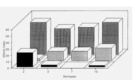
Figure 2 - Effect of supernatants of Ureaplasma urealyticum serotypes on the mitotic index of human lymphocytes in vitro. S1 ( ) = Supernatants of serotypes 2, 3, 7 and 10 at the
concentration of 105 CCU/ml; S
2 ( ) = supernatant duplicate; Cs ( ) =supernatant control.
Table 1 - Effect of serotype and concentration on U. urealyticum-induced modifications of human lymphocyte mitosis in vitro. Concentration is reported as CCU/ml (color changing units/ml).
U. urealyticum Total mitosis Partial mitosis Metaphases with Metaphases without
serotypes inhibition inhibition chromosome chromosome
alterations alterations
1 105 103, 104
2 103, 104, 105
3 103, 104, 105
5 103, 105 104
6 103, 105 104
7 103, 105 104
8 103, 104, 105, >105
9 105 103, 104
10 103, 104,105
11 103, 104, 105
classified into two clusters: A (serotypes 2, 4, 5, 7, 8, 9, 10, 11, 12 and 13) and B (serotypes 1, 3, 6 and 14). Among these, the B cluster serotypes are the most frequently involved in human pathologies, although serotypes 4, 8 and 10 from cluster A also play an impor-tant role in some processes (11,29,30).
The aim of the present study was to es-tablish a correlation between the serotypes studied and the structural chromosome alter-ations observed, and to determine (on the basis of the supernatants) if soluble products produced by the strains may be responsible for these alterations. The results indicated that serotypes 1, 2, 7, 10 and 12 from cluster A and serotype 3 from cluster B were re-sponsible for the partial or total inhibition of mitosis. Metaphases presenting morphologi-cal chromosome alterations were the main features among serotypes 8, 9, 11 and 12 from cluster A. Among these, serotype 8 presented a significantly higher frequency of these alterations. In addition, the superna-tants of serotypes 3 and 7 showed reduction in the mitotic index and clastogenic effects,
both possibly caused by ammonia release. Consequently, we observed that serotypes from both clusters had clastogenic effects on human chromosomes and on the mitotic pro-cess itself. Our results are consistent with the clinical analysis of some investigators who correlated positive cases of Ureaplasma urealyticum with the involvement of sero-types 3, 8 and 10 (12,29-32).
The isolation of mycoplasma from hu-man malignant tissues reported by some in-vestigators and the effects on human chro-mosomes demonstrated here suggest the im-portance of further studies on the role of these microorganisms in certain carcinogenic processes (33-35).
Acknowledgments
We would like to thank Carlos Malheiros, Difco Laboratories, for kindly providing the serotypes of Ureaplasma urealyticum, Suely Nonogaki for excellent technical assistance, and Prof. Dr. Ana Campa and Prof. Dr. Adelaide José Vaz for revising the text
References
1. Cunha RAF, Takimoto S & Takei K (1988). Micoplasmas: Isolamento e identificação em material cervical de gestantes. Revista de Microbiologia, 19: 379-384.
2. Kundsin RB, Driscoll SG & Ming PML (1967). Strain of mycoplasma associated with human reproductive failure. Science, 157: 1573-1574.
3. McGarrity GJ, Phillips DM & Vaydya AB (1990). Mycoplasmal infection of lympho-cyte cell cultures: Infection with Myco-plasma salivarium. In Vitro, 16: 346-356. 4. Ruth E & Praz F (1989). Interactions
be-tween mycoplasmas and the immune sys-tem. Immunological Reviews, 112: 133-160.
5. Aula P & Nichole W (1967). The cytoge-netic effect of mycoplasma in human leu-kocyte cultures. Journal of Cellular Physi-ology, 70: 281-290.
6. Cooperman R & Morton HE (1966). Re-versible inhibition of mitosis in lympho-cyte cultures by non-viable mycoplasma. Proceedings of the Society for Experimen-tal Biology and Medicine, 123: 790-795.
7. Ginsburg H & Nicolet J (1973). Extensive transformation of lymphocytes by a my-coplasma organism. Nature, 246: 143-146.
8. Naot Y & Ginsburg H (1978). Activation of B lymphocytes by mycoplasma mitogen. Immunology, 34: 715-720.
9. Naot Y (1982). In vitro studies on the mi-togenic activity of mycoplasmal species toward lymphocytes. Reviews of Infec-tious Diseases, 4: 205-209.
10. Quinn PA, Arshoff LV & Li HCS (1981). Serotyping of Ureaplasma urealyticum by immunoperoxidase assay. Journal of Clini-cal Microbiology, 13: 670-676.
11. Razin S & Yogeu D (1986). Genetic relat-edness among Ureaplasma urealyticum (serovars). Pediatric Infectious Diseases, 5: 300-304.
12. Robertson JA, Honoré LH, Kakulphimip J, Hill GB & Jenkins HJ (1990). Association of Ureaplasma urealyticum with sponta-neous abortion. In: Inums Congress Bac-teriology & Mycology. Osaka, 156 (Ab-stract).
13. Shepard MC (1974). Standard fluid medi-um U10 for cultivation and maintenance of
Ureaplasma urealyticum. International Journal of Systematic Bacteriology, 24: 160-171.
14. Shepard MC & Lunceford CD (1976). Dif-ferential agar medium (A7) for
identifica-tion of Ureaplasma urealyticum I (human T-mycoplasma) in primary cultures of clini-cal material. Journal of Cliniclini-cal Microbiol-ogy, 3: 625-631.
15. Moorhead PS, Noweel PC, Melmann WJ, Battips DM & Hungerford DA (1960). Chromosome preparations of leukocytes cultured from peripheral blood. Experi-mental Cell Research, 20: 613-616. 16. ISCN (1995). An International System for
Human Cytogenetic Nomenclature. Mitelman F (Editor). S. Karger, Basel, 27-29.
18. Richardson CR, Howard CA, Sheldon T, Wildgoose J & Thomas MG (1984). The human lymphocyte in vitro cytogenetic assay: positive and negative control ob-servations on approximately 30,000 cells. Mutation Research , 141: 59-64. 19. Evans HJ & ORiordan ML (1975). Human
peripheral blood lymphocytes for the a-nalysis of chromosome aberrations in mutagen tests. Mutation Research, 31: 135-148.
20. Matter BE (1976). Problems of testing drugs for potential mutagenicity. Muta-tion Research, 38: 243-258.
21. Gundy S & Varga LP (1983). Chromosomal aberrations in healthy persons. Mutation Research, 120: 187-191.
22. Littlefield GL & Goh KO (1973). Cytoge-netic studies in control men and women. Cytogenetics and Cell Genetics, 12: 17-34.
23. Lubs HA & Samuelson J (1967). Chromo-some abnormalities in lymphocytes from normal human subjects. Cytogenetics, 6: 402-411.
24. Mattei MG, Ayme S, Mattei JF, Aurran Y & Giraud F (1979). Distribution of sponta-neous chromosome breaks in man. Cyto-genetics and Cell Genetics, 23: 95-102.
25. Stalheim OHV, Proctor SJ & Gallagher JE (1976). Growth and effects of Ureaplasma urealyticum (T-mycoplasma) in bovine ovi-ductal organ cultures. Infection and Im-munity, 13: 915-925.
26. Taylor-Robinson D & Carney FG (1974). Growth and effect of mycoplasmas in Fal-lopian tube organ cultures. British Journal of Venereal Diseases, 50: 212-216. 27. Schneider E, Stanbridge EJ & Golbus CJE
(1974). Mycoplasma contamination of cul-tured amniotic fluid cells: Potential hazard to prenatal chromosomal diagnosis. Sci-ence, 184: 477-479.
28. Fogh J & Fogh H (1968). Karyotypic changes in mycoplasma modified lines of 11 human amnion cells. Proceedings of the Society for Experimental Biology and Medicine, 129: 944-950.
29. Robertson JA, Honoré LH & Stemke GW (1986). Serotypes of Ureaplasma urealyti-cum in spontaneous abortion. Pediatric Infectious Diseases, 5: 270 -272. 30. Zheng X, Watson HL, Waites KB & Cassel
GH (1992). Serotype diversity and antigen variation among invasive isolates of Urea-plasma urealyticum from neonates. Infec-tion and Immunity, 60: 3472-3474.
31. Cracea E, Botez DC & Braila SMG (1984). Ureaplasma urealyticum serotypes iso-lated from cases of female sterility. Yale Journal of Biology and Medicine, 57: 895. 32. Naessens A, Foulon W, Breynaert J & Lauers S (1988). Serotypes of Ureaplasma urealyticum isolated from normal preg-nant women and patients with pregnancy complications. Journal of Clinical Microbi-ology, 26: 319-322.
33. Barile MF (1967). Mycoplasma and leuke-mia. Annals of the New York Academy of Sciences, 143: 557-563.
34. Jansson E & Wager O (1967). Myco-plasma in collagen diseases and blood dyscrasia. Annals of the New York Acade-my of Sciences, 143: 535-543.