RETRACTI ON
ht t p: / / dx.doi.org/ 10.1590/ 1678- 77572016ret r001
The Edit orial Board of t he Journal Applied Oral Science com m unicat es t he form al ret ract ion of t he m anuscript :
Alvarez C, Benít ez A, Roj as L, Puj ol M, Carvaj al P, Díaz- Zúñiga, et al. Different ial expression of CC chem okines ( CCLs) and recept ors ( CCRs) by hum an T lym phocyt es in response t o diferent e Aggregat ibact er act inom ycet em com it ans serot ypes. J Appl Oral Sci. 2015; 23( 6) : 580- 90. ht t p: / / dx.doi.org/ 10.1590/ 1678-775720150285
Since it com prises a duplicat ed version of a m anuscript previously published in t he preceding edit ion of t he Journal Applied Oral Science:
Alvarez C, Benít ez A, Roj as L, Puj ol M, Carvaj al P, Díaz- Zúñiga, et al. Different ial expression of CC chem okines ( CCLs) and recept ors ( CCRs) by hum an T lym phocyt es in response t o diferent e Aggregat ibact er act inom ycet em com it ans serot ypes. J App. Oral Sci. 2015; 23( 5) : 536- 46. ht t p: / / dx.doi.org/ 10.1590/ 1678-775720150285
ABSTRACT
http://dx.doi.org/10.1590/1678-775720150285
Differential expression of CC chemokines (CCLs)
and receptors (CCRs) by human T lymphocytes
in response to different
A g g r e g a t i b a c t e r
act inom ycet em com it ans
serotypes
Carla ALVAREZ1, Alvaro BENÍTEZ1, Leticia ROJAS1, Myriam PUJOL1, Paola CARVAJAL2, Jaime DÍAZ-ZÚÑIGA2, Rolando VERNAL1,2
1- Universidad de Chile, Facultad de Odontología, Laboratorio de Biología Periodontal, Santiago, Chile. 2- Universidad de Chile, Facultad de Odontología, Departamento de Odontología Conservadora, Santiago, Chile.
Corresponding address: Rolando Vernal - Periodontal Biology Laboratory, Faculty of Dentistry, Universidad de Chile -
Sergio Livingstone Pohlhammer 943 - 838000 - Independencia - Santiago - Chile - Phone: +56 (2) 29781833 - Fax: +56 (2) 29781813 - e-mail: [email protected]
6XEPLWWHG-XQH0RGL¿FDWLRQ6HSWHPEHU$FFHSWHG6HSWHPEHU
I
n Aggregat ibact er act inom ycet em com it ans, different serotypes have been described based on LPS antigenicity. Recently, our research group has reported a differential immunogenicity when T lymphocytes were stimulated with these different serotypes. In particular, it was demonstrated that the serotype b of A. act inom ycet em com it ans has a stronger capacity to trigger Th1- and Th17-type cytokine production. Objective: This study aimed to quantify the expression of different CC chemokines (CCLs) and receptors (CCRs) in T lymphocytes stimulated with the different A. act inom ycet em com it ans serotypes. In addition, the expression of the transcription factors T-bet, GATA-3, RORC2, and Foxp3, master-switch genes implied in the Th1, Th2, Th17, and T-regulatory differentiation, respectively, was analy ed in order to determine T-cell phenotype-speci c patterns of CCL and CCR expression upon A. act inom ycet em com it ans stimulation. Material and Methods: Human naïve CD4+ T lymphocytes were obtained from healthy subjects and stimulated with autologous dendritic cells primed with the different A. act inom y cet em com it ansserotypes. The expression levels for the chemokines CCL1, CCL2, CCL3, CCL5, CCL11, CCL17, CCL20, CCL21, CCL25, and CCL28, as well as the chemokine receptors CCR1, CCR2, CCR3, CCR4, CCR5, CCR , CCR7, CCR8, CCR , and CCR10 were quanti ed by qPCR. Similarly, the expression levels for the transcription factors T-bet, GATA-3, RORC2, and Foxp3 were quanti ed and correlated with the CCL and CCR expression levels. Results: Higher expression levels of CCL2, CCL3, CCL5, CCL20, CCL21, CCL28, CCR1, CCR2, CCR5, CCR6, CCR7, and CCR9 were detected in T lymphocytes stimulated with the serotype b of
A. act inom ycet em com it ans compared with the other serotypes. In addition, these higher expression levels of CCLs and CCRs positively correlated with the increased levels of T-bet and RORC2 when T lymphocytes were stimulated with the serotype b. Conclusion: A T-lymphocyte response biased towards a Th1- and Th17-pattern of CCL and CCR expression was detected under stimulation with the serotype b of A. act inom ycet em com it ans.
Keywords:Aggregat ibact er act inom ycet em com it ans. Chemokines. Chemokine receptors. T-lymphocytes. Th1 cells. Th17 cells.
INTRODUCTION
In humans, CC chemokines (CCLs) and their speci c CC receptors (CCRs) play a central role in physiological and pathological recruitment of immune cells9,24. During infectious diseases,
the expression of CCLs and CCRs produces a chemotactic gradient between regional lymph nodes and infected tissues where, depending on the pattern of CCLs and/or CCRs expressed, specific dendritic cells and T lymphocytes are chemoattracted. Thus, it is established a cellular
pathway that goes both ways, in which 1) dendritic cells migrate toward lymphoid organs to present microbial antigens and 2) activated T helper (Th) lymphocytes migrate toward infected tissues to accomplish their speci c immunological function24.
Th lymphocytes play a central role in the pathogenesis of periodontitis, and a Th1 and Th17-dominated immuno-in ammatory response has been associated with periodontal tissue destruction, alveolar bone resorption, and teeth loss. In this context, the pattern of CCLs and CCRs expressed by Th lymphocytes is crucial in the establishment of the local Th-pattern of immuno-in ammatory response and in the outcome of the disease9,28. In
fact, greater levels of CCL3, CCL4, CCL5, CCL28, CCR1, CCR5, and CCR9 were detected in periodontal lesions of aggressive periodontitis patients, and increased levels of CCL2 and CCR4 were found in lesions of chronic periodontitis patients8,23. In
addition, increased production of IF - has been associated with both greater expression of CCR5 and differentiation of Th1 lymphocytes16. Similarly,
increased production of IL-6 and IL-23 has been associated with both greater expression of CCL2, CCR6, and CCR7, and subsequent differentiation, migration, and activation of Th17 lymphocytes13,15.
Conversely, increased production of IL-4 and IL-10 has been demonstrated to inhibit the production of CCR5 and to induce the expression of CCL11, CCR3, and CCR4, implied in the Th2 lymphocyte differentiation and function18.
Recently, our research group has reported a differential immunogenicity when dendritic cells and T lymphocytes were stimulated with the different serotypes of A. act in om y cet em com it an s2,3. In
particular, when T lymphocytes were stimulated with autologous monocyte-derived dendritic cells primed with the serotype b of A. act inom ycet em com it ans, higher levels of Th1- and Th17-associated transcription factors and cytokines were detected compared with similar experiments with the other serotypes, demonstrating that serotype b strains of A. a ct i n o m y cet em co m i t a n s have a higher capacity of triggering Th1 and Th17 phenotype and function. It is, therefore, the aim of this investigation to elucidate whether the different serotypes of A. act in om y cet em com it an s have a role on the differential expression of CCLs and CCRs. We hypothesized that the serotype b of A. act in om y cet em com it an s, when used to stimulate T lymphocytes, induces higher Th1- and Th17-associated CCL and CCR expression when compared with the other A. act inom ycet em com it ans serotypes.
MATERIAL AND METHODS
Experimental design
This experimental study consisted of cell cultures of peripheral naïve CD4+ T lymphocytes obtained
from healthy humans and infected in vit ro with A. act inom ycet em com it ans. The protocol of the study was clearly explained to all the participants, who agreed to participate in it by signing an institutional review board-approved informed consent (Protocol 2010/14). The study was conducted in accordance with the Helsinki Declaration of 1975, as revised in 2000.
A. a ct in om y ce t e m com it a n s strains
The A. act inom ycet em com it ans strains ATCC®
43717™ (serotype a), ATCC® 43718™ (serotype
b), and ATCC® 43719™ (serotype c) were cultured
on agar brain-heart infusion medium (Oxoid, Hampshire, UK) at 37°C and under capnophilic conditions (8% O2 and 12% CO2) using an appropriate microaerobic condition generator (CampyGen™; Oxoid, Hampshire, UK). Growth curves were obtained, and live bacteria, having their whole antigenic potentiality, were obtained at the exponential growth phase of the bacterial culture and used for in vit ro cell stimulation.
Blood donors
Blood cells were obtained during platelet-apheresis process from healthy donors consecutively enrolled at the Blood Bank of the Hospital Del Salvador in the Eastern Metropolitan Health Service. The study group consisted of 12 adult individuals (seven males and ve females, aged 21 to 38 years, mean age of 28.3±5.10 years) who did not have periodontal disease as determined by absence of gingival in ammation, clinical attachment loss, or increased probing depths (PD>3 mm). Further exclusion criteria were the positive test for HIV and hepatitis B or C virus, presence of manifest infections during the last month, fever, symptomatic allergies, abnormal blood cell counts, increased liver enzymes, or medication of any kind except vitamins and oral contraceptives.
Monocyte-derived dendritic cell generation For each subject, peripheral blood mononuclear cells (PBMCs) were isolated by density gradient centrifugation using standard procedures (Ficoll-Paque Plus; GE Healthcare, Uppsala, Sweden). For generating a puri ed population of immature dendritic cells, monocytes were purified from PBMCs by magnetic cell sorting separation (MACS; Miltenyi Biotec, Bergisch Gladbach, Germany). Brie y, PBMCs were incubated with an anti-CD14 monoclonal antibody conjugated to magnetic beads for 15 min at 4°C, loaded onto LS columns
and then separated in the magnetic eld of a cell separator (MACS; Miltenyi Biotec, Bergisch Gladbach, Germany). The retained CD14+ cells,
which correspond to monocytes, were then ushed out and washed twice in phosphate-buffered saline. Monocytes were then immediately differentiated to dendritic cells by culturing at 106 cells/mL in
RPMI-1640 medium supplemented with 10% foetal calf serum (Gibco Invitrogen Corp., Grand Island, NY, USA) and 20 ng/mL rhGM-CSF and rhIL-4 (R&D Systems Inc., Minneapolis, MN, USA) for 6 d at 37°C.
T-lympKocyte pXri¿cation
A puri ed population of naïve CD4+ T lymphocytes
was obtained from PBMCs by magnetic cell sorting depletion (MACS; Miltenyi Biotec, Bergisch Gladbach, Germany). Brie y, both non-T helper and memory T helper cells were depleted using a cocktail of biotin-conjugated monoclonal antibodies (CD8, CD14, CD15, CD16, CD19, CD25, CD34,
CD36, CD45RO, CD56, CD123, TCR / , HLA-DR, and CD235a) and anti-biotin monoclonal antibodies conjugated to magnetic beads. The magnetically labelled cells were retained within LD columns in the magnetic eld of a cell separator (MACS; Miltenyi Biotec, Bergisch Gladbach, Germany), while the unlabelled naïve CD4+ T lymphocytes ran through.
Cell stimulation
M o n o c y t e - d e r i ve d d e n d r i t i c c e l l s w e r e primed at a multiplicity of infection MOI=102
(bacteria/cells ratio) with different serotypes of A. a ct i n o m y ce t e m co m i t a n s, and then 106
T-lymphocytes/mL were activated with primed autologous dendritic cells (50:1) in culture with RPMI-1640 containing 10% foetal calf serum (Gibco Invitrogen Corp., Grand Island, NY, USA) for 5 d at 37°C. Previous to each cell co-culture, dendritic cells were washed twice with RPMI-1640 supplemented with 50 IU/mL penicillin and 50 g/ mL streptomycin (Sigma Chemical Co., St. Louis,
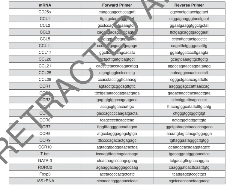
mRNA Forward Primer Reverse Primer
CD25D caagcgagccttccagatt ggccactgctacctggtact
CCL1 ttgctgctagctgggatgt ctggagaagggtacctgcat
CCL2 gcctccagcatgaaagtctc ggaatgaaggtggctgctat
CCL3 cagcagacagtggtcagtcc ttctgagcaggtgacggaat
CCL5 ggtgtggtgtccgaggaata cctcattgctactgccctct
CCL11 cccttcagcgactagagagc cagctttctggggacatttg
CCL17 ggcttctctgcagcacatc ggaatggctcccttgaagta
CCL20 gctgctttgatgtcagtgct gcagtcaaagttgcttgctg
CCL21 caccctctaccacagacatgg aggccagaaccaggataagg
CCL25 ctgagttggtcctccctctg aatcaggccaactccctctt
CCL28 ccacctacctggttcaaacg cgggctgacacagattcttc
CCR1 agtacctgcggcagttgttc aaggggagccatttaaccag
CCR2 tttctgataaaccgagaacgaga gagacaagccacaagctgaa
CCR3 gagtgtgtggccagaagaca cttcctggattcagccctct
CCR4 accgcgtgcacaattgc tttacagtggcatattctttgtcatg
CCR5 gaccagccccaagatgacta cttgggtggtggctgtgt
CCR6 tcagccccttcagctcac actgtggctgttggtttgtg
CCR7 ttggtttaggggacaatagcc ggctgataagctaacaccagaca
CCR8 atgcctagggagagctgtga aaaatgtagtctacgctggagga
CCR9 tttccccagacactgagagc tgttaggaatagggcttgtgg
CCR10 agtaggtgggggaacactga gcaaggcacagaggtagtcc
T-bet tccaagtttaatcagcaccaga tgacaggaatgggaacatcc
GATA-3 ctcattaagcccaagcgaag tctgacagttcgcacaggac
RORC2 agaaggacagggagccaag caagggatcacttcaatttgtg
Foxp3 acctacgccacgctcatc tcattgagtgtccgctgct
18S rRNA ctcaacacgggaaacctcac cgctccaccaactaagaacg
Figure 1- Forward and reverse primers used for CD25D, CCL, CCR, and transcription factor mRNA and 18S rRNA DPSOL¿FDWLRQVE\T3&5
MI, USA). T-lymphocyte cultures devoid of dendritic cells or exposed to non-induced autologous dendritic cells were used for comparisons. In each experimental step, dendritic cell and T-lymphocyte counting was performed with a hemocytometer and using a phase contrast microscopy (Axiovert 100; eiss Co., G ttingen, Germany), and cell viability equal to or higher than 95% was calculated by Trypan blue dye exclusion. For each individual, the experiment was performed separately.
Phenotypic cell analysis
T-lymphocyte puri cation and their subsequent activation were analyzed by ow cytometry (BD FACSCanto™; Becton Dickinson Immunocytometry Systems, San José, CA, USA). Cells were stained using the following monoclonal antibodies conjugated with uorescein isothiocyanate (FITC) or phycoerythrin (PE): anti-CD4 (CD4+ T-lymphocytes), anti-CD25
(activated CD4+ T-lymphocytes), anti-CD45RA
(n aïv e CD4+ T-lymphocytes), and anti-CD45RO
(memory CD4+ T-lymphocytes) following the
manufacturer’s recommendations (BD Biosciences Pharmingen, San José, CA, USA). Isotype-matched control monoclonal antibodies were used to determine the negative cell populations.
Expression of CD25D, CCR, CCL, and
transcription factor mRNAs
From activated T lymphocytes, total cytoplasmic RNA was isolated using 400 μl of ice-cold lysis buffer containing 0.5% Igepal® CA-630 (Sigma-Aldrich,
Saint Louis, MO, USA), 50 mM Tris-HCl pH8, 100 mM NaCl, 5 mM MgCl2, and 10 mM VRC-40 (Gibco Invitrogen, Carlsbad, CA, USA). Isolated RNA was quanti ed using a spectrophotometer (Synergy HT; Bio-Tek Instrument Inc., Winooski, VT, USA), and the rst-strand cDNA was synthesized using 5 μg of total RNA with a SuperScrip™III reverse
transcription kit, following the manufacturer’s instructions (Invitrogen, Grand Island, NY, USA). The mRNA expression levels for the chemokines CCL1, CCL2, CCL3, CCL5, CCL11, CCL17, CCL20, CCL21, CCL25, and CCL28, the chemokine receptors CCR1, CCR2, CCR3, CCR4, CCR5, CCR6, CCR7, CCR8, CCR9, and CCR10 and the transcription factors T-bet (Th1), GATA-3 (Th2), RORC2 (Th17), and Foxp3 (T-regulatory), as well as for the activated T-lymphocyte marker CD25D, were quanti ed by qPCR using the appropriate primers (Figure 1). Brie y, 50 ng of cDNA were ampli ed using a KAPA™ SYBR® Fast qPCR reagent (KAPA
Biosystems, Woburn, MA, USA) in a StepOnePlus®
equipment (Applied Biosystems, Singapore) as follows: 95°C for 3 min, followed by 40 cycles of 95°C for 3 s, and 60°C for 30 s, and nally a melt curve of 95°C for 15 s, 60°C for 1 min, and 95°C for 15 s, for detection of non-speci c product
formation and false positive ampli cation. As an endogenous control, 18S rRNA expression levels were determined.
Data analysis
The ow cytometry data were analyzed using the WinMDi 2.9 software (The Scripps Research Institute, La Jolla, CA, USA), represented as histograms, and expressed as the percentage of positive cells over the total. The qPCR data were analyzed using the StepOne Software 2.2.2 (Applied Biosystems, Singapore) and presented in fold-change of relative quantities by normalizing the CD25D, CCR, CCL, or transcription factor mRNA expression to 18S rRNA expression using the 2- Ct method. Data were statistically analyzed
using the SPSS 15.0 software (Lead Technologies Inc., Charlotte, NC, USA). The normality of data distribution was determined using the Kolmogorov-Smirnov test. Differences regarding CD expression levels analyzed by ow cytometry were determined using the chi-square test. Differences among groups and within each group regarding the CD25D, CCR, CCL, and transcription factor mRNA expression were analyzed using the Kruskal-Wallis test or ANOVA and Tukey post - hoc tests. Correlation coef cients were obtained using the Pearson or Spearman tests. Asterisks were used to graphically indicate statistical signi cance. A value of p 0.05 was considered statistically signi cant.
RESULTS
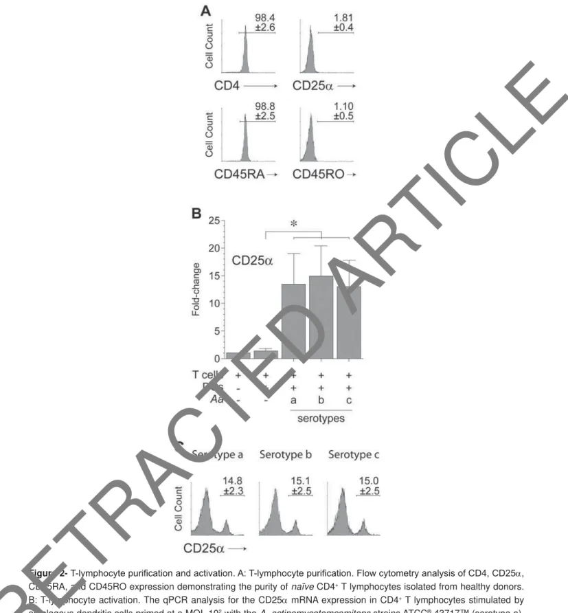
T-lymphocyte puri¿cation and activation For this study, highly puri ed (>98%) populations of n a ïv e CD4+ T lymphocytes (CD4+CD25D
-CD45RA+CD45RO-), devoid of activated or memory
CD4+ T lymphocytes, were isolated from peripheral
blood of healthy donors (Figure 2A). These T lymphocytes activated at a similar extent upon stimulation with dendritic cells primed with the different serotypes of A. act inom ycet em com it ans, as shown by the similar signi cant over-expression in CD25D mRNA levels (p<0.001) compared with T lymphocytes exposed to non-induced dendritic cells (Figure 2B). These similar levels of T lymphocyte activation were con rmed at a protein level when the cell-surface expression of CD25D was determined by ow cytometry. In fact, the frequency of CD25D
expression (~15%) in T lymphocytes exposed to the different serotypes of A. act inom ycet em com it ans did not differ signi cantly (Figure 2C).
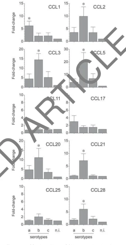
E x p r e s s i o n o f C C L s b y A . a c t i n o m y c e t e m c o m i t a n s- i n d u c e d T lymphocytes
The mRNA expression for the analyzed chemokines was determined by qPCR and represented as
Figure 2- 7O\PSKRF\WHSXUL¿FDWLRQDQGDFWLYDWLRQ$7O\PSKRF\WHSXUL¿FDWLRQ)ORZF\WRPHWU\DQDO\VLVRI&'&'D, CD45RA, and CD45RO expression demonstrating the purity of naïve CD4+ T lymphocytes isolated from healthy donors. B: T-lymphocyte activation. The qPCR analysis for the CD25D mRNA expression in CD4+ T lymphocytes stimulated by autologous dendritic cells primed at a MOI=102 with the A. actinomycetemcomitans strains ATCC® 43717™ (serotype a), ATCC® 43718™ (serotype b), and ATCC® 43719™ (serotype c). C: T-lymphocyte activation. Flow cytometry analysis of WKH&'ĮH[SUHVVLRQGHPRQVWUDWLQJWKHOHYHOVRIDFWLYDWLRQRI&'+ T lymphocytes after 5-day stimulation under the VDPHFRQGLWLRQVGHVFULEHGLQ)LJXUH%7KHÀRZF\WRPHWU\GDWDIURPHDFKH[SHULPHQWZHUHH[SUHVVHGDVSHUFHQWDJH of positive cells over the total, and shown as mean±SD from 4 independent experiments. For relative expression, the CD25D mRNA expression in T lymphocytes cultured in the absence of dendritic cells was considered as 1, as a reference for fold-change in expression. Data are represented as fold-change for 8 independent experiments. Each experiment was performed in duplicate. Comparisons were done versus T lymphocytes exposed to non-induced dendritic cells (*p<0.05).
Aa, Aggregatibacter actinomycetemcomitans; CD, cluster of differentiation; DCs, dendritic cells
change for each condition (Figure 3). When the strain ATCC® 43718™ belonging to the serotype
b of A. act in om y cet em com it an s was used for T-lymphocyte activation, higher expression levels of CCL2 (p=0.025 and p=0.024), CCL3 (p=0.003 and p=0.005), CCL5 (p=0.004 and p=0.013), CCL20 (p=0.05 and p=0.02), CCL21 (p=0.001 and p=0.001), and CCL28 (p=0.004 and p=0.008) were detected, when compared with the strains ATCC® 43717™ and ATCC® 43719™ belonging to
the serotypes a or c, respectively. Conversely, when the serotype a of A. act inom ycet em com it ans was used for T-lymphocyte activation, higher expression levels of CCL1 (p=0.008 and p=0.009) and CCL17 (p>0.05 and p>0.05) were detected, when compared with the serotypes b or c, respectively. CCL11 and CCL25 were not over-expressed in any experimental condition.
E x p r e s s i o n C C R s b y A . a c t i n o m y c e t e m c o m i t a n s- i n d u c e d T lymphocytes
The mRNA expression for the analyzed chemokine receptors was determined by qPCR and represented as fold-change for each condition (Figure 4). When the serotype b of A. act inom ycet em com it ans was used for T-lymphocyte activation, higher expression levels of CCR1 (p=0.036 and p=0.026), CCR2 (p=0.041 and p=0.042), CCR5 (p=0.029 and p=0.035), CCR6 (p=0.045 and p=0.044), CCR7 (p=0.039 and p=0.020), CCR9 (p=0.040 and p=0.035), and CCR10 (p=0.018 and p=0.022) were detected compared with the serotypes a or c, respectively. Conversely, when the serotype a of A. act in om y cet em com it an s was used for T-lymphocyte activation, higher expression levels of CCR3 (p=0.006 and p=0.007), CCR4 (p>0.05 and p>0.05), and CCR8 (p>0.05 and p>0.05) were detected, when compared with the serotypes b or c, respectively.
Expression of T-bet, GATA-3, RORC2, and Foxp3 by A . a c t i n o m y c e t e m c o m i t a n s -induced T lymphocytes
The mRNA expression for T-bet, GATA-3, RORC2, and Foxp3 was determined by qPCR in T lymphocytes stimulated by dendritic cells primed at a MOI=102 with the different serotypes of A.
act inom ycet em com it ans (Figure 5). Similarly to our previous experiments2, T lymphocytes stimulated
with the serotype b showed a higher relative expression of T-bet (p<0.001 and p<0.001) and RORC2 (p<0.001 and p=0.001) mRNAs than the same cells stimulated with the serotypes a or c, respectively.
Figure 3- CC chemokines (CCL) expression by A.
actinomycetemcomitans-induced T lymphocytes.
CCL mRNA expression in T lymphocytes activated by dendritic cells primed at a MOI=102 with the A.
actinomycetemcomitans strains ATCC® 43717™
(serotype a), ATCC® 43718™ (serotype b), and ATCC® 43719™ (serotype c). For relative expression, the CCL mRNA expression in T lymphocytes exposed to non-induced dendritic cells was considered as 1, as a reference for fold-change in expression (n.i). Data are represented as fold-change for 8 independent experiments. Each experiment was performed in duplicate. Comparisons were done between the different
A. actinomycetemcomitans serotypes (*p<0.05)
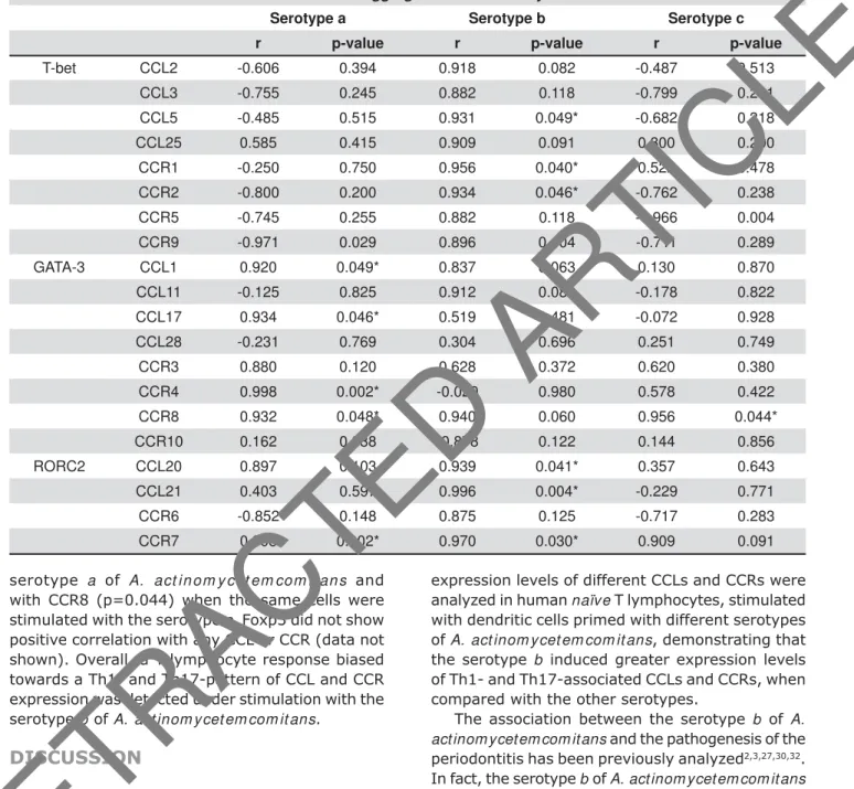
Correlation analysis between T-bet, GATA-3, RORC2, and Foxp3 ve r su s CCL and CCR expression levels
The correlation analyses between the expression of the transcription factors and the CCLs and CCRs on each activation condition tested (Table 1) yielded positive correlation between T-bet and CCL2, CCL3, CCL5, CCL25, CCR1, CCR2, CCR5, and CCR9, being statistically signi cant for CCL5 (p=0.049), CCR1 (p=0.040), and CCR2 (p=0.046), when T lymphocytes were stimulated with the serotype b of A. act inom ycet em com it ans. Under the same condition, a positive correlation was also observed between RORC2 and CCL20, CCL21, CCR6, and CCR7, being statistically significant for CCL20 (p=0.041), CCL21 (p=0.004), and CCR7 (p=0.030). In contrast, GATA-3 showed positive correlation with CCL1, CCL17, CCR3, CCR4, and CCR8, being statistically signi cant for CCL1 (p=0.049), CCL17 (p=0.046), CCR4 (p=0.002), and CCR8 (p=0.048) when T lymphocytes were stimulated with the
Figure 4- CC receptors (CCR) expression by A.
actinomycetemcomitans-induced T lymphocytes.
CCR mRNA expression in T lymphocytes activated by dendritic cells primed at a MOI=102 with the A.
actinomycetemcomitans strains ATCC® 43717™
(serotype a), ATCC® 43718™ (serotype b), and ATCC® 43719™ (serotype c). For relative expression, the CCR mRNA expression in T lymphocytes exposed to non-induced dendritic cells was considered as 1, as a reference for fold-change in expression (n.i). Data are represented as fold-change for 8 independent experiments. Each experiment was performed in duplicate. Comparisons were done between the different
A. actinomycetemcomitans serotypes (*p<0.05)
Figure 5- Transcription factor expression by A. actinomycetemcomitans-induced T lymphocytes. T-bet (Th1), GATA-3 (Th2), RORC2 (Th17), and Foxp3 (T-regulatory) mRNA expression in T lymphocytes stimulated by dendritic cells primed at a MOI=102 with the A. actinomycetemcomitans strains ATCC® 43717™ (serotype a), ATCC® 43718™ (serotype b), and ATCC® 43719™ (serotype c). For relative expression, the transcription factor mRNA expression in T lymphocytes exposed to non-induced dendritic cells was considered as 1, as a reference for fold-change in expression (n.i.). Data are represented as fold-change for 8 independent experiments. Each experiment was performed in duplicate. Comparisons were done between the different
A. actinomycetemcomitans serotypes (*p<0.05)
serotype a of A. act in om y cet em com it an s and with CCR8 (p=0.044) when the same cells were stimulated with the serotype c. Foxp3 did not show positive correlation with any CCL or CCR (data not shown). Overall, a T-lymphocyte response biased towards a Th1- and Th17-pattern of CCL and CCR expression was detected under stimulation with the serotype b of A. act inom ycet em com it ans.
DISCUSSION
There is strong evidence suggesting that variations in the host immuno-inflammatory response, in particular, in the T lymphocyte phenotype and function, play an important role in the susceptibility, onset, and severity of periodontitis6,10.
In particular, a Th1 and Th17-dominated immuno-in ammatory response has been associated with the pathogenesis of periodontitis, and an increased expression of Th1- and Th17-related transcription factors and pro-in ammatory mediators have been reported in active periodontal lesions, where alveolar bone resorption is occurring6,17. In this study, the
expression levels of different CCLs and CCRs were analyzed in human naïve T lymphocytes, stimulated with dendritic cells primed with different serotypes of A. act inom ycet em com it ans, demonstrating that the serotype b induced greater expression levels of Th1- and Th17-associated CCLs and CCRs, when compared with the other serotypes.
The association between the serotype b of A. act inom ycet em com it ans and the pathogenesis of the periodontitis has been previously analyzed2,3,27,30,32.
In fact, the serotype b of A. act inom ycet em com it ans triggers a greater immunogenic and pathogenic response when in contact with different host cells compared with the other serotypes. For instance, the serotype b induces increased resistance to phagocytosis and to killing by macrophages and neutrophils32, higher expression of IL-8 and ICAM-1
in gingival epithelial cells27, greater production of
IL-1 in macrophages30, and stronger induction of
Th1- and Th17-type of response in dendritic cells and CD4+ T lymphocytes2,3.
To our knowledge, this is the first report a s s o c i a t i n g t h e d i f f e r e n t s e r o t y p e s o f A .
Aggregatibacter actinomycetemcomitans
Serotype a Serotype b Serotype c
r p-value r p-value r p-value
T-bet CCL2 -0.606 0.394 0.918 0.082 -0.487 0.513
CCL3 -0.755 0.245 0.882 0.118 -0.799 0.201
CCL5 -0.485 0.515 0.931 0.049* -0.682 0.318
CCL25 0.585 0.415 0.909 0.091 0.800 0.200
CCR1 -0.250 0.750 0.956 0.040* 0.522 0.478
CCR2 -0.800 0.200 0.934 0.046* -0.762 0.238
CCR5 -0.745 0.255 0.882 0.118 -0.966 0.004
CCR9 -0.971 0.029 0.896 0.104 -0.711 0.289
GATA-3 CCL1 0.920 0.049* 0.837 0.063 0.130 0.870
CCL11 -0.125 0.825 0.912 0.088 -0.178 0.822
CCL17 0.934 0.046* 0.519 0.481 -0.072 0.928
CCL28 -0.231 0.769 0.304 0.696 0.251 0.749
CCR3 0.880 0.120 0.628 0.372 0.620 0.380
CCR4 0.998 0.002* -0.020 0.980 0.578 0.422
CCR8 0.932 0.048* 0.940 0.060 0.956 0.044*
CCR10 0.162 0.838 -0.878 0.122 0.144 0.856
RORC2 CCL20 0.897 0.103 0.939 0.041* 0.357 0.643
CCL21 0.403 0.597 0.996 0.004* -0.229 0.771
CCR6 -0.852 0.148 0.875 0.125 -0.717 0.283
CCR7 0.998 0.002* 0.970 0.030* 0.909 0.091
Table 1- Correlation analysis of transcription factors versus CCL and CCR mRNA expressions. The Pearson’s correlation FRHI¿FLHQWUEHWZHHQWKHWUDQVFULSWLRQIDFWRUV7EHW7K*$7$7KDQG525&7KDQGWKH7K7KRU Th17-associated CCLs and CCRs were calculated using T lymphocytes stimulated by autologous dendritic cells primed at a MOI=102 with the different A. actinomycetemcomitans serotypes. *p<0.05
act inom y cet em com it ans with a Th1- and Th17-pattern of immuno-inflammatory response by analyzing the CC chemokines and receptors involved in the selective chemo-attraction of Th lymphocytes. These ndings are suggestive of the pathogenic role of A. act inom ycet em com it ans in the aetiology of periodontitis, which may differ among serotypes and speci cally let us propose that serotype b could play a role in the pathogenesis of the disease by the induction of a local in ammatory environment that favors the speci c recruitment of Th1 and Th17 lymphocytes towards the infected periodontal tissues.
In rheumatoid arthritis, which is an in ammatory disease characterized by the development of a Th1 and Th17-dominated immuno-in ammatory response in the affected articular tissues14, it
has been established that the chronicity of the in ammation is associated with a Th1- and Th17-pattern of CCL and CCR expression. In fact, in chronic in amed joints, over-expressed levels of CCL2, CCL3, CCL5, CCR1, CCR2, and CCR5, related to a Th1-type response26,29, and CCL20, CCL21, CCR6,
and CCR7, related to a Th17-type response14,22,
have been detected compared with non-in amed joints. In addition, increased expression of CCL20 has been associated with predominant Th17 lymphocyte in ltration in the affected articular tissues11 and, when the chemokine receptors CCR1,
CCR2, CCR5, and CCR6 were blocked, a decreased Th1 lymphocyte migration towards the in amed joints was detected, promoting the resolution of the disease11,26.
The results of the present study are consistent with the available scientific evidence, and demonstrate that a Th1- and Th17-pattern of immuno-in ammatory response develops during the periodontal infection, at least during an in vit ro mono-infection with A. act inom y cet em com it ans. Thus, the Th1 and Th17-dominated immuno-in ammatory response described immuno-in periodontitis could be explained by the increased chemo-attraction of recently activated and differentiated Th1 and Th17 lymphocytes from regional lymph nodes and/or by the activation of n aïv e and memory Th1 and Th17 lymphocytes residing in the periodontal tissues12,25. In this context, the serotype
b of A. act inom ycet em com it ans could be associated with local periodontal tissue destruction by the over-production of Th1- and Th17-associated cytokines, such as IL1- , IL-6, IL-17A, and TNF- 2,3, and the
over-expression of Th1- and Th17-associated CCLs and CCRs, such as CCL2, CCL3, CCL5, CCR1, CCR2, CCR5, and CCR7, as demonstrated in the present study, which are involved in the differentiation and activation of osteoclasts and the resorption of tooth-supporting alveolar bone6-9,28. In fact,
a more frequent and higher expression of CCL2
and its speci c receptor CCR4 have been reported in chronic periodontitis, and a more prevalent and higher expression of CCL3 and its speci c receptor CCR5 have been reported in aggressive periodontitis9. Thus, the A. act inom ycet em com it ans
serotype b-induced Th1 and/or Th17 lymphocytes could migrate towards infected tissues following the CCL2 and/or CCL3 chemotactic gradient, resulting in a Th1 and/or Th17-associated cytokine production at the periodontal lesion that locally promote the alveolar bone resorption.
The A. act inom y cet em com it ans strains used in the present study are phenotypically smooth. In fact, these different serotypes correspond to structurally distinct O-polysaccharide components of their respective LPS that function as immuno-dominant antigens. The O-polysaccharide from the LPS produced by the serotype b of A. act in om y cet em com it an s is structurally distinct from the O-polysaccharide produced by the other serotypes. In particular, the O-polysaccharide from serotype b consists of a repeating trisaccharide unit composed of -D-fucose, D-L-rhamnose and -D-N-acetyl-galactosamine residues, and the O-polysaccharide from serotypes a and c is composed of 6-deoxy-D-D-talose and 6-deoxy-D -L-talose, respectively19,20.
Interestingly, our data also show that serotype b of A. a ct i n o m y ce t e m co m i t a n s induces an increment in the CCR10 expression in stimulated T lymphocytes, a chemokine receptor associated with Th2 lymphocyte differentiation and function31.
In this context, it has been demonstrated that CCR10 is also expressed by Th22 lymphocytes, a recently described T lymphocyte population that complies pro-in ammatory activities5; however,
this T-cell phenotype has not yet been described in periodontitis. Accordingly, it could be hypothesized that Th22 lymphocytes may be associated with the pathogenesis of periodontitis1, which would clarify,
at least to a certain degree, the over-expressed levels of CCR10 and its speci c chemokine ligand CCL28 detected in the present study. In fact, higher levels of CCL28 have been detected in the gingival crevicular uid of chronic and aggressive periodontitis patients compared with gingivitis and healthy individuals4.
During periodontitis, antigen presentation may occur both in the regional lymph nodes that drain the periodontal tissues and locally in the infected periodontal tissues, in a periodontal site-speci c manner. In fact, formation of periodontal lymphoid clusters in which dendritic cells present bacterial antigens to naïve or memory T lymphocytes has been reported, promoting the activation and selective differentiation of Th1 and Th17 lymphocytes12,25.
In this scenario, CCR7 and its speci c chemokine ligands CCL19 and CCL21 could play a role in the
organization of these cellular clusters, as an ectopic lymphoid-like structure21, promoting the alveolar
bone resorption characteristic of the periodontitis.
CONCLUSIONS
In T lymphocytes, the serotype b of A . act inom ycet em com it ans induces higher expression levels of chemokines CCL2, CCL3, CCL5, CCL20, CCL21, and CCL28 and of chemokine receptors CCR1, CCR2, CCR5, CCR6, CCR7, and CCR9, when compared with the other serotypes. These increased levels associated with the expression of the transcription factors master-switch genes that trigger the Th1 and Th17 lymphocyte differentiation. Considered together, these data let us propose that variability in the Th1 and Th17 immuno-in ammatory response induced by the different serotypes of A. act inom ycet em com it ans is associated, at least to a certain degree, with the CC chemokines and receptors that they express; however, functional studies are necessary to con rm our hypothesis.
ACKNOWLEDGMENTS
The authors declare no competing nancial interests. This study was supported by grants FONDECYT 11100298 and 1140904.
REFERENCES
1- Araujo-Pires AC, Francisconi CF, Biguetti CC, Cavalla F, Aranha AM, Letra A, et al. Simultaneous analysis of T helper subsets (Th1, Th2, Th9, Th17, Th22, Tfh, Tr1 and Tregs) markers expression in periapical lesions reveals multiple cytokine clusters accountable for lesions activity and inactivity status. J Appl Oral Sci. 2014;22(4):336-46.
2- Díaz-Zúñiga J, Melgar-Rodríguez S, Alvarez C, Monasterio G, Benítez A, Ciuchi P, et al. T-lymphocyte phenotype and function triggered by Aggregat ibact er act inom ycet em com it ans is serotype-dependent. J Periodontal Res. 2015;50(6):824-35.
3- Díaz-Zúñiga J, Yáñez JP, Alvarez C, Melgar-Rodríguez S, Hernández M, Sanz M, et al. Serotype-dependent response of human dendritic cells stimulated with Ag g r eg a t i b a ct er act inom ycet em com it ans. J Clin Periodontol. 2014;41(3):242-51. 4- Ertugrul AS, Sahin H, Dikilitas A, Alpaslan N, Bozoglan A. Comparison of CCL28, interleukin-8, interleukin-1beta and tumor necrosis factor-alpha in subjects with gingivitis, chronic periodontitis and generalized aggressive periodontitis. J Periodontal Res. 2013;48(1):44-51.
5- Fujita H. The role of IL-22 and Th22 cells in human skin diseases. J Dermatol Sci. 2013;72(1):3-8.
6- Garlet GP. Destructive and protective roles of cytokines in periodontitis: a re-appraisal from host defense and tissue destruction viewpoints. J Dent Res. 2010;89(12):1349-63. 7- Garlet GP, Avila-Campos MJ, Milanezi CM, Ferreira BR, Silva JS. Act inobacillus act inom y cet em com it ans-induced periodontal disease in mice: patterns of cytokine, chemokine, and chemokine receptor expression and leukocyte migration. Microbes Infect. 2005;7(4):738-47.
8- Garlet TP, Fukada SY, Saconato IF, Avila-Campos MJ, Silva TA, Garlet GP, et al. CCR2 de ciency results in increased osteolysis in experimental periapical lesions in mice. J Endod. 2010;36(2):244-50.
9- Garlet GP, Martins W Jr, Ferreira BR, Milanezi CM, Silva JS. Patterns of chemokines and chemokine receptors expression in different forms of human periodontal disease. J Periodontal Res. 2003;38(2):210-7.
10- Gemmell E, Yamazaki K, Seymour GJ. The role of T cells in periodontal disease: homeostasis and autoimmunity. Periodontol 2000. 2007;43:14-40.
11- Hirota K, Yoshitomi H, Hashimoto M, Maeda S, Teradaira S, Sugimoto N, et al. Preferential recruitment of CCR6-expressing Th17 cells to in amed joints via CCL20 in rheumatoid arthritis and its animal model. J Exp Med. 2007;204(12):2803-12.
12- Hjelmstrom P. Lymphoid neogenesis: de novo formation of lymphoid tissue in chronic in ammation through expression of homing chemokines. J Leukoc Biol. 2001;69(3):331-9.
13- Kabashima H, Yoneda M, Nagata K, Hirofuji T, Maeda K. The presence of chemokine (MCP-1, MIP-1alpha, MIP-1beta, IP-10, RANTES)-positive cells and chemokine receptor (CCR5, CXCR3)-positive cells in in amed human gingival tissues. Cytokine. 2002;20(2):70-7.
14- Kochi Y, Suzuki A, Yamamoto K. Genetic basis of rheumatoid arthritis: a current review. Biochem Biophys Res Commun. 2014;452(2):254-62.
15- Kuwabara T, Ishikawa F, Yasuda T, Aritomi K, Nakano H, Tanaka Y, et al. CCR7 ligands are required for development of experimental autoimmune encephalomyelitis through generating IL-23-dependent Th17 cells. J Immunol. 2009;183(4):2513-21. 16- Loetscher P, Uguccioni M, Bordoli L, Baggiolini M, Moser B, Chizzolini C, et al. CCR5 is characteristic of Th1 lymphocytes. Nature. 1998;391(6665):344-5.
17- Ohyama H, Kato-Kogoe N, Kuhara A, Nishimura F, Nakasho K, Yamanegi K, et al. The involvement of IL-23 and the Th17 pathway in periodontitis. J Dent Res. 2009;88(7):633-8.
18- Perros F, Hoogsteden HC, Coyle AJ, Lambrecht BN, Hammad H. Blockade of CCR4 in a humanized model of asthma reveals a critical role for DC-derived CCL17 and CCL22 in attracting Th2 cells and inducing airway in ammation. Allergy. 2009;64(7):995-1002. 19- Perry MB, MacLean LM, Brisson JR, Wilson ME. Structures of the antigenic O-polysaccharides of lipopolysaccharides produced by Act inobacillus act inom ycet em com it ans serotypes a, c, d and
e. Eur J Biochem. 1996;242(3):682-8.
20- Perry MB, MacLean LL, Gmür R, Wilson ME. Characterization of the O-polysaccharide structure of lipopolysaccharide from
Act inobacillus act inom ycet em com it ans serotype b. Infect Immun. 1996;64(4):1215-9.
21- Pitzalis C, Jones GW, Bombardieri M, Jones SA. Ectopic lymphoid-like structures in infection, cancer and autoimmunity. Nat Rev Immunol. 2014;14(7):447-62.
22- Ren Y, Yang B, Yin Y, Leng X, Jiang Y, Zhang L, et al. Aberrant CD200/CD200R1 expression and its potential role in Th17 cell differentiation, chemotaxis and osteoclastogenesis in rheumatoid arthritis. Rheumatology. 2015;54(4):712-21.
23- Repeke CE, Ferreira SB Jr, Vieira AE, Silveira EM, Avila-Campos MJ, Silva JS, et al. Dose-response met-RANTES treatment of experimental periodontitis: a narrow edge between the disease severity attenuation and infection control. PLoS One. 2011;6(7):e22526.
24- Rossi D, Zlotnik A. The biology of chemokines and their receptors. Annu Rev Immunol. 2000;18:217-42.
25- Schroeder HE, Graf-de Beer M. Stereologic analysis of chronic lymphoid cell in ltrates in human gingiva. Arch Oral Biol. 1976;21(9):527-37.
26- Shadidi KR, Aarvak T, Henriksen JE, Natvig JB, Thompson KM. The chemokines CCL5, CCL2 and CXCL12 play signi cant roles in the migration of Th1 cells into rheumatoid synovial tissue. Scand J Immunol. 2003;57(2):192-8.
27- Shimada T, Sugano N, Nishihara R, Suzuki K, Tanaka H, Ito K. Differential effects of ve Aggregat ibact er act inom ycet em com it ans
strains on gingival epithelial cells. Oral Microbiol Immunol. 2008;23(6):455-8.
28- Silva TA, Garlet GP, Fukada SY, Silva JS, Cunha FQ. Chemokines in oral in ammatory diseases: apical periodontitis and periodontal disease. J Dent Res. 2007;86(4):306-19.
29- Subasinghe NL, Lanter J, Markotan T, Opas E, McKenney S, Crysler C, et al. A novel series of N-(azetidin-3-yl)-2-(heteroarylamino)acetamide CCR2 antagonists. Bioorg Med Chem Lett. 2013;23(4):1063-9.
30- Takahashi T, Nishihara T, Ishihara Y, Amano K, Shibuya N, Moro I, et al. Murine macrophage interleukin-1 release by capsularlike serotype-specific polysaccharide antigens of Act in ob acillu s act inom ycet em com it ans. Infect Immun. 1991;59(1):18-23.
31- Xia M, Hu S, Fu Y, Jin W, Yi Q, Matsui Y, et al. CCR10 regulates balanced maintenance and function of resident regulatory and effector T cells to promote immune homeostasis in the skin. J Allergy Clin Immunol. 2014;134(3):634-44.
32- Yamaguchi N, Kawasaki M, Yamashita Y, Nakashima K, Koga T. Role of the capsular polysaccharide-like serotype-speci c antigen in resistance of Act inobacillus act inom ycet em com it ans
to phagocytosis by human polymorphonuclear leukocytes. Infect Immun. 1995;63(12):4589-94.