Patterns of cerebral activation during
lexical and phonological reading in
Portuguese
1Grupo de Neurologia Cognitiva e do Comportamento,
Departamento de Neurologia, 2Instituto de Radiologia, 3Instituto do Coração, Faculdade de Medicina,
Universidade de São Paulo, São Paulo, SP, Brasil M.L.H. Senaha1,
M.G.M. Martin2,
E. Amaro Jr.2,
C. Campi3
and P. Caramelli1
Abstract
According to the concepts of cognitive neuropsychology, there are two principal routes of reading processing: a lexical route, in which global reading of words occurs and a phonological route, responsible for the conversion of the graphemes into their respective phonemes. In the present study, functional magnetic resonance imaging (fMRI) was used to investigate the patterns of cerebral activation in lexical and phonological reading by 13 healthy women with a formal educational level greater than 11 years. Participants were submitted to a silent reading task containing three types of stimuli: real words (irregular and foreign words), nonwords and illegitimate graphic stimuli. An increased number of activated voxels were identified by fMRI in the word reading (lexical processing) than in the nonword reading (pho-nological processing) task. In word reading, activation was greater than for nonwords in the following areas: superior, middle and inferior frontal gyri, and bilateral superior temporal gyrus, right cerebellum and the left precentral gyrus, as indicated by fMRI. In the reading of nonwords, the activation was predominant in the right cerebellum and in the left superior temporal gyrus. The results of the present study suggest the existence of differences in the patterns of cerebral activa-tion during lexical and phonological reading, with greater involve-ment of the right hemisphere in reading words than nonwords.
Correspondence
M.L.H. Senaha Rua Montesquieu, 371/62 Chácara Klabin 04116-190 São Paulo, SP Brasil
Fax: +55-11-3251-3698 E-mail: [email protected]
M.L.H. Senaha held a scholarship from FAPESP (No. 99-1067-2) from 1999 to 2001. M.G.M. Martin received financial support from CAPES (No. BEX1341/04-9) from 2004 to 2005.
Received March 3, 2004 Accepted August 5, 2005
Key words
•Functional magnetic
resonance imaging
•Reading
•Lexical processing •Phonological processing •Cerebral activation
Introduction
Systematic studies of reading disturbances began at the end of the 19th century with the investigations conducted by the neurologists Wernicke, Charcot and Déjerine. This pe-riod was marked by the search for the neuro-anatomic substrates responsible for the men-tal functions and the characterization of syn-dromes through the description and
functional neuroimaging has been illustrated in several studies (11-16).
Cognitive neuropsychology aims to un-derstand the function of a normal or dam-aged brain through models of information processing. Cognitive models of reading postulate the existence of two main routes of processing, a lexical one and a phonological one. In the phonological route, also called perilexical route, the segmentation from the graphic stimuli (e.g., word) to sublexical components (e.g., letters) occurs and each letter/grapheme is converted to its respec-tive sound/phoneme. This type of reading may occur for low-frequency and low-fa-miliar regular words. This kind of reading process is essential for nonword reading, for instance, “ced”. In the lexical route, the word is read without the decomposition from the written stimuli to sublexical components, the word is read as a whole and the access to the semantic system may happen. Only pre-viously known words can be read by the lexical route. Frequent and familiar words tend to be processed by the lexical route and the appropriate pronunciation of irregular words like “pint” depends on the lexical reading. Therefore, the linguistic variables of the stimuli determine or facilitate the type of reading processing to be done. Irregular words must be read by the lexical route, and nonwords by the phonological route.
The central, linguistic disturbances of reading can be divided into two groups: the first includes patients who predominantly read using the phonological route through the rules of grapheme-phoneme conversion, and the second includes those who read the words globally, that is, using the lexical route. Surface dyslexia belongs to the first group and the deep, phonological and se-mantic dyslexias belong to the second (17-19).
Phonological dyslexia is characterized by the relative preservation of the lexical route with respect to a disturbance of the phonological reading responsible for the
grapheme-phoneme conversion. For this rea-son, the individuals with phonological dys-lexia can read frequent irregular words lexi-cally whereas they are not capable of read-ing nonwords, unknown or low-frequency words. In contrast, surface dyslexia preserves reading ability by the phonological route, while the lexical route is impaired. The indi-viduals with surface dyslexia read nonwords and regular words through the grapheme-phoneme conversion route, but they have difficulties in reading irregular words. These individuals, due to damage to the lexical mechanisms involved in reading, read the irregular words through the phonological route, applying the grapheme-phoneme con-version rules, and consequently presenting regularizations, i.e., they produce /saδofone/ when trying to read the written word <saxo-phone>. The verification of these two differ-ent types of dyslexia is considered to be an example of double dissociation. Thus, from the observation of patients with surface and phonological dyslexias, it is concluded that the phonological and lexical routes involve different cognitive mechanisms and it is as-sumed that the neural mechanisms respon-sible for these processes are at least partially different.
silent reading of real words, suggesting that the prefrontal area on the left could be in-volved in the semantic processing of words. Rumsey et al. (14), in another PET study, examined the orthographic (lexical) and pho-nological processing of 14 right-handed men. Even in reading aloud tasks as well as in lexical decision, activation was lateralized to the left hemisphere, including the follow-ing areas: lfollow-ingual and fusiform gyri, peri-Rolandic cortex, thalamus, and anterior cin-gulate. Reading aloud real words and nonwords activated the left superior tempo-ral gyrus with a more significant activation during the phonological than the lexical (or-thographic) reading. The authors concluded that their results supported the connectionist models of reading since they did not reveal differences in the activation during the read-ing of real words and nonwords, and sug-gested that the processing of real words as well as of nonwords occurs inside a common neural network.
Different results were obtained in an-other study (20) using functional magnetic resonance imaging (fMRI), in which 38 in-dividuals were submitted to four different tasks involving graphic stimuli. One of the tasks involved phonological processing (non-word rhyme judgment) and another involved lexical-semantic processing (semantic cat-egory judgment). The researchers observed activation during phonological processing in the inferior frontal gyrus and temporal lobe (with unilateral activation of the infe-rior frontal gyrus in men, but bilateral acti-vation in women). During the lexical-se-mantic processing, the important areas were the middle and superior temporal gyri of both hemispheres.
Considering the Cognitive Neuropsychol-ogy approach regarding the different reading processes in patients with brain lesions -the existence of double dissociation (phono-logical dyslexia versus surface dyslexia) sug-gests that the phonological and lexical routes involve different cognitive mechanisms and,
probably, different neural substrates are re-sponsible for these processes. The objective of the present study was to investigate the patterns of cerebral activation by fMRI dur-ing lexical and phonological readdur-ing in 13 healthy females. We hypothesized that the cerebral activation patterns differ according to the different types of reading process.
Subjects and Methods
Subjects
Thirteen healthy right-handed women aged 18-36 years (mean = 25.2 years) par-ticipated in the study. All individuals were readers of the Portuguese language, with a minimum of 12 years of education and with the maximum possible mark in the Edinburgh Handedness Questionnaire (21). No history of neurological disease or of learning diffi-culties was reported and the subjects were not using any drug with action on the central nervous system. Subjects were selected through the application of a questionnaire that contained questions to determine if they fulfilled the inclusion criteria mentioned above. A brief reading task was applied for the exclusion of non-fluent readers. The study was approved by the Ethics Committee of the University Hospital and the individuals selected gave written informed consent to participate.
Tasks
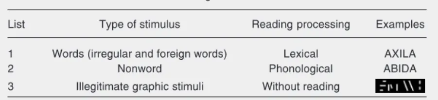
(non-readable graphic stimuli; see Table 1 for an example).
The first list (list 1) was composed of 50 real words, half of them consisting of irregu-lar words in Portuguese and the other half consisting of foreign words. The words ranged from four to nine letters in length and all were concrete nouns. The stimuli of this list containing irregular words and foreign words, should be read by lexical processing. The second list (list 2) consisted of 50 nonwords written in the alphabetic system and was elaborated from the stimuli of the first list. The consonants, but not the vowels, of each real word were modified, with nonwords with the same structure and syl-labic extension thus being obtained. For in-stance: starting from the irregular word axila,
the nonword abida was generated. The
read-ing of nonwords should be accomplished by phonological processing, that is, by graph-eme-phoneme conversion processing.
The third list (list 3) had 50 illegitimate graphic stimuli. These stimuli were not read-able because they were formed by pseudo-letters obtained through distortions of the letters. The illegitimate graphic stimuli were elaborated from the second list, with the lines of the letters being modified and served as controls for the stimuli of lists 1 and 2. Therefore, in the analysis of the images from fMRI, these illegitimate graphic stimuli were used as a baseline condition, containing ele-ments of visual and pre-linguistic processing. As described above, the silent reading task resulted from the combination of three different lists, each containing 50 stimuli, involving real words, nonwords and illegiti-mate graphic stimuli. The 50 stimuli of each
list were distributed into five blocks of 10 stimuli each. Therefore, the task was divided into 15 blocks that were randomly presented. To minimize possible effects from habitua-tion to the type of stimulus, each epoch contained 80% of the stimuli belonging to one condition and 10% of the stimuli be-longing to each of the remaining conditions.
Preparation of the stimuli for the reading task
The graphic stimuli written in the alpha-betic system were typed using the Microsoft Word program (Microsoft, Seattle, WA, USA), in capital letters of the Arial font type, size 90. All stimuli were transported from the Word to the Paint program where they appeared against a black background. The illegitimate graphic stimuli generated from the nonword letter distortions were elabo-rated in the Paint program using the “eraser” feature to fade part of the letters. Stimuli were presented as bitmap files to be used by the Visual Basic program version 4.0 (Mi-crosoft).
The duration of stimulus presentation and the interval between stimuli were 1000 ms. Stimulus presentation and image acquisition were synchronized via a TTL pulse (Zurc & Zurc DataSystems, São Paulo, SP, Brazil) and the stimuli were projected by a multime-dia projector. The subjects visualized the stimuli through a mirror (each letter size occupied4" degrees in the visual field) at-tached to the head coil, and were instructed to read them silently.
After the silent reading task during the acquisition of the images, in order to verify if the subjects had adhered to the experi-ment, they were asked if they remembered the stimuli they had read. To confirm the capacity of lexical and phonological read-ing, a post-test of word and nonword reading aloud was applied. All individuals had ad-hered to the experiment and were perfectly capable of reading through lexical and pho-nological processes.
Table 1. Stimuli used in the silent reading task.
List Type of stimulus Reading processing Examples
1 Words (irregular and foreign words) Lexical AXILA
2 Nonword Phonological ABIDA
Image acquisition and data processing
The images were obtained in a Horizon 1.5 Tesla magnet of the General Electric Medical Systems (Milwaukee, MN, USA), equipped with a 40 mT/m gradient, using the blood oxygen level-dependent (BOLD) con-trast technique with echo planar imaging. Fifteen slices were collected. The images were oriented on the anterior commissure-posterior commissure line, with 7-mm thick, 0.7-mm gap slices covering the whole brain. The time of repetition was 2000 ms, the time to echo was 40 ms, the field of view was 400 x 200 mm, with a 90º flip angle and a matrix of 128 x 64 voxels. A total of 2250 images were collected for each subject. Analysis was performed with a personal computer running Linux.
The data were first realigned (22) to mini-mize motion-related artifacts and smoothed with a Gaussian filter (full-width half-maxi-mum - 7.2 mm). Responses to the experi-mental paradigms were then detected by time-series analysis using gamma variate functions (peak responses at 4 and 8 s) to model the BOLD response. The analysis was set up as follows. First, each experimen-tal condition was convolved separately with the 4- and 8-s Poisson functions to yield two models of the expected hemodynamic re-sponse to that condition. The weighted sum of these two convolutions that gave the best fit to the time series at each voxel was then computed. This weighted sum effectively allows voxel-wise variability in time to peak hemodynamic response. Following this fit-ting operation, a ratio of the sum of squares of deviations from the mean intensity value due to the model (fitted time series) divided by the sum of squares due to the residuals (original time series minus model time se-ries) was calculated. This statistic is called the SSQratio. The percent change of the BOLD signal at each voxel was also calcu-lated. This was done by ((fitmax – fitmin)/ mean signal intensity) x 100, where fitmax
and fitmin were the maximum and minimum values of the fitted response for the time series in question. In order to sample the distribution of the SSQratio under the null hypothesis that observed SSQratio values were not determined by experimental design (with minimal assumptions), the time series at each voxel was permuted using a wavelet-based resampling method described in detail by Bullmore et al. (23). This process was repeated 10 times at each voxel and the data were combined over all voxels, resulting in 10 permuted parametric maps of the SSQratio on each plane for each participant. The same permutation strategy was applied to each voxel to preserve spatial correlational struc-ture in the data during randomization. Com-bining the randomized data over all voxels yields the distribution of the SSQratio under the null hypothesis. A test that any given voxel is activated at any required type I error can then be carried out by obtaining the appropriate critical value of the SSQratio from the null distribution. For example, SSQratio values in the observed data lying above the 99th percentile of the null distri-bution have a probability of ≤0.01 under the null hypothesis. We have shown that this permutation method gives very good type I error control with minimal distributional as-sumptions (23,24).
After these processes, two activation maps were created for each individual, one of them comparing real words to illegitimate graphic stimuli (A x C), and the other com-paring nonwords to illegitimate graphic stimuli (B x C). The objective of this com-parison was to study the word and nonword silent reading by eliminating (subtracting) the activations of the neural structures re-sponsible for the primary and pre-linguistic visual processing. Consequently, the A x C activation map represents the activation dur-ing the silent readdur-ing of words and the B x C map represents activation during the silent reading of nonwords.
slices (Figure 1). To identify the anatomical localization of the activated areas, each slice was analyzed visually, determining which areas were activated using as reference the Atlas of Duvernoy (25). The activated areas were identified independently using an Atlas by two examiners (MLHS and PC). The points of disagreement between the two analyses were discussed by the examiners, who then reached a consensus. Based on these data, two tables were elaborated, one for each comparison (A x C and B x C) for the areas activated in each subject. The num-ber of activated voxels was recorded in all active clusters resulting from the A x C and B x C comparisons.
Initially we determined the frequency of activation in different brain areas for each individual in each specific condition. This procedure provided an overview of all pos-sible areas detected in the subjects. Since not all areas were present in all subjects, the frequency of activation detected in each area provides a measure of the commonalities in the neural correlates of each task found in the participants. Areas displaying activation
in more than 50% of the individuals were judged to represent relevant activations. This approach has been used in past publications by others, and we believe it represents a compromise between subject variability and group-shared activation foci (26,27).
Results
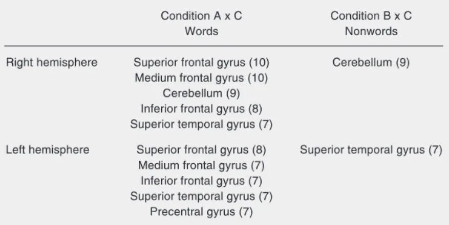
The comparison of the number of acti-vated voxels in all individuals in the condi-tions A x C (words x illegitimate graphic stimuli) and B x C (nonwords x illegitimate graphic stimuli) showed a larger number of voxels activated in the first condition, indi-cating a greater activation in word than in nonword reading. Furthermore, in both condi-tions the left cerebral hemisphere was more activated than the right hemisphere (Table 2). Table 3 shows the areas that were acti-vated in more than 50% of the individuals in the conditions A x C and B x C in the right and left hemispheres. A larger number of activated common areas can be observed in the silent reading of words than of nonwords in both hemispheres. In the first condition, i.e., word silent reading, a mirror correspon-dence of activations was observed when com-paring the left and right hemispheres. The following areas were activated: superior, middle and inferior frontal gyri and the su-perior temporal gyrus in both cerebral hemi-spheres. The cerebellum was activated in the right hemisphere and the precentral gyrus in the left. In the condition B x C, the right cerebellum and the left superior temporal gyrus were activated in more than 50% of the individuals.
Discussion
Differences in the cerebral activation patterns of lexical and phonological reading
The activation patterns detected in sub-jects during word and nonword reading were different. In the word reading task, we iden-Figure 1. Brodmann areas (BA) are shown in circles for two different analysis from the same
tified ten different activated areas in more than 50% of the individuals, five of them in the left hemisphere and five in the right. In the reading of nonwords, only two activated areas were found in more than 50% of the individuals. During word reading, a similar pattern of activation was observed in the left and right hemispheres. In other words, of the five areas activated in each hemisphere, four were activated bilaterally: superior, middle and inferior frontal gyri, and superior tem-poral gyrus. In addition to these areas, the right cerebellum and the left pre-central gy-rus were also activated. In the reading of nonwords, only the right cerebellum and the left superior temporal gyrus were activated. The mirror pattern of activation found in word reading was not observed in nonword reading. The areas activated during the read-ing of nonwords, the right cerebellum and the left superior temporal gyrus, were also activated during the reading of words.
Petersen et al. (11) and Pugh et al. (20) have also reported different patterns of cere-bral activation when comparing the mechan-isms involved in word and nonword reading. Petersen et al. (11), in a study with PET, observed that silent reading of words acti-vated the left frontal cortex. The medial extrastriate visual cortex in the left hemi-sphere was activated during the silent read-ing of both words and nonwords. In the study of Pugh et al. (20), using fMRI, 38 individuals completed a judgment task of phonological processing (nonword rhyme judgment) and lexical-semantic processing (semantic category judgment). The research-ers observed activation during phonological processing in the inferior frontal gyrus and in the temporal lobe (with lateralization of the activation of the inferior frontal gyrus to the left in men, and bilateral activation in women) and activation of the superior and middle temporal gyri in both hemispheres during the lexical-semantic processing. In contrast, in a PET study on 14 right-handed men, Rumsey et al. (14) did not find
differ-ences in activation during tasks of lexical and phonological reading. The reading of irregular words as well as of nonwords acti-vated the following areas: left lingual and left fusiform gyri, cerebellum, superior tem-poral gyrus bilaterally, left thalamus, peri-Rolandic cortex (pre- and post-central gyri) bilaterally, anterior/mid cingulate gyrus on the left, and left insula. Price et al. (13), in another study with PET and reading of real words, found activation during silent read-ing in the followread-ing areas: medial extrastri-ate cortex on the left, middle temporal cortex on the left, middle frontal cortex on the left, and middle temporal cortex and inferior fron-tal cortex on the right.
The comparison of our data with those reported by other researchers should be made with caution. One of the reasons for this is related to the use of different tasks to probe
Table 2. Number of voxels activated in the word and nonword silent reading.
Condition A x C Condition B x C
Words Nonwords
Right cerebral hemisphere 794 voxels 446 voxels
Left cerebral hemisphere 874 voxels 526 voxels
Condition A = list 1; condition B = list 2; condition C = list 3. See Table 1 for explanation of lists.
Table 3. Areas activated in more than 50% of the individuals during the silent word and nonword reading in the right and left hemispheres.
Condition A x C Condition B x C
Words Nonwords
Right hemisphere Superior frontal gyrus (10) Cerebellum (9)
Medium frontal gyrus (10) Cerebellum (9) Inferior frontal gyrus (8) Superior temporal gyrus (7)
Left hemisphere Superior frontal gyrus (8) Superior temporal gyrus (7) Medium frontal gyrus (7)
Inferior frontal gyrus (7) Superior temporal gyrus (7)
Precentral gyrus (7)
the same function. In the investigations men-tioned above, the activation during reading was confirmed through similar but not iden-tical tasks, i.e., silent reading, reading aloud, lexical decision, and judgment. Probably, the execution of these tasks involves similar and different mechanisms. In this way, the cerebral areas activated during the perfor-mance of these tasks should be different. Even the sensitivity of the paradigm can influence the data. Price et al. (13) varied the duration of stimulus presentation (150 and 981 ms) in tasks of silent reading, reading aloud and lexical decisions and showed that the time of exhibition, significantly influ-enced the correlation between blood flow and word reading. The divergence of the results obtained in the different studies is an important issue to be considered in studies that involve functional neuroimaging and mental functions and should be addressed by additional studies in the field.
Right cerebral hemisphere and word reading
An interesting aspect of the results ob-tained in the present study is the mirror correspondence of the areas that were acti-vated. This pattern was only observed for the silent reading by the lexical route, that is, for the processing of irregular and foreign words. From these findings the following question could be raised: does the right cerebral hemi-sphere play a role in lexical reading?
In reality this issue has already been dis-cussed in the literature for some years (28-30). Some researchers defend the “right hemi-sphere hypothesis”, which proposes that the right cerebral hemisphere plays a role in the orthographic and lexical processes of read-ing, permitting the processing of high imaginability and high frequency written words. This hypothesis is supported by case studies of left hemispherectomy, split-brain cases and patients with deep dyslexia and pure alexia.
Patterson et al. (31) described the case of
patient NI, who, after a normal childhood, developed refractory epilepsy, needing the surgical removal of the left cerebral hemi-sphere. After the hemispherectomy, NI pre-sented reading residual abilities characteris-tic of deep dyslexia. She was capable of reading some concrete and high frequency words, making some semantic mistakes. Zaidel and Peters (32) described two split-brain patients who were able to associate with objects the written word presented to the right hemisphere. Coslett and Saffran (33) reported four patients with pure alexia whose performance in tasks of lexical deci-sion and semantic categorization was con-sistent with the right hemisphere hypothesis. Some investigators relate the residual read-ing abilities of patients with deep dyslexia to the orthographic and semantic aspects of reading by the right hemisphere. In this con-dition, the patients present an extensive le-sion of the left hemisphere and are able to read some frequent words through lexical processing, which would be an indication that in these patients with deep dyslexia reading is being accomplished by the right hemisphere.
Coslett and Monsul (29) conducted a study using transcranial magnetic stimulation (TMS) in a case of pure alexia (JG) to test the “right hemisphere hypothesis”. The authors compared the reading performance of JG in three different situations: without TMS, with TMS on the right parietal lobe and with TMS in the same area on the left. Having as a base the performance in reading without TMS, the authors compared the results of the influ-ence of TMS on the right and the left parietal lobe and verified that the reading perfor-mance of JG was impaired only when the right cerebral hemisphere was stimulated, supporting the idea that the residual reading of that patient was due to a linguistic ability of the non-dominant hemisphere.
dur-ing real word readdur-ing support this idea that the right cerebral hemisphere has a role in lexical reading.
The role of the cerebellum
In this study, the right cerebellar hemi-sphere was activated in both the word and nonword reading tasks in more than half the subjects. These data suggest the participa-tion of the cerebellum in reading, corrobo-rating the findings from the study of Fulbright et al. (34). The contribution of the cerebel-lum to cognitive processing was discussed by these researchers, who studied the cer-ebellar activation by fMRI during reading and concluded that the cerebellum is en-gaged not only in the ability of phonologic assembly but also in semantic processing.
Limitations and perspectives
As in other studies that use fMRI, one of the limitations of the present study was re-lated to the small number of subjects evalu-ated. We tried to minimize this problem by selecting right-handed female volunteers with a high educational level to obtain a more homogeneous sample.
Another limitation refers to individual differences in the performance of cognitive tasks. The acquisition of reading, differently from the acquisition of the oral language, is not an innate ability of humans. Since read-ing is a culturally biased function it might suffer a larger inter-individual variability than the other innate and primary cognitive abilities. In the present study, we chose the silent reading task to determine the pattern of cerebral activation because of our interest in examining the complete process of read-ing from the input of visual information to phonological codification, without the re-quirement of speech articulation that could provoke the appearance of artifacts in the fMRI images. Some studies of reading use other tasks, such as lexical decision and
semantic categorization of written words, that test some isolated mechanisms involved in reading. Leff et al. (35) consider silent reading to be a natural function, different from tasks of lexical decision or of choice, that imply the activation of other associated mental processes difficult to control for each individual. If, on the one hand, it seems to have advantages over the choice of the silent reading task, it also has disadvantages. When using the silent reading task during image acquisition by fMRI we cannot be certain of the volunteer’s collaboration at the moment of scanning. In the present study, to confirm that the silent reading task was actually per-formed, soon after the experiment, individu-als had to mention some stimuli that had been presented.
Some studies have investigated cerebral activation using a limited number of slices, without a global vision of the brain. In our study, the 15 slices covered the whole brain. This type of technical approach became avail-able recently. It facilitates not only the obser-vation of the actiobser-vation of areas of interest, but also the inspection of all the areas of the brain. In spite of the limitations and controver-sies generated from the cerebral activation maps related to the cognitive abilities, stud-ies with the use of fMRI contribute and will continue to contribute to the understanding of the cognitive and neural mechanisms re-lated to the mental functions. fMRI, together with other experimental approaches using advanced techniques like PET and TMS, as well as lesion studies, should provide rel-evant information about reading processes.
Acknowledgments
We wish to thank Antônio Cesário Mon-teiro da Cruz Junior for providing the
elec-tronic instruments necessary for synchro-nizing stimulus presentation and data acqui-sition, and for the network configuration.
References
1. Hécaen H & Albert ML (1977). Human Neuropsychology. Wiley-Interscience, New York.
2. Black SE & Behrmann M (1994). Localization in alexia. In: Kertesz A (Editor), Localization and Neuroimaging in Neuropsychology. Aca-demic Press, San Diego, CA, USA, 331-376.
3. Lecours AR, Delgado AP & Pimenta MAM (1993). Distúrbios adquiridos da leitura e da escrita. In: Mansur LL & Rodrigues N (Editors), Temas em Neurolingüística. Tec Art, São Paulo, SP, Brazil, 45-62.
4. McCarthy RA & Warrington EK (1990). Cognitive Neuropsychology:
A Clinical Introduction. Academic Press, San Diego, CA, USA.
5. Lesser R & Milroy L (1993). Linguistics and Aphasia:
Psycholinguis-tic and PragmaPsycholinguis-tic Aspects of Intervention. Longman, New York,
52-80.
6. Ellis AW & Young AW (1988). Human Cognitive Neuropsychology. Lawrence Erlbaum, London, UK.
7. Price CJ (2000). Functional imaging studies of aphasia. In: Mazziotta JC, Toga AW & Frackowiak RS (Editors), Brain Mapping: the Disor-ders. Academic Press, San Diego, CA, USA, 181-200.
8. Basso A (2000). The aphasias: fall and renaissance of the neuro-logical model? Brain and Language, 71: 15-17.
9. Marshall JC & Newcombe M (1973). Patterns of paralexia: a psycholinguistic approach. Journal of Psycholinguistic Research, 2: 175-200.
10. Caramelli P, Parente MAMP, Hosogi ML et al. (1994). Unexpected reading dissociation in a Brazilian “nisei” with crossed aphasia.
Behavioural Neurology, 7: 165-170.
11. Petersen SE, Fox PT, Snyder AZ et al. (1990). Activation of extra-striate and frontal cortical areas by visual words and word like stimuli. Science, 249: 1041-1044.
12. Small SL, Flores DK & Noll DC (1998). Different neural circuits subserve reading before and after therapy for acquired dyslexia.
Brain and Language, 62: 298-308.
13. Price CJ, Wise RJ, Watson DG et al. (1994). Brain activity during reading. The effects of exposure duration and task. Brain, 117: 1255-1269.
14. Rumsey JM, Horwitz B, Donohue BC et al. (1997). Phonological and ortographic components of word recognition: a PET-rCBF study.
Brain, 120: 739-759.
15. Henson RNA, Burgess N & Frith CD (2000). Recoding, storage, rehearsal and grouping in verbal short-term memory: an fMRI study.
Neuropsychologia, 38: 426-440.
16. Price CJ, Gorno-Tempini ML, Graham KS et al. (2003). Normal and pathological reading: converging data from lesion and imaging stud-ies. Neuroimage, 20: S30-S41.
17. Coltheart M, Patterson KE & Marshall JC (Editors) (1980). Deep
Dyslexia. Routledge, Londres, UK.
18. Coltheart M, Masterson J, Byng S et al. (1983). Surface dyslexia.
Quarterly Journal of Experimental Psychology, 35A: 469-495.
19. Lecours AR & Parente MAMP (1997). Dislexia: Implicações do
Sistema de Escrita do Português. Artes Médicas, Porto Alegre, RS,
Brazil.
20. Pugh KR, Shaywitz BA, Shaywitz SE et al. (1996). Cerebral organi-zation of component process in reading. Brain, 119: 1221-1238. 21. Oldfield RC (1971). The assessment and analysis of handedness:
the Edinburgh Inventory. Neuropsychologia, 9: 97-113.
22. Bullmore E, Brammer MJ, Rabe-Hesketh S et al. (1999). Methods for diagnosis and treatment of stimulus-correlated motion in generic brain activation studies using fMRI.Human Brain Mapping, 7: 38-48. 23. Bullmore E, Long C, Suckling J et al. (2001). Colored noise and computational inference in neurophysiological (fMRI) time series analysis: resampling methods in time and wavelet domains.Human
Brain Mapping, 12: 61-78.
24. Bullmore E, Fadili J, Breakspear M et al. (2003). Wavelets and statistical analysis of functional magnetic resonance images of the human brain.Statistical Methods in Medical Research, 12: 375-399. 25. Duvernoy HM (1991). The Human Brain-Surface,
Three-Dimen-sional Sectional Anatomy and MRI. Springer-Verlag, Vienna,
Aus-tria.
26. Uchida I, Kikyo H, Nakajima K et al. (1999). Activation of lateral extra-striate areas during orthographic processing of Japanese char-acters studied with fMRI. Neuroimage, 9: 208-215.
27. Wood AG, Harvey AS, Wellard RM et al. (2004). Language cortex activation in normal children. Neurology, 63: 1035-1044.
28. Coltheart M (2000). Deep dyslexia is right-hemisphere reading.
Brain and Language, 71: 299-309.
29. Coslett HB & Monsul N (1994). Reading with the right hemisphere: evidence from transcranial magnetic stimulation. Brain and
Lan-guage, 46: 198-211.
30. Ellis AW (1995). Leitura, Escrita e Dislexia: Uma Análise Cognitiva. Artes Médicas, Porto Alegre, RS, Brazil.
31. Patterson K, Vargha-Khadem F & Polkey C (1988). Reading with one hemisphere. Brain, 112: 39-63.
32. Zaidel E & Peters AM (1981). Phonological encoding and ideo-graphic reading by the disconnected right hemisphere: two case studies. Brain and Language, 14: 205-234.
33. Coslett HB & Saffran EM (1989). Evidence for preserved reading in “pure alexia”. Brain, 113: 327-329.
34. Fulbright RK, Jenner AR, Mencl WE et al. (1999). The cerebellum’s role in reading: a functional MR imaging study. American Journal of
Neuroradiology, 20: 1925-1930.
35. Leff AP, Crewes H, Plant GT et al. (2001). The functional anatomy of single-word reading in patients with hemianopic and pure alexia.