Effects of sedation during upper
gastrointestinal endoscopy on
endocrine response and
cardiorespiratory function
1Department of Surgery and Surgical Endoscopy Unit, 2Department of Anesthesiology, 3Department of Biochemistry,
Ôi
Õli Etfal Training and Research Hospital, Istanbul, Turkey G. Yetkin1, S. Oba2,M. Uludag1, I. Paksoy2,
I. Akgün1 and N. Eren3
Abstract
Upper gastrointestinal endoscopy is often accompanied by tachycar-dia which is known to be an important pathogenic factor in the development of myocardial ischemia. The pathogenesis of tachycar-dia is unknown but the condition is thought to be due to the endocrine response to endoscopy. The purpose of the present study was to investigate the effects of sedation on the endocrine response and cardiorespiratory function. Forty patients scheduled for diagnostic upper gastrointestinal endoscopy were randomized into 2 groups. While the patients in the first group did not receive sedation during upper gastrointestinal endoscopy, the patients in the second group were sedated with intravenous midazolam at the dose of 5 mg for those under 65 years or 2.5 mg for those aged 65 years or more. Midazolam was administered by slow infusion.In both groups, blood pressure, ECG tracing, heart rate, and peripheral oxygen saturation (SpO2) were
monitored during endoscopy. In addition, blood samples for the determination of cortisol, glucose and C-reactive protein levels were obtained from patients in both groups prior to and following endos-copy. Heart rate and systolic arterial pressure changes were within normal limits in both groups. Comparison of the two groups regarding the values of these two parameters did not reveal a significant differ-ence, while a statistically significant reduction in SpO2 was found in
the sedation group. No significant differences in serum cortisol, glucose or C-reactive protein levels were observed between the se-dated and non-sese-dated group. Sedation with midazolam did not reduce the endocrine response and the tachycardia developing during upper gastrointestinal endoscopy, but increased the reduction in SpO2.
Correspondence G. Yetkin
Ataköy 3. kisim O/8 blok. Daire 7 Bakirköy
Istanbul 34158 Turkey
E-mail: [email protected] GSM: 0 532 613 94 71
Received January 31, 2007 Accepted July 31, 2007
Key words
•Stress response •Oxygen saturation •Sedation
•Upper gastrointestinal endoscopy
•Tachycardia
Introduction
The value of diagnostic and therapeutic endoscopic applications in gastrointestinal diseases is increasing progressively.
tachy-cardia during upper gastrointestinal endos-copy (1-3).
Although the use of sedation during up-per gastrointestinal endoscopy is prevalent (4,5), there is much debate about the admin-istration of sedatives. While some investiga-tors propose that sedation-related complica-tions may develop (6,7), many have reported that conscious sedation increases comfort during upper gastrointestinal endoscopy and reduces cardiac stress (8,9).
In the present study, we investigated the effects of sedation on the hemodynamic pa-rameters and classical endocrine stress re-sponse that may cause tachycardia during routine upper gastrointestinal endoscopy.
Patients and Methods
Forty volunteer patients with ASA scores of 1 or 2, who were referred to the endos-copy laboratory for diagnostic upper gas-trointestinal endoscopy were included in the study, which was approved by the local Eth-ics Committee. The patients gave written informed consent to participate and were randomized to two groups by means of sealed envelopes.
The two groups of patients of 20 each did not receive premedication prior to
gastros-copy. Lidocaine spray (Xylocaine® pump
spray, Astra Zeneca, Lund, Sweden) was used for topical pharyngeal anesthesia. None of the patients received supplementary
oxy-gen during gastroscopy. All patients were investigated by the same endoscopist (G.Y.) using the same endoscope (Olympus GIF XQ 230 videoendoscope, 9.8 mm in diam-eter, Olympus Optical Co. Ltd., Tokyo, Ja-pan). The patients in group 1 did not receive sedation, whereas the patients in group 2 were sedated with intravenous midazolam at the dose of 2.5 mg for subjects aged 65 years or older, or at the dose of 5 mg for subjects younger than 65 years. Midazolam was ad-ministered by slow infusion into the antecu-bital vein at a single dose.
Continuous monitoring of blood pres-sure, ECG tracing, heart rate, and peripheral oxygen saturation (SpO2) was performed using a Nikon-Kohden BSM 4113-K (To-kyo, Japan) standard monitor in both groups. Values were recorded 10 min prior to, dur-ing, and 20 min following the endoscopic examination. Also, blood samples were ob-tained from both groups prior to and 10 min and 1 h after endoscopy for the determina-tion of cortisol, glucose, and C-reactive pro-tein (CRP).
Data were analyzed statistically by the Mann-Whitney U-test for comparisons be-tween groups and by the Tukey test for comparisons within groups, with the level of significance set at P < 0.05.
Results
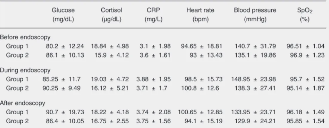
All 40 patients tolerated and successfully completed the procedure. The mean dura-tion of endoscopy was 6.54 min (range: 4.57-10.38 min) for group 1 (non-sedated) and 6.85 min (range: 4.38-9.9 min) for group 2 (sedated), with no significant difference between groups (P = 0.4382). Mean age and sex distribution were similar in both groups (Table 1). The effects of sedation on the endocrine response and cardiorespiratory function prior to, during and following en-doscopy are shown in Table 2.
The values of heart rate, systolic blood pressure, and SpO2 obtained for both groups Table 1. Physical characteristics of the patients
and duration of endoscopy.
Group 1 Group 2 (N = 20) (N = 20)
Age (years) 43.85 46.35
Sex ratio (male:female) 11:9 12:8 Mean endoscopy time (min) 6.54 7.25
Figure 1. Changes in cardiopulmonary parameters (heart rate (A), systolic blood pressure (B), and peripheral oxygen saturation (SpO2, C) detected
before (1), during (2), and after (3) endoscopy. Circles = non-sedated group; triangles = sedated group.
Table 2. Effects of sedation on the endocrine response and cardiorespiratory function before, during and after endoscopy.
Glucose Cortisol CRP Heart rate Blood pressure SpO2
(mg/dL) (µg/dL) (mg/L) (bpm) (mmHg) (%)
Before endoscopy
Group 1 80.2 ± 12.24 18.84 ± 4.98 3.1 ± 1.98 94.65 ± 18.81 140.7 ± 31.79 96.51 ± 1.04 Group 2 86.1 ± 10.13 15.9 ± 4.12 3.6 ± 1.61 93 ± 13.43 135.1 ± 19.86 96.9 ± 1.23
During endoscopy
Group 1 85.25 ± 11.7 19.03 ± 4.72 3.88 ± 1.95 98.5 ± 15.73 148.95 ± 23.98 95.7 ± 1.52 Group 2 90.25 ± 9.49 16.12 ± 5.21 3.71 ± 1.7 100.8 ± 12.6 138.3 ± 27.41 95.14 ± 1.87
After endoscopy
Group 1 90.7 ± 19.73 18.22 ± 4.18 3.74 ± 2.08 100.65 ± 12.85 133.95 ± 23.71 96.18 ± 1.49 Group 2 86.4 ± 10.05 16.75 ± 2.55 3.75 ± 1.56 94.1 ± 15.19 129.9 ± 24.21 95.85 ± 1.54
Data are reported as mean ± SD for 20 patients in each group. See Table 1 for definition of groups. CRP = C-reactive protein; SpO2 = peripheral oxygen saturation. There were no statistical differences between groups (Mann-Whitney
U-test).
are displayed in Figure 1A,B and C, respec-tively. Heart rate and systolic arterial pres-sure increased in both groups during the endoscopic procedure, but arrhythmias and signs of myocardial ischemia were not ob-served in any of the patients. The increases in heart rate during endoscopy were nonsig-nificant in both the non-sedated group and the sedated group. In addition, no statisti-cally significant difference was found be-tween groups regarding the simultaneous values of heart rate both during and after endoscopy. Similar results were also ob-tained in the statistical analysis of the alter-ations in systolic arterial pressure. The in-crements in systolic arterial pressure ob-served during endoscopy were nonsignifi-cant in both group 1 and group 2.
Compari-son of the simultaneous values of systolic blood pressure between groups also revealed no significant difference either during or following endoscopy.
SpO2 levels decreased during endoscopy in both groups, but while this reduction was nonsignificant in the group without seda-tion, a statistically significant reduction was found in the sedation group (P = 0.002). In
both groups, SpO2 levels increased to
pre-endoscopy values following pre-endoscopy, and comparison of the pre- and post-endoscopy SpO2 levels revealed no statistically signifi-cant difference in either group. Also, no statistically significant difference was found between groups regarding the simultaneous
SpO2 levels during and following
Serum cortisol, glucose and CRP levels increased following endoscopy in both groups; however, none of the increments in these parameters was statistically signifi-cant. Also, comparison of the groups regard-ing the simultaneous levels of these param-eters revealed no statistically significant dif-ference (Figure 2A-C).
Discussion
An organism undergoing trauma initiates a systemic response against the trauma in order to maintain homeostasis. Secretion of the mediators, and the intracellular and in-tercellular metabolic alterations that initiate the neuroendocrine response developing af-ter trauma depend on the duration and type of trauma (10,11). Regardless of its dura-tion, gastroscopic examination is also a trauma which elicits a metabolic endocrine response in the body. The temporary endo-crine response occurring in the body during gastroscopic examination resembles the well-described classical stress response develop-ing against surgery (12). Afferent vagal, sym-pathetic and phrenic mechanoreceptors ex-ist in the esophagus and stomach. It has been proposed that mechanical autonomic stimu-lation of these organs during gastroscopy may initiate the endocrine response devel-oping during endoscopy. The symptoms of intolerance to endoscopy such as hypoxia, pain and coughing that develop during the
endoscopic procedure may cause a stronger response. The endocrine hormones that are secreted may play a role in the development of tachycardia which emerges during endo-scopy, by affecting the target organs includ-ing the cardiovascular system (13,14).
Tachycardia is known to be an important pathogenic factor in the development of myocardial ischemia that may occur during endoscopy (15). Therefore, it has been sug-gested that ß-adrenergic blockade of the heart, which is one of the target organs of the endocrine stress, might prevent the develop-ment of myocardial ischemia, as observed in a study in which ß-adrenergic blockade in-duced by metoprolol was shown to reduce the incidences of tachycardia and myocar-dial ischemia during endoscopy (16). Simi-lar to the target organ blockade seen here, reduction of afferent stimulation has been proposed to be effective in reducing the stress response emerging during upper gas-trointestinal endoscopy (12). Particularly, upper gastrointestinal endoscopy performed with induction of sedation has been reported to be more comfortable than endoscopy with-out sedation, and sedation has been pro-posed to reduce the stress response (8). Thus, in the present study, we investigated whether sedation reduces the stress response and tachycardia occurring in upper gastrointesti-nal endoscopy.
In several studies, the classical metabolic endocrine response has been obtained with
upper gastrointestinal endoscopy (12,17). In the present study, serum cortisol, glucose and CRP levels increased during endoscopy, although the increments were not statisti-cally significant. In addition, comparison of the groups with and without sedation re-vealed that the increments in serum cortisol, glucose and CRP levels were similar in both groups and that the values of the parameters obtained prior to and 10 min and 1 h after endoscopy were not statistically different between groups. These results agree with those reported by Yazawa et al. (18), and suggest the view that sedation is not effec-tive in reducing the endocrine response oc-curring during upper gastrointestinal endos-copy.
Hypoxia developing during upper gas-trointestinal endoscopy is a well-recognized complication; however, its cause has not been well clarified. Sedation might prima-rily cause hypoxia by inducing respiratory depression, while the presence of an endo-scope may also cause hypoxia by narrowing the upper airway (19). In a study conducted on 330 patients, Banks et al. (20) demon-strated that the arterial oxygen pressure was considerably decreased in patients with se-dation compared to those without, while Wang et al. (19) observed hypoxia in both patients with and without sedation, but stressed that sedation significantly increased hypoxia. Yazawa et al. (18) found a reduc-tion in arterial oxygen pressure in the group with sedation compared to that without, but the reduction was not statistically signifi-cant. In another study, Kýlýc et al. (21) reported that sedation with midazolam does not have unfavorable effects on arterial oxy-gen saturation, but might show unfavorable effects in patients who smoke or patients who have pulmonary problems. In the pres-ent study, the patipres-ents in both groups were healthy individuals with ASA scores of 1 or 2. Unlike Kýlýc et al. (21), we found signifi-cant reductions in SpO2 levels in the sedated group, which was composed of healthy
indi-viduals. We think that it would be important to study groups of healthy individuals with ASA scores of 1 or 2 to explore the effects of sedation on cardiopulmonary function while performing upper gastrointestinal endoscopy so the effect of pulmonary diseases on the results of the study was prevented.
Arterial oxygen pressure was reduced in both the groups with and without sedation during endoscopy, with the reduction being statistically significant in the group with sedation, but not in the non-sedated group. However, comparison of the simultaneous arterial oxygen pressures in the sedated and non-sedated groups revealed no statistically significant difference. This situation sup-ports the view that both gastrointestinal en-doscopy and sedation might be effective in reducing SpO2 levels.
Ristikankare et al. (22) pointed out that tachycardia plays a more important role than hypoxia in the pathogenesis of cardiorespi-ratory complications that may develop dur-ing upper gastrointestinal endoscopy, and sedation with midazolam causes reduced SpO2 levels, but also prevents the increase in systolic blood pressure and tachycardia ob-served during endoscopy. Our findings are similar to those of Ristikankare et al. (22) in
that we found reductions in SpO2 levels in
the group sedated with midazolam. How-ever, we did not observe the favorable influ-ences of sedation on systolic blood pressure and tachycardia observed by these investi-gators. The absence of a favorable effect of sedation on these two parameters suggests that, because these parameters increase as a result of the endocrine stress response to the endoscope, and because sedation is ineffec-tive against the stress response mediators, it is also ineffective against tachycardia and increased systolic blood pressure.
References
1. Hart R, Classen M. Complications of diagnostic gastrointestinal endoscopy. Endoscopy 1990; 22: 229-233.
2. Schenck J, Muller CH, Lubbers H, Mahlke R, Lehnick D, Lankisch PG. Does gastroscopy induce myocardial ischemia in patients with coronary heart disease? Endoscopy 2000; 32: 373-376.
3. Wilcox CM, Faibicher M, Wenger NK, Shalek KA. Prevalence of silent myocardial ischemia and arrhythmias in patients with coro-nary heart disease undergoing gastrointestinal tract endoscopic procedures. Arch Intern Med 1993; 153: 2325-2330.
4. Cohen LB, Wecsler JS, Gaetano JN, Benson AA, Miller KM, Durkalski V, et al. Endoscopic sedation in the United States: results from a nationwide survey. Am J Gastroenterol 2006; 101: 967-974. 5. Lazzaroni M, Bianchi PG. Preparation, premedication, and
surveil-lance. Endoscopy 2005; 37: 101-109.
6. Leslie K, Stonell CA. Anaesthesia and sedation for gastrointestinal endoscopy. Curr Opin Anaesthesiol 2005; 18: 431-436.
7. Chak A, Rothstein RI. Sedationless upper endoscopy. Rev Gastro-enterol Disord 2006; 6: 13-21.
8. Froehlich F, Schwizer W, Thorens J, Kohler M, Gonvers JJ, Fried M. Conscious sedation for gastroscopy: patient tolerance and cardio-respiratory parameters. Gastroenterology 1995; 108: 697-704. 9. Abraham NS, Fallone CA, Mayrand S, Huang J, Wieczorek P,
Barkun AN. Sedation versus no sedation in the performance of diagnostic upper gastrointestinal endoscopy: a Canadian random-ized controlled cost-outcome study. Am J Gastroenterol 2004; 99: 1692-1699.
10. Kohl BA, Deutschman CS. The inflammatory response to surgery and trauma. Curr Opin Crit Care 2006; 12: 325-332.
11. Kehlet H. Manipulation of the metabolic response in clinical practice.
World J Surg 2000; 24: 690-695.
12. Tonnesen H, Puggaard L, Braagaard J, Ovesen H, Rasmussen V, Rosenberg J. Stress response to endoscopy. Scand J Gastroenter-ol 1999; 34: 629-631.
13. Lynn RB. Mechanisms of esophageal pain. Am J Med 1992; 92: 11S-19S.
14. Adachi W, Yazawa K, Owa M, Koide N, Hanazaki K, Kajikawa S, et al. Quantification of cardiac stress during EGD without sedation.
Gastrointest Endosc 2002; 55: 58-64.
15. Holm C, Rosenberg J. Pulse oximetry and supplemental oxygen during gastrointestinal endoscopy: a critical review. Endoscopy
1996; 28: 703-711.
16. Rosenberg J, Overgaard H, Andersen M, Rasmussen V, Schulze S. Double blind randomised controlled trial of effect of metoprolol on myocardial ischaemia during endoscopic cholangiopancreatogra-phy. BMJ 1996; 313: 258-261.
17. Oei-Lim VL, Kalkman CJ, Bartelsman JF, Res JC, van Wezel HB. Cardiovascular responses, arterial oxygen saturation and plasma catecholamine concentration during upper gastrointestinal endos-copy using conscious sedation with midazolam or propofol. Eur J Anaesthesiol 1998; 15: 535-543.
18. Yazawa K, Adachi W, Owa M, Koide N, Hanazaki K, Kajikawa S, et al. Can sedation reduce the cardiac stress during gastrointestinal endoscopy? A study with non-invasive automated cardiac flow measurement by color Doppler echocardiography. Scand J Gastro-enterol 2002; 37: 602-607.
19. Wang CY, Ling LC, Cardosa MS, Wong AK, Wong NW. Hypoxia during upper gastrointestinal endoscopy with and without sedation and the effect of pre-oxygenation on oxygen saturation. Anaesthe-sia 2000; 55: 654-658.
20. Banks MR, Kumar PJ, Mulcahy HE. Pulse oximetry saturation levels during routine unsedated diagnostic upper gastrointestinal endos-copy. Scand J Gastroenterol 2001; 36: 105-109.
21. Kýlýc M, Bayan K, Yýlmaz S, Tüzün Y, Dursun M, Canoruc F. Changes in pulse oximetry levels and factors affecting oxygen satu-ration during upper gastrointestinal endoscopy with or without seda-tion. Turk J Gastroenterol 2006; 17: 279-282.
22. Ristikankare M, Julkunen R, Heikkinen M, Mattila M, Laitinen T, Wang SX, et al. Sedation, topical pharyngeal anesthesia and cardio-respiratory safety during gastroscopy. J Clin Gastroenterol 2006; 40: 899-905.
In conclusion, we propose that sedation performed with midazolam does not reduce the endocrine stress developing against