Postprandial symptoms in dysmotility-like
functional dyspepsia are not related to
disturbances of gastric myoelectrical activity
Departamentos de 1Clínica Médica, Faculdade de Medicina de Ribeirão Preto,
and 2Física e Matemática, Faculdade de Filosofia, Ciências e Letras de Ribeirão Preto,
Universidade de São Paulo, Ribeirão Preto, SP, Brasil A.S. Oba-Kuniyoshi1,
J.A. Oliveira Jr.1,
E.R. Moraes2
and L.E.A. Troncon1
Abstract
Gastric dysrhythmias, such as tachy- or bradygastria, have been re-ported in patients with functional dyspepsia (FD), but their role in symptom production is uncertain. It is also not known whether gastric dysrhythmias in these patients can be elicited by physiological gastric distension with a meal. We investigated the relationships between symptoms after ingestion of different volumes of water following a test meal and gastric dysrhythmias in FD patients. Fourteen patients with dysmotility-like FD and 13 healthy volunteers underwent paired electrogastrography (EGG) studies. Fasted subjects ingested 150 ml of yoghurt with either 150 ml (low volume) or 300 ml (high volume) water in random order. Fasting and fed EGGs with monitoring of symptoms were performed in both studies. Ten FD patients (71.4%) reported upper abdominal discomfort and bloating after the low volume meal, but only one (7.1%) presented an abnormal EGG (dominant frequency in the 2-4-cpm range: 58%). Following the high volume meal, 7 patients (50%) had symptoms, but none had EGG abnormalities. No significant differences were found between FD patients and controls for any of the EGG variables, in any test. In FD patients with postprandial symptoms, the percentage of the EGG dominant frequency in the normal range (median, 84.6%; range, 76.0-100.0%) was similar (P > 0.20) to that in those without symptoms (88.5%; 75.0-100.0%). We conclude that disturbances of gastric myoelectrical activity are unlikely to play a role in the origin of postprandial upper abdominal discomfort and bloating in dysmotility-like FD.
Correspondence L.E.A. Troncon
Departamento de Clínica Médica Hospital das Clínicas, FMRP, USP 14048-900 Ribeirão Preto, SP Brasil
Fax: +55-16-633-6695 E-mail: ledatron@fmrp.usp.br
The present address of E.R. Moraes is Instituto de Pesquisa e Desenvolvimento, Universidade do Vale do Paraíba, Av. Shishima Hifumi, 2911, 12244-000 São José do Campos, SP, Brasil.
Research supported by PRONEX/ FINEP/CNPq and FAEPA/HCFMRP.
Publication supported by FAPESP.
Received March 17, 2003 Accepted October 30, 2003
Key words
•Functional dyspepsia •Gastric myoelectrical
activity
•Electrogastrography •Postprandial symptoms
Introduction
Functional dyspepsia is a common clini-cal condition defined by pain or discomfort centered in the upper abdomen, which is not explained by any identifiable structural or biochemical abnormality (1,2). A number of studies have shown that patients with this condition may have disordered gastric
motil-ity (3-11), but the relationships between these gastrointestinal motor abnormalities and symptoms in functional dyspepsia are poorly understood.
cycles per minute (12). Abnormally high (“tachygastria”) or low (“bradygastria”) fre-quencies of gastric electrical oscillations have been detected by surface electrode recording (electrogastrography, EGG) in a variety of clinical conditions, usually in association with antral hypomotility and delayed gastric emptying (12), such as in idiopathic (13) and diabetic (14) gastroparesis. Disturbances of gastric myoelectrical activity have also been described in patients with functional dys-pepsia presenting symptoms suggestive of motility disorders of the upper gastrointesti-nal tract (3-5,15-17). However, the origin of these disturbances and the role of gastric dysrhythmias in symptom production in func-tional dyspepsia are unclear.
In a previous study, we demonstrated that graded distension of the gastric fundus with a plastic bag elicited greater instability in gastric myoelectrical rhythm in patients with functional dyspepsia (18). We there-fore determined whether physiological in-gestion of increasing volumes of water fol-lowing a standard test meal would induce disturbances of gastric myoelectrical activ-ity in functional dyspepsia patients, which could be related to symptom production.
Patients and Methods
Fourteen patients with dysmotility-like functional dyspepsia and 13 healthy volun-teers participated in the study after giving informed consent. The protocol for the study was approved by the Ethics Committee (State-ment number 7244/98) of our University Hospital.
Patients
The group of patients with functional dys-pepsia consisted of 9 women and 5 men with a median age of 44 years (range: 20-54 years). Body weight ranged from 48 to 85 kg (median: 59.5 kg), and body mass index ranged from 20.19 to 34.91 kg/m2 (median: 24.24 kg/m2).
All patients complained of chronic, persistent, or recurrent non-painful discomfort in the up-per abdomen after meals as the predominant symptom and fulfilled Rome II criteria for dysmotility-like functional dyspepsia (2). All patients had chronic, severe postprandial full-ness in the absence of any abnormality on upper gastrointestinal endoscopy and upper abdominal ultrasonography. On the basis of the answers to a structured questionnaire whose objective was to characterize dyspeptic symp-toms, it was found that all patients had upper abdominal discomfort following meals, 11 pa-tients had early satiety, defined as the inability to complete a normal size meal, bloating was present in 10 cases, and nausea was reported by 6 of the 14 patients. As a whole, symptoms were regarded by all patients as severe enough to interfere with both their usual activities and feeding habits.
None of the patients had previous sur-gery of the gastrointestinal tract, except for appendectomy, or had evidence of any sys-temic disease, which was additionally ex-cluded by common laboratory tests and sero-logical reactions for Chagas’ disease. None were regularly using any medication, but all were specifically instructed not to take any drug for at least 72 h before the studies.
Control group
The control group consisted of 13 healthy asymptomatic volunteers (8 women and 5 men), with a median age of 40 years (range: 22-51 years), who were selected from the medical staff and student population of the University Hospital. In this group, body weight ranged from 48 to 80 kg (median: 63.0 kg) and body mass index ranged from 20.86 to 30.51 kg/m2 (median: 24.44 kg/m2). None had a
his-tory of any gastrointestinal or systemic disease or previous digestive operations.
Study protocol
two EGG studies on two different days. In each study, subjects drank either 150 ml (low volume) or 300 ml (high volume) water in random order after the ingestion of a standard test meal consisting of 150 ml of yoghurt (carbohydrates: 21 g, lipids: 12 g, protein: 12 g, total calorie content: 240 kcal). All studies were performed in the morning after a fasting period of at least 6 h. Just before and every 5 min after the ingestion of the test meal, upper gastrointestinal symp-toms were evaluated. Women from both the dyspepsia and control groups were studied only during the first period of the menstrual cycle, so as to avoid undesirable influences on EGG results (19). The interval between the two EGG studies ranged from 1 to 30 days (median: 14 days).
Electrogastrography
EGG recordings were carried out for 30 min in the fasting state and for another 30 min after test meal ingestion. Subjects were placed in a comfortable recumbent position and were instructed to remain as quiet as possible for the entire study. The abdominal skin was cleaned with water and ordinary soap and shaved when necessary, after which gentle skin abrasion was performed using an appropriate gel (Omni-Prep, D.O. Weaver & Co., Aurora, CO, USA). A conductive cream (Parker Laboratories, Inc., Orange, NJ, USA) was then applied to the skin and 3 Ag-AgCl electrodes (Anamed Medical Instruments, São Paulo, SP, Brazil) were affixed to the abdomen at standard positions. A first elec-trode connected to one of the active leads was positioned in the midline, halfway be-tween the xiphoid process and the umbili-cus. The second electrode was placed on the patient’s left side, approximately 1 cm be-low the bottom rib, and 5 cm above the first electrode at a 45º angle. A third electrode, which was connected to the reference lead, was positioned below the right bottom rib, so as to form an equilateral triangle with the
other electrodes.
EGG recordings were performed with a commercially available recorder (PC Poly-graph, Synetics Medical AB, Stockholm,
Sweden) containing pre-amplifiers and fil-ters for recording of electrical oscillations with frequencies ranging from 0.0 to 18 cycles per min (cpm). The EGG signal was captured and digitized using a sampling fre-quency of 4 Hz and stored on the hard disk of a personal computer. All recordings were inspected for gross motion artifacts, which were removed digitally before data analysis.
Data analysis
Data were processed using specific soft-ware (Matlab-Mathworks, Version 4.2c1; MathWorks, Inc., Natick, MA, USA, which allowed digital filtering using second-order Butterworth filters with cut-off adjusted for high- and low-pass frequencies of 1 and 12 cpm, respectively. Spectral analysis was per-formed using the Fast Fourier Transform, with a 256-s window applied to overlapping stretches of the signal (12,18). The follow-ing variables were obtained for both fastfollow-ing and postprandial recording periods: a) domi-nant frequency (DF), defined as the fre-quency at which the highest power spectrum was observed; b) DF in the various fre-quency ranges, the percentages of time dur-ing which gastric myoelectrical activity was recorded in the normal (2-4 cpm), bradygastria (1-1.9 cpm) or tachygastria (4-9 cpm) ranges; c) DF instability coefficient, the ratio between the standard deviation and the mean value for the frequencies with the highest power in the various spectrum lines. Additionally, the power ratio, defined as the ratio between the values for the highest power spectrum observed before and after the test meal was calculated for every EGG study.
volunteers previously studied using the same test meal and similar equipment (3).
Statistical analysis
Data are reported as medians and range. All comparisons between groups and sub-groups were performed using two-tailed non-parametric tests. The Mann-Whitney test was used to assess the differences regarding EGG variables between control and functional dys-pepsia groups, as well as those between sub-groups of patients with and without symp-toms. Comparisons within-groups were made using the Wilcoxon test for paired data. The proportions of patients and controls report-ing postprandial symptoms were compared using Fisher’s exact probability test. Differ-ences were regarded as significant for P values of less than 0.05.
Results
Postprandial symptoms
None of the subjects presented any symp-tom before test meal ingestion. After the meal, none of the controls presented any
symptoms. In contrast, in the group with functional dyspepsia, symptoms of upper abdominal discomfort and bloating were re-ported by 10 patients (71.4%; P < 0.002 vs
controls) after the ingestion of the low vol-ume meal, and by 7 patients (50.0%; P < 0.005 vs controls) after the high volume
meal. However, the difference between the proportions of patients presenting symptoms after the low and the high volume meal was not statistically significant (P = 0.44). In all cases, symptoms arose immediately after meal ingestion and persisted for at least half of the recording period.
Electrogastrography
Five of the 14 patients with functional dyspepsia presented abnormal fasting EGG recordings in at least one study. Neverthe-less, only one patient showed an abnormal EGG (DF in the 2-4 cpm range: 58%), with predominance of tachygastria (26% of the DF), after the low volume, but not after the high volume test meal.
Data for the postprandial EGG variables of patients and controls are shown in Table 1. There were no significant differences be-tween EGG data for the low and the high meal volume within any group. There were also no significant differences between pa-tients and controls concerning the various postprandial variables for either the low or the high volume test meal.
Relationships between symptoms and EGG findings
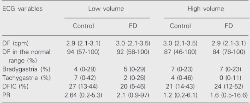
The only functional dyspepsia patient with an abnormal EGG after the low volume test meal reported symptoms of upper abdomi-nal discomfort and bloating in the two stud-ies, even when presenting an entirely normal EGG after the high volume test meal. After the low volume meal, only 1 of the 10 pa-tients with symptoms presented an abnormal EGG, and none of the 7 patients with symp-Table 1. Postprandial electrogastrography (EGG) variables in 14 patients with
dysmotility-like functional dyspepsia (FD) and 13 healthy volunteers (control) after a standard test meal (150 ml of yoghurt), followed by the ingestion of either a low (150 ml) or high (300 ml) volume of water.
ECG variables Low volume High volume
Control FD Control FD
DF (cpm) 2.9 (2.1-3.1) 3.0 (2.1-3.5) 3.0 (2.1-3.5) 2.9 (2.1-3.1) DF in the normal 94 (57-100) 92 (58-100) 87 (46-100) 84 (76-100)
range (%)
Bradygastria (%) 4 (0-29) 5 (0-29) 7 (0-23) 7 (0-23) Tachygastria (%) 7 (0-42) 2 (0-26) 4 (0-46) 0 (0-11) DFIC (%) 27 (13-44) 20 (5-46) 21 (14-43) 24 (12-52) PR 2.64 (0.2-5.3) 2.1 (0.9-97) 1.2 (0.2-6.1) 1.6 (0.5-16.6)
toms after the high meal volume had EGG abnormalities.
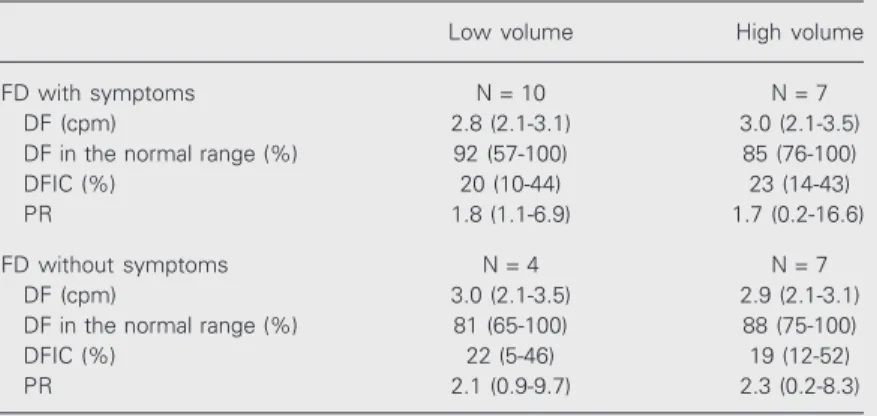
Data for postprandial EGG variables in the subgroups of patients with and without symptoms are shown in Table 2. There were no significant differences between the sub-groups of functional dyspepsia patients with and without symptoms after meal ingestion concerning any postprandial EGG variable.
Discussion
The present study shows that the post-prandial symptoms of upper abdominal dis-comfort and bloating elicited by the inges-tion of a relatively low calorie test meal with increasing volumes of water in patients with dysmotility-like functional dyspepsia are not associated with abnormalities in gastric myo-electrical activity revealed by cutaneous EGG.
We have reported that controlled disten-sion of the stomach with a plastic bag posi-tioned in the gastric fundus produced insta-bility of gastric myoelectrical rhythm at high distension volumes (480-600 ml), which were associated with symptoms of upper abdomi-nal discomfort and nausea (18). We there-fore aimed to determine whether in more physiological settings, with subjects drink-ing a test meal instead of havdrink-ing a barostat bag inside their stomach, symptoms and gas-tric dysrhythmias would be produced in func-tional dyspepsia patients. Since the ingested meal would distribute more fully to the gas-tric antrum (8,9), we suggested that overdis-tension of the gastric antrum in functional dyspepsia patients would induce gastric dysrhythmias. However, we were not able to confirm it in the present study.
It could be argued that ingestion of dif-ferent volumes of water would result in un-predictable distension of the different ana-tomical portions of the stomach. Neverthe-less, an ultrasound study (20) has shown that ingestion of stepwise increasing amounts of water produces linear increases in both
proxi-mal and distal stomach dimensions. In pa-tients with functional dyspepsia, the distal stomach size was significantly greater than in control subjects with ingested volumes equal or greater than 500 ml (20).
Although a substantial proportion of pa-tients (5 of 14) had EGG abnormalities in the fasting state, we did not find any consistent postprandial EGG abnormality in dysmotil-ity-like functional dyspepsia. This finding disagrees with those from a number of other studies (3-5,15-17). On the other hand, our data agree with those reported by Jebbink et al. (21), who did not find any difference between functional dyspepsia patients and healthy volunteers concerning the incidence of gastric dysrhythmias. It is possible that such discrepancies may be related to differ-ences in either patient selection or the defini-tion of funcdefini-tional dyspepsia, or both, since there is no biological marker for the condi-tion. The patients included in the present study fulfilled strict contemporary criteria (Rome II) for dysmotility-like functional dyspepsia (2), whereas other studies show-ing high proportions of EGG abnormalities in functional dyspepsia (3-5,15-17) might
Table 2. Postprandial electrogastrography (EGG) variables in patients with dysmotility-like functional dyspepsia (FD), with and without symptoms, after a standard test meal (150 ml of yoghurt), followed by the ingestion of either a low (150 ml) or high (300 ml) volume of water.
Low volume High volume
FD with symptoms N = 10 N = 7
DF (cpm) 2.8 (2.1-3.1) 3.0 (2.1-3.5)
DF in the normal range (%) 92 (57-100) 85 (76-100)
DFIC (%) 20 (10-44) 23 (14-43)
PR 1.8 (1.1-6.9) 1.7 (0.2-16.6)
FD without symptoms N = 4 N = 7
DF (cpm) 3.0 (2.1-3.5) 2.9 (2.1-3.1)
DF in the normal range (%) 81 (65-100) 88 (75-100)
DFIC (%) 22 (5-46) 19 (12-52)
PR 2.1 (0.9-9.7) 2.3 (0.2-8.3)
also have included patients with more severe involvement of gastric motility, such as that found in idiopathic gastroparesis (13).
Differences between studies in terms of technical aspects, such as test meal composi-tion and volume and recording time, might also contribute to the differences in results. Nevertheless, low volume, low calorie test meals (3), as well as small volumes of water (22) have been shown to induce EGG abnor-malities in patients with functional dyspep-sia. Furthermore, even with short length (30 min) postprandial recordings an association between nausea and tachygastria was dem-onstrated (23).
Although in the present study postpran-dial symptoms of upper abdominal discom-fort and bloating were recorded in up to 70% (10/14) of patients with functional dyspep-sia, no patient complained of nausea, a symp-tom that has been consistently associated with gastric dysrhythmias in a number of experimental conditions, both in healthy vol-unteers (24) and in patients (3-5,13-16). It is therefore uncertain whether the utilization of a test meal more likely to induce nausea might have produced different results and yield a stronger association between gastric dysrhythmias and symptoms.
It is also important to consider the fact that the signal captured by surface electrodes in EGG actually represents the summation of several electrical vectors corresponding to the propagation throughout the stomach of waves arising from the gastric pacemaker (12). It is therefore plausible that electrical vectors corresponding to occasional distur-bances originating in one region may indeed be annulled by another similar vector gener-ated in an opposite region. This may theo-retically be responsible for the relatively low accuracy of the EGG in detecting gastric motor abnormalities (25), and also for the conflicting results (3-5,15-17) obtained for this heterogeneous clinical entity named
func-tional dyspepsia (1,2).
The origin of postprandial symptoms in dysmotility-like functional dyspepsia is un-clear. Although a number of physiological abnormalities have been well demonstrated in this situation, their relationship to symp-toms has not been definitely established (26). Nevertheless, various mechanisms may ex-plain why symptoms following a meal may arise independently of either the appearance of gastric dysrhythmias or their potential consequences, such as antral hypomotility and delayed gastric emptying. Upper ab-dominal discomfort, bloating, and nausea may be caused by impaired fundal relaxation (10,11), leading to increased intragastric pres-sure (10) and antral overdistention produced by displacement of food from the proximal to the distal stomach (8,9) Furthermore, in-creased sensitivity to gastric distention due to visceral hyperalgesia (27) may also be involved in symptom production. The eluci-dation of the roles of each of these physi-ological abnormalities in the pathogenesis of functional dyspepsia demands further stud-ies, which will probably require multiple and sophisticated methods to approach sev-eral mechanisms.
The observations made in the present study do not support the view that distur-bances of gastric myoelectrical activity play a role in the origin of postprandial symptoms of upper abdominal discomfort and bloating, which were found in substantial proportions of patients with dysmotility-like functional dyspepsia, even after the ingestion of low volume, low calorie meals.
Acknowledgments
References
1. Heading RC (1991). Definitions of dyspepsia. Scandinavian Journal of Gastroenterology, 26 (Suppl 182): 1-6.
2. Talley NJ, Stanghellini V, Heading RC & Koch KL (1999). Functional gastroduodenal disorders. Gut, 45 (Suppl II): II-37-II-42.
3. Rezende Filho J (1995). Estudo da atividade mioelétrica gástrica por eletrogastrografia cutânea - Eletrogastrograma.Arquivos de Gastro-enterologia, 32: 54-65.
4. Pfaffenbach B, Adamek RJ, Bartholomaus C & Wegener M (1997). Gastric dysrhythmias and delayed gastric emptying in patients with functional dyspepsia. Digestive Diseases and Sciences, 42: 2094-2099.
5. Lin Z, Eaker EY, Sarosiek I & McCallum RW (1999). Gastric myo-electrical activity and gastric emptying in patients with functional dyspepsia. American Journal of Gastroenterology, 94: 2384-2389. 6. Stanghellini V, Ghidini C, Ricci MM, Paparo GF, Corinaldesi R &
Barbara L (1992). Fasting and postprandial gastrointestinal motility in ulcer and nonulcer dyspepsia. Gut, 33: 184-190.
7. Corinaldesi R, Stanghellini V, Raiti C, Rea E & Salgemini R (1987). Effect of chronic administration of cisapride on gastric emptying of a solid meal and on dyspeptic symptoms in patients with idiopathic gastroparesis. Gut, 28: 300-305.
8. Troncon LEA, Bennett RJM, Ahluwalia NK & Thompson DG (1994). Abnormal intragastric distribution of food during gastric emptying in functional dyspepsia patients. Gut, 35: 327-332.
9. Hausken T & Berstad A (1992). Wide gastric antrum in patients with non-ulcer dyspepsia. Scandinavian Journal of Gastroenterology, 27: 427-432.
10. Troncon LEA, Thompson DG, Ahluwalia NK, Barlow J & Heggie LJ (1995). Relations between upper abdominal symptoms and gastric distension abnormalities in dysmotility like functional dyspepsia and after vagotomy. Gut, 37: 17-22.
11. Tack J, Piessevaux H, Coulie B, Caenepeel P & Janssens J (1998). Role of impaired gastric accommodation to a meal in functional dyspepsia. Gastroenterology, 115: 1346-1352.
12. Smout AJPM, Van Der Schee EJ & Grashuis JL (1980). What is measured in electrogastrography? Digestive Diseases and Sci-ences, 25: 179-187.
13. Telander RL, Morgan KG, Kreulen DL, Scmalj PF, Kelly KA, Szurzewsky JH (1978). Human gastric atony with tachygastria and gastric retention. Gastroenterology, 75: 497-501.
14. Koch KL, Stern RM, Stewart WR, Vasey MW & Sullivan ML (1989). Gastric emptying and gastric myoelectrical activity in patients with diabetic gastroparesis: Effect of long-term domperidone treatment.
American Journal of Gastroenterology, 84: 1069-1075.
15. Geldof H, van der Schee EJ, van Blankstein M & Grashis JL (1986). Electrogastrographic study of gastric myoelectrical activity in
pa-tients with unexplained nausea and vomiting. Gut, 27: 799-808. 16. Chen J, Lin Z, Pan J & McCallum RW (1996). Abnormal gastric
myoelectrical activity and delayed gastric emptying in patients with symptoms suggestive of gastroparesis. Digestive Diseases and Sciences 41: 1538-1545.
17. Parkman H, Miller M, Trate D, Knight LC, Urbain JL, Maurer AH & Fisher RS (1997). Electrogastrography and gastric emptying scintig-raphy are complementary for assessment of dyspepsia. Journal of Clinical Gastroenterology, 24: 214-219.
18. Taylor EJ & Troncon LEA (1993). The use of Fourier transform and spectral analysis in the detection of distension-induced gastric ar-rhythmias in dyspeptic patients. Physiological Measurements, 14: 137-144.
19. Parkman HP, Harris AD, Miller MA & Fisher RS (1996). Influence of age, gender, and menstrual cycle on the normal electrogastrogram.
American Journal of Gastroenterology, 91: 127-133.
20. Marzio L, Falcucci M, Grossi L, Ciccaglione FA, Malatesta MG, Castellano A & Ballone E (1998). Proximal and distal gastric disten-sion in normal subjects and H. pylori-positive and -negative dyspep-tic patients and correlation with symptoms. Digestive Diseases and Sciences: 43: 2757-2763.
21. Jebbink HJA, Van Berg-Henegouwen GP, Bruijs PPM, Akkermans L & Smout AJPM (1995). Gastric myoelectrical activity and gas-trointestinal motility in patients with functional dyspepsia.European Journal of Clinical Investigation, 25: 429-437.
22. Koch KL, Hong SP & Xu L (2000). Reproducibility of gastric electrical activity and the water load test in patients with dysmotility-like dyspepsia symptoms and control subjects. Journal of Clinical Gas-troenterology, 31: 125-129.
23. Koch KL, Stern RM, Vasey M, Botti JJ, Creasy GW & Dwyer A (1990). Gastric dysrhythmias and nausea of pregnancy. Digestive Diseases and Sciences, 35: 961-968.
24. Stern RM, Koch KL, Stewart WR & Lindblad IM (1987). Spectral analysis of tachygastria recorded during motion sickness. Gastroen-terology, 92: 92-97.
25. Bortolotti M (1998). Electrogastrography: a seductive promise, only partially kept. American Journal of Gastroenterology, 93: 1791-1794.
26. Malagelada JR (2001). Review article: the continuing dilemma of dyspepsia. Alimentary Pharmacology and Therapeutics, 15 (Suppl 1): 6-9.