INTRODUCTION
In the last decades, there has been a signiicant evolution in
the knowledge of rheumatoid arthritis (RA) physiopathogeny, leading to changes in disease’s approach and therapeutics. The concept of “early or initial rheumatoid arthritis” was
deined by the majority of authors as the initial phase of
disease symptoms up to 12 months, period of time in which
Distinct pattern of rheumatoid factor
serotypes in serial evaluation of patients
with early rheumatoid arthritis
Licia Maria Henrique da Mota1, Leopoldo Luiz dos Santos Neto2, Rufus Burlingame3, Ieda Maria Magalhães Laurindo4
Received on 11/03/2008. Approved on 02/18/2009.
Rheumatology Service at the Hospital Universitário de Brasília da Universidade de Brasília,1,2 INOVA Diagnostics Inc., San Diego, California, USA,3
Rheumatol-ogy Service at the Hospital das Clínicas da Faculdade de Medicina da Universidade de São Paulo.4
1. Rheumatologist Physician of the Rheumatology Service, Hospital Universitário de Brasília – Universidade de Brasília (UnB), Postgraduation student (Doctor-ate) in Medical Sciences at the Faculdade de Medicina da Universidade de Brasília (UnB)
2. Associate Professor of Clinical Medicine and at the Rheumatology Service at the Hospital Universitário de Brasília (HUB) – Universidade de Brasília (UnB) 3. Senior Scientist of INOVA Diagnostics Inc., San Diego, California, USA
4. Collaborating Professor at the Hospital das Clínicas da Faculdade de Medicina da Universidade de São Paulo (HC-FMUSP)
Correspondence to: Licia Maria Henrique da Mota, SHIS QI 23 conjunto 02 casa 09, Lago Sul, Zip code: 71660-020, Brasília - DF - Brasil, phone: 55 (61) 3208-5568, fax: 55 (61) 3245-1966. E-mail: [email protected], [email protected]
ABSTRACT
Introduction: Rheumatoid factor (RF), despite its limitations, is still the most applied serological marker for diagnosis of early rheumatoid arthritis (RA). Sensitivity, speciicity, correlation with prognosis and radiological progression, as well as variation over time of serotypes titers IgG, IgM and IgA, are yet controversial. Objective: To evaluate the pattern of the different RF serotypes (IgG, IgM, and IgA) in serial evaluations during the irst 36 months after RA diagnosis and their correlation with occurrence of radiographic erosions. Patients and methods: Forty patients with diagnosis of RA (less than 12 months of symptoms) were evaluated during 3 years of follow-up. Titers of RF serotypes were analyzed by ELISA at the initial evaluation and after 12, 24 and 36 months of follow-up. A mixed-effect regression model was applied, considering the presence of radiographic erosions as outcome (annual radiography of hands and wrists, feet and ankles). Results: At the initial evaluation, 30%, 42.5% and 50% of the patients were positive for IgG, IgA and IgM RF, respectively. The titers of IgA and IgM RF were higher for patients who had radiographic erosions during the follow-up period (10-220 IU/dL versus 0 to 10 IU/dL in patients without erosions, P < 0,05). The titers of IgM and IgG RF remained unchanged over the three years of follow-up. On the other hand, there was a positive linear increasing trend for titers of IgA RF (P = 0.0013) only in the group with radiographic erosions. Conclusion: 1) Search of RF serotypes IgA and IgG does not increase the frequency of RF positivity and therefore it does not contribute to the RA diagnosis; 2) IgM RF stability observed over time does not justify repeated requests of RF during RA follow-up; 3) higher titers of IgA and IgM RF are observed in more severe patients, with radiographic erosions; 4) IgA RF presents a clearly distinct behavior in patients with or without radiographic erosions, which may have implications for pathophysiology and prognostic evaluation of the disease.
Keywords: rheumatoid factor, serotypes, early rheumatoid arthritis, diagnosis, prognosis.
establishing of adequate therapy would result in increased
clinical improvement. It has also been possible to conirm
that early treatment and diagnosis can modify the disease
course.1 Laboratorial and image methods were developed,
contributing to an earlier diagnosis and prognostic
determination,2 and, above all, there have been great changes
in therapeutic approach of the disease, with the use of novel
Despite the description of new diagnostic and prognostic markers for RA, most of all in its early phase, rheumatoid factor (RF) is still the most used serological marker for early
RA diagnosis.4
RF is found in about 70% of patients with established RA, and it is of great importance for the diagnosis and prediction
of disease prognosis.4 The presence of RF is one of the
seven 1987 classiication criteria of the American College of
Rheumatology (ACR)5 and its detection is variable according
to the dosage method and to the isotype.4 Only the IgM isotype
is examined in routine tests.
RF, nevertheless, presents a series of limitations as a marker
for RA initial phase diagnosis:4 there is controversy regarding
sensitivity, speciicity, correlation with radiographic prognosis
and variation over time of usually investigated serotypes titers – IgG, IgM and IgA.
The objective of this work was to examine the pattern of different RF serotypes (IgG, IgM, and IgA), in serial evaluations over 36 months, and its correlation with the occurrence of radiographic erosions, in a large group of patients with early RA diagnosis.
PATIENTS AND METHODS
As a prospective observational study, measurements of IgA, IgG and IgM RF were registred over time (36 months) in patientes with early RA diagnosis.
Forty patients were evaluated with early RA diagnosis.
They were followed up in the Clínica de Artrite Reumatoide
Inicial do Hospital Universitário de Brasília over three years and included in the study consecutively.
Initial RA was defined as the occurrence of articular symptoms compatible with the disease, lasting more than 6 weeks and less than 12 months. The criterion used was the clinical diagnosis of RA, performed by the rheumatologist,
independently of the ACR classiication criteria fulillment.
Titration of RF serotypes (IgG, IgM, and IgA) was performed
by ELISA6 (Quanta lite of INOVA Diagnostics Inc.), in baseline
and serially over 36 months (evaluations in 3, 6, 12, 18, 24, and 36 months). The detection test was semiquantitative, and values superior to 15 IU/mL (RF IgM and IgA) and 20 IU/mL (RF IgG) were considered as positivity cut off.
Simple radiographies of hands and wrist, feet and ankles were performed annually (in baseline and at 12, 24, and 36 months), and the reports were issued by only one radiologist, who did not know the results of the patients’ serologies and the sequence of the tests. The report was about the presence or absence of erosion.
During the whole follow-up, patients received the standard treatment protocol used in the service, including traditional disease-modifying antirheumatic drugs (DMARDs) and/or biological therapy, according to necessity.
For the purpose of analysis, a regression model of mixed effects was applied, considering as outcome the occurrence of radiographic erosions and using the following statistic model:
yijk = µ + αi + bij + γk + (αγ)ik + εijk
where: yijkis the measure of the titers IgA, IgG or IgM RF
in k time, over the jth patient from group i
µ + αi + γk + (αγ)ik is the mean of the titers of IgA, IgG or
IgM RF from group i in k time
bij is the random effect associated with the patient j in
group i,N(0, σ2
B)
εijk is the random error associated with the patient j from
group i in k time, with variance and covariance matrix with
random intercept (RI).
We chose to use this new statistic model, mixed effects regression, for it is a study in which the longitudinal data present a hierarchic structure (repeated measures for the same individual). The mixed effects regression model allows the analysis of unbalanced longitudinal data (measures obtained in each individual observed in different times) in hierarchical structure, incorporating the dependency and the structure of errors correlation.
The signiicance level of 5% was considered.
The work was approved by the Comitê de Ética em Pesquisa
da Faculdade de Medicina da Universidade de Brasília.
RESULTS
Characteristics of the studied population
Among the 40 patients followed-up with early RA diagnosis, there was a predominance of female gender (36 patients, 90%) and the mean age was 45.9 years (21 to 71). The mean duration of articular symptoms in the moment of diagnosis was 20.8 weeks (4 to 48), and 19 patients (47.9%) had less than 12 weeks
of symptoms before the diagnosis. Although the fulilling of ACR criteria has not been considered as deinition criterion of initial RA in this study, 33 patients (82.5%) fulilled at least four ACR criteria in the irst evaluation, and 100% after 12
There was no loss of follow-up of any patient during the three years of the study.
Presence of radiographic erosions
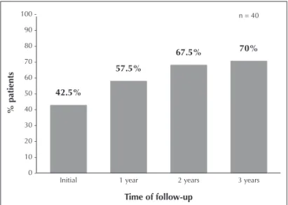
At baseline, 17 patients (42.5%) presented at least one erosion in the radiographs of hands and wrist, feet and ankles. During follow-up, there was a change in this percentage (Figure 1), so that, after 36 months of follow-up, 28 (70%) patients presented at least one erosion.
Pattern of the different serotypes of RF (IgG, IgM, and IgA), in the serial evaluations, and correlation with occurrence of radiographic erosions
At baseline, 21 patients (52.5%) were positive for at least one of the RF serotypes – 17 patients (42.5%) for IgA RF, 12 (30%) for IgG RF, and 20 (50%) for IgM RF, respectively. Sixteen patients (40% of the total sample and 76.1% of those positive for at least one of the RF serotypes) were positive for more than one serotype. Just two patients (5% of the total) were negative for IgM RF and positive for IgA RF.
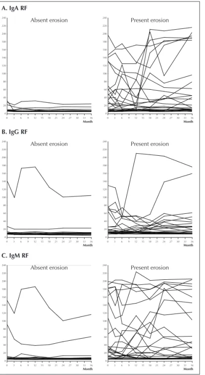
Profile descriptive analysis per group
The pattern of IgA, IgG and IgM RF was analyzed over time (months) according to the presence or absence of erosion, as demonstrated in Figure 2.
Among those patients with “present erosion” in the initial moment or over the evolution (28 individuals), 18 (64.2%) were positive for at least one serotype of RF in the initial evaluation – 15 (53.5%) being positive for IgA RF, 8 (28.5%)
for IgG RF, and 17 (60.7%) for IgM RF. Sixteen patients (57.1% of the total of patients with present “erosion”) were positive for more than one RF serotype.
In the “absent erosion” group during follow-up (12 individuals), three patients (25%) presented initially positive RF. In one of the cases, only the IgA serotype was found, and in the others the three serotypes of RF were present.
As shown in Figure 2 (descriptive analysis), a uniform distribution in the titration of IgA, IgM and IgG RF (from 0 to 10 IU/mL) was observed in patients of “absent erosion” group, exception made to two cases that presented values of all of the RF isotypes above that level (from 0 to 10 IU/mL) and to one individual who presented values of IgG RF discrepant from the group.
In the “present erosion” group, the titers of IgA RF and IgG RF ranged from 10 to 200 IU/mL (Figures 2A and 2B) and the IgM RF ones varied from 10 to 220 IU/mL (Figure 2C). The IgA and IgM serotypes presented values statistically superior to patients in “absent erosion” group (P < 0.05 for both).
Analysis of estimated tendency compared to observed means
To better evaluate the pattern of RF serotypes over time, in relation to the occurrence of radiographic erosion, the adjusting of estimated tendencies model was done compared to means observed in the titers of IgA, IgG and IgM RF by group (“absent
erosion” versus “present erosion”), over 36 months of
follow-up, as shown in Figure 3.
For the IgA RF (Figure 3A), the model adjustment revealed that the linear effect for “absent erosion” group was not
signiicant (P = 0.8048), while for “present erosion” group there was a signiicant linear tendency (P = 0.0013); the
titers of IgA RF tended to increase over time in patients who presented erosions. The difference between titers of IgA RF
of the two groups was statistically signiicant in all periods
analyzed, except in the 12 months analysis, due to one isolated (discrepant) value of a patient who lowered the mean for the group with erosion.
In the case of IgG RF (Figure 3B), the pattern of behavior was similar for patients with or without radiographic erosion
and the linear effect was not signiicant (P = 0.2007 and P
= 0.5833, respectively), it represents the mean values of IgG RF tend to vary very little over time in patients with or without erosion. There was not statistic difference (P > 0.05) regarding the titers of RF IgG in the two groups (presence and absence of radiographic erosions) during the 36 months of follow-up.
Figure 1. Evolution of radiographic erosions proile – occurrence of at least one
radiographic erosion (radiograph of hands and wrists, feet and ankles) in initial evaluation and serial follow-up.
Initial 1 year 2 years 3 years n = 40 100
90 80 70 60 50
40 30 20 10 0
-42.5%
57.5%
67.5% 70%
% patients
Figure 2. Chart of model adjustment of estimated tendencies compared to obser-ved means in the titers of rheumatoid factor (RF) IgA, IgG and IgM per group (“absent erosion” versus “present erosion”), over 36 months of follow-up. For IgA RF (Figure 3A), the model adjustment reveals that the linear effect for “absent
erosion” group is not signiicant (P = 0.8048), while for “present erosion” group there is a signiicant linear tendency (P = 0.0013), it means that IgA RF titers tend to increase over time. The difference between the two groups is signiicant (P <
0.05) in all periods analyzed, except in the analysis of 12 months. In the case of IgG RF (Figure 3B), the behavior pattern is similar for patients with or without
radiographic erosion: the linear effect is not signiicant (P = 0.2007 and P = 0.5833,
respectively), which represents mean values of IgG tend to vary very little over time. The model adjustment for IgM RF (Figure 3C) also shows that, for the group of patients without erosion, mean values of IgM RF tend to vary very little over time
(P = 0.4728), while for the group with erosion there is a positive, almost signiicant
tendency of linear effect (P = 0.0568). The difference between IgM RF titers of the
two groups is signiicant (P < 0.05) from the 12th month of follow-up. (Regression
of mixed effects analysis). . gA Erosão ausente . IgG . gM
ente Erosão presente
C. IgM RF B. IgG RF A. IgA RF
Present erosion Absent erosion
3
0 6 9 12 15 18 21 24 27 30 33 36 240 220 200 180 160 140 120 100 80 60 40 20 0 -3
0 6 9 12 15 18 21 24 27 30 33 36 240 220 200 180 160 140 120 100 80 60 40 20 0 -Present erosion Absent erosion 3
0 6 9 12 15 18 21 24 27 30 33 36 240 220 200 180 160 140 120 100 80 60 40 20 0 -3
0 6 9 12 15 18 21 24 27 30 33 36 240 220 200 180 160 140 120 100 80 60 40 20 0 -Present erosion Absent erosion 3
0 6 9 12 15 18 21 24 27 30 33 36 240 220 200 180 160 140 120 100 80 60 40 20 0 -3
0 6 9 12 15 18 21 24 27 30 33 36 240 220 200 180 160 140 120 100 80 60 40 20 0 -Month Month Month Month Month Month
Figure 3. Chart of proile per group – behavior of patients over time (months –
m) in relation to measurements of rheumatoid factor (RF) IgA, IgG and IgM per group (“absent erosion” versus “present erosion”). It is observed that the patients of “absent erosion” group are uniformly distributed regarding the titration of IgA, IgM and IgG RF (from 0 to 10 IU/mL), while in the “present erosion” group patients presented different behaviors regarding the titration of RF. The titers of IgA RF (Figure 2A) varied from 10 to 200 IU/mL, and values superior to the patients in the absent erosion group were presented. The IgG RF titers (Figure 2B) also ranged from 10 to 200 IU/mL in the “present erosion” group, but did not tend to present superior values in relation to “absent erosion” group. The IgM RF titers (Figure 2C) varied from 10 to 220 IU/mL in patients with erosions presenting superior values in relation to patients without erosion. (Descriptive analysis).
Initial P = 0.0640 3m P = 0.0518 6m P = 0.0932 12m P = 0.0495 18m P = 0.0354 24m P = 0.0076 36m P = 0.0190
IgM - Mean
C. IgM RF
3
0 6 9 12 15 18 21 24 27 30 33 36 80 70 60 50 40 30 20 10 0
-Initial P = 0.5750 3m P = 0.4578 6m P = 0.9802 12m P = 0.7649 18m P = 0.5308 24m P = 0.2225 36m P = 0.2546
IgG - Mean
B. IgG RF
3
0 6 9 12 15 18 21 24 27 30 33 36 40
30
20
10
0
-Initial P = 0.0270 3m P = 0.0254 6m P = 0.0218 12m P = 0.0585 18m P = 0.0030 24m P = 0.0034 36m P = 0.0008 Present erosion P = 0.0013
Absent erosion P = 0.8048
Present erosion P = 0.2007
Absent erosion P = 0.5833
Present erosion P = 0.0568
Absent erosion P = 0.4728
IgA - Mean
A. IgA RF
3
0 6 9 12 15 18 21 24 27 30 33 36 70 60 50 40 30 20 10 0 -Month Month Month
The model adjustment for IgM RF (Figure 3C) also shows that for the group of patients without erosion, mean values of IgM RF tend to vary very little over time (P = 0.4728), while for the group with erosions, there is a positive tendency of linear
effect, almost signiicant (P = 0.0568). Starting from the 12th
month of follow-up, the difference between the titers of RF
IgM of the two groups is signiicant (P < 0.05).
DISCUSSION
The validity of the RF isotypes research in the evaluation of initial RA remains questionable. For example, the existence of correlation between the titers of different isotypes of RF and the
diagnosis of RA is not deined, as well as the relation between the presence of some speciic serotype (or more than one) and
a worst radiologic prognostic, or the behavior of different RF isotypes over time.
Although there is controversy, it was suggested IgM, IgA
and IgG RF are signiicantly associated to RA diagnosis.4
The isotypes positivity seems to be variable according to
the studied population.8 In the work by Visser et al.,9 ELISA
sensitivity for IgG, IgA and IgM RF was 72%, 44%, and 69%,
respectively, and the speciicity was 52%, 84%, and 86%. The
metanalysis of Nishimura et al.10 concludes that, for IgM RF,
sensitivity and speciicity (with the respective conidence
intervals – CI) were 69% (CI: 65% to 73%) and 85% (CI: 82% to 88%). The results for RF IgG and IgA were similar.
In our study, IgM RF was observed in 49,23%, IgA in 43% and IgG in 29,2% of the patients with RA diagnosis and less than 12 months of symptoms duration – rates similar to the
ones referred in other works, such as the one by Vittecoq et
al.,8 which described the presence of IgM RF in 51%, IgA RF
in 36% and IgG RF in 32% of the patients with RA diagnosis and less than two years of duration.
IgM RF is a useful marker for discriminating patients
with polyarthritis who will evolve or not to RA.8,11-14 On
the other hand, diagnostic properties of IgA and IgG RF are
questioned.8,12,15,16,17
Some authors have reported conlicting results regarding
the fact that IgG and IgA RF are possible better prognostic
markers than IgM RF for RA.18-22 In the metanalysis of
Nishimura et al.,11 greater differences between the three
serotypes were not found.
Rantapää-Dahlqvist et al.6 demonstrated in blood samples
obtained before the beginning of RA (blood donors) that the presence of the three serotypes, especially IgA RF, was a predictor of RA. IgA and IgM RF were detected in serum
storaged to 18 years before the diagnosis of RA.19
In our study, the search of RF serotypes IgA and IgG did not increase the frequency of RF’s positivity; therefore, it did not seem to contribute to initial RA diagnosis.
RF is known as one of serologic markers associated to
a worst prognostic in RA,20 including a serious radiological
evolution of the disease.21-24 On the other hand, the studies are
not as clear regarding the importance of other RF isotypes as predictors of the occurrence of erosion in patients with early RA diagnosis.
Di Franco et al.25 correlated the levels of RF IgA, IgG and IgM
with the occurrence of erosions at magnetic resonance, but not in
plain radiographs. Visser et al.9 demonstrated that the speciicity
of all RF isotypes in the discrimination between nonerosive or erosive RA after two years of evolution was low (RF IgG: 41%, IgA: 44%, and IgM: 47%) and that the IgG and IgA isotypes were not useful for diagnosing RA and predicting erosive disease.
Most authors agree that the presence of RF IgM is an independent predictor of the occurrence of radiographic
erosion.26,27 Other works suggest that elevated serum
concentrations of RF IgA can be an early and reliable marker
of evolution to erosive forms of RA.28,29 Elevated titers of
RF IgG have also been considered determinative of a worst
radiographic prognosis.27
Regarding the possible association between the RF serotypes titers and the radiologic prognostic, our results demonstrated that, in a purely descriptive analysis higher titers of the three serotypes are observed in critical patients, with radiographic erosions. Nevertheless, over the follow-up,
only the most elevated mean values of RF IgA (since the irst evaluation) and RF IgM titers (from the irst year of follow-up)
are statistically correlated to the occurrence of erosions. The presence of IgG RF could not predict the occurrence of the erosive form of RA neither on the initial moment nor during the follow-up.
Regarding the behavior of different RF isotypes titers over time, we tried to determine if the variations in its titers could predict a better or worse radiographic prognosis.
Variations in the titers of different RF isotypes during the patients with RA follow-up were observed, especially in
works evaluating the inluence of speciic therapeutics over
RF titers.16,30-32
Swedler et al.16 demonstrated that there was a continuous
decline of the three RF serotypes in patients being treated
with gold salts. Previously, Lemm et al.30 had published that
the titers of IgG RF seemed to be a good parameter for the evolution of RA control under therapy with gold salts.
Bobbio-Pallavicini et al.31 observed that in patients being
individuals for RF stabilizes over time, the mean of RF titers suffers a progressive reduction. The same authors later
reported that, in patients using inliximab and DMARDs, the treatment resulted in an early and signiicant reduction of the
RF IgA and IgM titers, but not RF IgG, and the decrease of
IgM was sustained.32
On the other hand, we observed in our work that RF IgA showed a clearly distinct evolutive behavior in patients who presented or not radiographic erosions, which had not been previously reported. RF IgA titers increased over time among patients who presented erosions and stabilized in the remaining, although both groups have received treatment with DMARDs since the early diagnosis, including biological therapy.
The behavior of RF IgM was similar to RF IgA, but there
was not statistic signiicance. Maybe the increase of the
studied casuistic or of the time of follow-up allows greater conclusions about the matter. The titers of RF IgG did not suffer variation over time.
These changes in RF titers during patients follow-up have a yet uncertain meaning. It is possible that the increase in RF IgA titers during the three years of follow-up, observed only in patients who presented radiographic erosions, is a marker of bad prognostic in a population with severe evolution. The presence of radiographic erosions evolved from 42,5% in the initial evaluation to 70% in the third year, despite early treatment (47,9% of the patients begun follow-up and treatment with up to 12 weeks of articular symptoms).
This behavior of RF IgA, distinct of the remaining serotypes, can have possible implications in the physiopathogenesis and in the prognostic evaluation of the disease.
Due to the limited number of individuals evaluated, and to the various subschemes of treatment used, it was not
possible to evaluate the inluence of DMARDs use over
the radiographic evolution. It is known that the early use of the adequate therapy for RA, whether it is conventional or biological, could inhibit the appearance of erosions. This could be a factor of confusion in the evaluation of radiographic evolution outcome in the evaluated population in this study.
CONCLUSIONS
The results of this work allow us to conclude that: 1) RF IgA and RF IgG serotypes research does
not increase the positivity frequency of RF and, therefore, does not contribute to diagnosis of RA; 2) RF IgM stability observed over time does not justify
repeated solicitations of RF during RA evolution;
3) Higher titers of the three serotypes are observed in critical patients with radiographic erosions, but only the most elevated mean values of RF
IgA (since the irst evaluation) and RF IgM titers (from the irst year of follow-up) correlated
statistically to the occurrence of erosions;
4) RF IgA presents a clearly distinct behavior in patients who presented or not radiographic erosions, which can have implications in the physiopathogenesis and in the prognostic evaluation of the disease.
Investigations in a larger number of patients with initial RA and longer follow-up are necessary to evaluate the characteristics of RF isotypes titers variations during the disease evolution, its correlation with radiological prognostic,
and inluence of speciic therapeutics.
CONFLICTS OF INTEREST
The author RB works for INOVA Diagnostics Inc., where the serologic tests were performed. RB did not have access to the clinical data of patients previously to the tests results. This study was supported by FINATEC (Santos-Neto LL:
129/2008 – Universidade de Brasília). The remaining authors
declare no conlicts of interest.
ACKNOWLEDGMENT
We thank Dr. Francisco Aires Corrêa Lima, Dr. Rodrigo Aires Corrêa Lima, Dr. Ana Patrícia de Paula, Professor Cezar Kozak Simaan, Dr. José Antonio Braga da Silva, Dr. Hermes Matos Filho, Dr. Regina Alice von Kircheheim, Dr. Luciana Alves Almeida, Dr. Talita Yokoy Souza, Dr. Jamille Nascimento Carneiro and Dr. Francieli Sousa Rabelo, for forwarding the evaluated patients, and Dr. Paulo Sérgio Mendlovitz, for performing the radiographic tests.
REFERÊNCIAS REFERENCES
Lard LR, Visser H, Seyer I, Horst-Bruinsma IEV, Zwinderman AH, 1.
Breedveld FC et al. Early versus delayed treatment in patients with recent onset rheumatoid arthritis: comparison of two cohorts who received different treatment strategies. Am J Med 2001;111:446-51. Hoving JL, Bucbinder R, Hall S, Lawler G, Coombs P, McNealy S 2.
et al. A comparison of magnetic resonance imaging, sonography and radiographic of the hand in patients with early rheumatoid arthritis. J Rheumatol 2004;31:663-75.
Haque UJ, Bathon JM. The role of biological in early rheumatoid 3.
arthritis. Best Pract & Res Clin Rheum 2005;19:179-89. Renaudineau Y,
Arnett FC, Edworty SM, Bloch DA, McShane DJ, Fries JF, Cooper 5.
NS et al. The American Rheumatism Association 1987 revised
criteria for the classiication of rheumatoid arthritis. Arthritis Rheum
1988;31:315-24. Rantapää-Dahlqvist
6. S, de Jong BA, Berglin E, Hallmans G, Wadell G, Stenlund H et al. Antibodies against cyclic citrullinated peptide and IgA rheumatoid factor predict the development of rheumatoid arthritis. Arthritis Rheum 2003;48:2741-9.
Fitzmaurice GM, Ravichandran C. A primer in longitudinal data 7.
analysis. Circulation 2008;118:2005-10. Vittecoq
8. O, Pouplin S, Krzanowska K, Jouen-Beades F, Menard JF, Gayet A et al. Rheumatoid factor is the strongest predictor of radiological progression of rheumatoid arthritis in a three-year prospective study in community-recruited patients. Rheumatology 2003;42:939-46.
Visser H,
9. Gelinck LB, Kampfraath AH, Breedveld FC, Hazes JM. Diagnostic and prognostic characteristics of the enzyme linked immunosorbent rheumatoid factor assays in rheumatoid arthritis. Ann Rheum Dis 1996;55:157-61.
Nishimura K,
10. Sugiyama D, Kogata Y, Nishimura K, Sugiyama D, Kogata Y et al. Meta-analysis: diagnostic accuracy of anti-cyclic citrullinated peptide antibody and rheumatoid factor for rheumatoid arthritis. Ann Intern Med 2007;146:797-808.
Wolfe F, Cathey MA, Roberts FK. The latex test revised rheumatoid 11.
factor testing in 8,287 rheumatic disease patients. Arthritis Rheum 1991;34:951-60.
Saraux A,
12. Berthelot JM, Chalès G, Le Henaff C, Mary JY, Thorel JB et al. Value of laboratory tests in early prediction of rheumatoid arthritis. Arthritis Rheum 2002;47:155-65.
Vallbracht
13. I, Rieber J, Oppermann M, Förger F, Siebert U, Helmke K. Diagnostic and clinical value of anti-cyclic citrullinated peptide antibodies compared with rheumatoid factor isotypes in rheumatoid arthritis. Ann Rheum Dis 2004;63:1079-84.
Greiner A,
14. Plischke H, Kellner H, Gruber R. Association of anti-cyclic citrullinated peptide antibodies, anti-citrullin antibodies, and IgM and IgA rheumatoid factors with serological parameters of disease activity in rheumatoid arthritis. Ann N Y Acad Sci 2005;1050:295-303. Procaccia S,
15. Gasparini A, Colucci A, Lanzanova D, Bianchi M, Forcellini P et al. ELISA determined IgM, IgG and IgA rheumatoid factors in rheumatoid arthritis and in other connective tissue diseases. Clin Exp Rheumatol 1987;5:335-42.
SwedLer W, Wallman J, Froelich CJ, Teodorescu M. Routine 16.
measurement of IgM, IgG, and IgA rheumatoid factors: high
sensitivity, speciicity, and predictive value for rheumatoid arthritis.
J Rheumatol 1997;24:1037-44.
van Leeuwen MA, Westra J, van Riel PL, Limburg PC, van Rijswijk 17.
MH. IgM, IgA, and IgG rheumatoid factors in early rheumatoid arthritis predictive of radiological progression? Scand J Rheumatol 1995;24:146-53.
Jansen A
18. L, van der Horst-Bruinsma I, van Schaardenburg D, van de Stadt RJ, de Koning MH, Dijkmans BA. Rheumatoid factor and antibodies to cyclic citrullinated Peptide differentiate rheumatoid arthritis from undifferentiated polyarthritis in patients with early arthritis. J Rheumatol 2002;29:2074-6.
Jørgensen K
19. T, Wiik A, Pedersen M, Hedegaard CJ, Vestergaard BF, Gislefoss RE et al. Cytokines, autoantibodies and viral antibodies
in premorbid and postdiagnostic sera from patients with rheumatoid arthritis: case-control study nested in a cohort of Norwegian blood donors. Ann Rheum Dis 2008;67:860-6.
Morel J, Cobe B. How to predict prognosis in early rheumatoid 20.
arthritis. Best Pract & Res Clin Rheum 2005;19:137-46. Combe B, Dougados M, Gouille P,
21. Cantagrel A, Eliaou JF, Sibilia J
et al. Prognostic factors for radiographic damage in early rheumatoid arthritis: a multiparameter prospective study. Arthritis Rheum 2001;44:1736-43.
Young A, Corbett M, Winield J,
22. Jaqueremada D, Williams P,
Papasavvas G et al. A prognostic index for erosive changes in the hands, feet, and cervical spines in early rheumatoid arthritis. Br J Rheumatol 1988;27:94-101.
Paimela L, Alosuo T, Leirisalo-Repo M,
23. Helve T, Aho K. Prognostic
value of quantitative measurement of rheumatoid factor in early rheumatoid arthritis. Br J Rheumatol 1995;34:1146-50.
Brennan P, Harrison B, Barrett E,
24. Chakravarty K, Scott D, Silman A
et al. A simple algorithm to predict the development of radiological erosions in patients with early rheumatoid arthritis: prospective cohort study. BMJ 1996;313:471-6.
Di Franco
25. M, Spadaro A, Mauceri MT, Cortese A, Guerrisi R, Ciocci A. Relationship of rheumatoid factor isotype levels with joint lesions detected by magnetic resonance imaging in early rheumatoid arthritis. Rev Rhum Engl Ed 1999;66:251-5.
Syversen S
26. W, Gaarder PI, Goll GL, Ødegård S, Haavardsholm EA, Mowinckel P et al. High anti-cyclic citrullinated peptide levels and an algorithm of four variables predict radiographic progression in patients with rheumatoid arthritis: results from a 10-year longitudinal study. Ann Rheum Dis 2008;67:212-7.
Wagner
27. E, Ammer K, Kolarz G, Wagner E, Ammer K, Kolarz G et al. Predicting factors for severity of rheumatoid arthritis: a prospective multicenter cohort study of 172 patients over 3 years. Rheumatol Int 2007;27:1041-8.
Ates A, Kinikli G, Turgay M, Akay G, Tokgöz G. Effects of 28.
rheumatoid factor isotypes on disease activity and severity in patients with rheumatoid arthritis: a comparative study. Clin Rheumatol 2007;26:538-45.
Berglin E,
29. Johansson T, Sundin U, Berglin E, Johansson T, Sundin U et al. Radiological outcome in rheumatoid arthritis is predicted by presence of antibodies against cyclic citrullinated peptide before and at disease onset, and by IgA-RF at disease onset. Ann Rheum Dis 2006;65:453-8.
Lemm G,
30. Ruschen S, Warnatz H. An ELISA for IgA-IgG and IgM-RF measurement. II. RF in several disease and control groups and under gold therapy in RA. Scand J Rheumatol Suppl 1988;75:256-60.
Bobbio
31. -Pallavicini F, Alpini C, Caporali R, Avalle S, Bugatti S, Montecucco C. Autoantibody proile in rheumatoid arthritis during
long-term inliximab treatment. Arthritis Res Ther 2004;6:R264-72.
Bobbio-Pallavicini F,