Imagem em Medicina
382
Rev Assoc Med Bras 2009; 55(4): 382-3leukoplakia
of
the
anal
verge
with
bleeding
hemorrhoids
-
a
case
report
pravin J gupta Clinical Head Nagpur, Índia
c
aser
eportOn 14th August 2002, a 46-year- old male, an ofice clerk,
reported with bleeding and prolapse per rectum for since 6 months, however without any complaint of any pain, pruritus or discharge per anus..
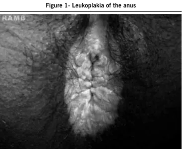
Upon anal and intra anal examination a circumferential area
of thickening and whitish discoloration of the anal verge, which was extending up to the dentate line, was found. Perianal skin was macerated, excoriated and thickened, suggestive of leuko
-plakia. (Figure 1) Anoscopy showed prolapsing hemorrhoids. The patient gave no history of previous anal surgery, homose-xual contact or exposure to radiation. He had been using various hemorrhoids ointments in and around the anus for the last four months. The test for HIV virus was negative.
The hemorrhoids were ablated using a Ellman radiowave generator [Ellman International Inc., Oceanside, NY, USA] under local anesthesia with the patient in a lithotomy position. A biopsy
was taken from the anal verge and the patient was discharged after 2 hours. He was asked to return after 4 weeks.
The microscopic picture of the biopsied anal skin showed pronounced corniication of the anoderm. Acanthosis was
prominent, but rete cells were orderly and the basal layer was
well deined. The dermis showed profuse chronic inlammation,
with lymphocytes, plasma cells and large mononuclear cells in the papillary layer.
At the 4 week follow-up, the patient was asymptomatic, the
biopsy wound was healed and the hemorrhoids were not visible.
The patient was asked to report each year or earlier if symptoms
appeared. At the last follow-up in October 2007, the patient had
no speciic anal symptoms except occasional Dyschezia. The leukoplakic area remained unchanged.
d
iscussionLeukoplakic lesions are most commonly seen in the oral
cavity, more frequently in men than in women at that site. . They
are somewhat less common on the vulva. The bladder, kidney
pelvis, ureters, larynx, esophagus, cervix and glans penis are occasionally involved.1 Extensions of primary vulvar leukoplakic
lesions with involvement of the perineum and perianal skin have been described in literature. Primary leukoplakia on the
anoderm of the anal canal has been occasionally reported. The lesions developed on the so-called “mucosa” or anoderm of the anal canal, with some extension to the adjacent rectal mucosa.
Leukoplakia is considered to be a precancerous dermatosis of mucous membranes analogous to senile keratosis of exposed skin
surfaces2. Several patients with carcinoma in situ had a discrete
area of leukoplakia in the anal canal or a pigmented plaque of
the anus and anal canal 3. However, in this case, leukoplakia
was absolutely innocuous with no change in the color, texture
or extension of the lesion noticed over a period of ive years. The cause of leukoplakia is still controversial 4. Earlier
inves-tigators indicated syphilis as a prominent factor in oral lesions.
Deiciencies of vitamin A, hydrochloric acid, vitamin C and
estrogens have been mentioned as causative factors in vulvar
leukoplakia. Excessive excretion of irritating organic urinary acids
has also been considered 5. Other investigators consider that
prolonged local irritation from any cause, acting on a deicient,
aging epithelium is a fundamental cause of this lesion6. The
author considers that, probably, continuous use of hemorrhoids
cream was the cause of leukoplakia in this patient. A correctly
interpreted preliminary biopsy serves as a guide for a proper therapeutic approach.
Follow-up of our case shows little evidence that leukoplakia
of the anal canal is premalignant, but patients should be followed carefully, since the natural history of this rare lesion remains
unknown.
r
eferences1. Bender MD, Lechago J. Leukoplakia of the anal canal. Am J Dig Dis.
1976;21:867-72.
2. Templeton JL, Brennen MD. Anal leukoplakia: management using a staged plastic procedure. J R Coll Surg Edinb. 1991;36:333-4.
leukoplakiaoftheanalvergewithbleedinghemorrhoids- acasereport
383
Rev Assoc Med Bras 2009; 55(4): 382-3
3. Jongen J, Reh M, Bock JU, Rabenhorst G. Perianal precancerous conditions (Bowen disease, Paget disease, Carcinoma in situ, Buschke-Lowenstein tumor) Kongressbd Dtsch Ges Chir Kongr. 2001;118:79-86.
4. Riedler L. “Bowenoid” leukoplakia in the anal region. Klin Wochenschr.
1988;66:271-3.
5. Foust RL, Dean PJ, Stoler MH, Moinuddin SM. Intraepithelial neoplasia of
the anal canal in hemorrhoidal tissue: a study of 19 cases. Hum Pathol. 1991;22:528-34.