Ophthalmology Clinic, Hospital das Clínicas, São Paulo University Medical School - São Paulo/SP, Brazil.
Email: [email protected]
Received for publication on December 06, 2005. Accepted for publication on March 21, 2006.
ORIGINAL RESEARCH
PEDIATRIC AND ADOLESCENT POPULATION WITH
VISUAL IMPAIRMENT: STUDY OF 385 CASES
Maria Aparecida Onuki Haddad, Frederico José Correa Lobato, Marcos Wilson Sampaio, and Newton Kara-José
Haddad MAO, Lobato FJC, Sampaio MW, Kara-José N. Pediatric and adolescent population with visual impairment: study of 385 cases. Clinics. 2006;61(3):239-46.
OBJECTIVE: To analyze data on the pediatric population attending the Ophthalmologic Clinic’s Low Vision Service at the São Paulo University Medical School.
METHODS: Low vision ophthalmologic assessment, from April 1998 to December 2003, of 385 children and adolescents with mean age of 7 years; 51.7% males and 48.3% females. The main data analyzed were age, diagnosis, anatomic site of the ocular injury, visual acuity, and prescription of optical aids.
RESULTS: 45.4% were below 6 years, and 54.6% were between 6 and 16 years. 35.5% experienced moderate visual impairment, 26% had severe visual impairment, 8.6% had profound visual impairment, 10.6% were near blind, and 1.6% were blind. The main causes of visual impairment included congenital glaucoma (30.6%), macular retinochoroiditis due to congenital toxoplasmosis (16.7%), congenital cataract (12.8%), retinal and macular inherited disorders (11.7%), and optic atrophy (9.8%). Among school-age children, 52.9% received a prescription of optical aids. The most widely used optical aids for distance were 2.8 X 26 (34.4%); 4.2 X 12 (30.3%); and 6 X 17 (26.8%) telescopic systems. The most frequently prescribed optical aid for near vision was the 2x magnifying bar (33.3%).
CONCLUSIONS: There is a need for prevention of primary (congenital infections), secondary (congenital glaucoma and retinopathy of prematurity), and tertiary (congenital cataract) visual impairment. The prescription of optical aids for school-age children will help them perform better at school and contribute to their social inclusion.
KEYWORDS: Blindness. Low vision. Rehabilitation. Congenital glaucoma. Congenital toxoplasmosis.
INTRODUCTION
Visual impairment in childhood has an impact on mo-tor, cognitive, and affective development. Etiological fac-tors, age of onset, presence of other impairments, environ-mental aspects, and the interactions among these will de-termine the child’s difficulties and possible delay in devel-opment.1
According to the 10th revised International
Classifica-tion of Diseases and Health-related Problems, a person has
low vision when his/her best-corrected visual acuity is be-tween 6/18 and 3/60 (visual impairment grade categories 1 and 2), and the person is considered blind when this value falls below 3/60 (categories 3, 4, and 5 ).2
The World Health Organization (WHO) estimates that, worldwide, 140 million people have low vision and 45 mil-lion are blind.3 Visual impairment in childhood is
under-estimated, and its prevalence is only partially known,4
rang-ing from 0.3 to 1.5/1,000, dependrang-ing on the regional so-cioeconomic development (between 80 and 100 blind chil-dren per 1 million in industrialized countries and 400 blind children per 1 million in the poorest areas of the world).5,6
There are an estimated 1.5 million blind children in the world, with three fourths of this total living in the poorest areas of Asia and Africa5; of these, 40% of the cases could
world-wide incidence of blindness in childhood is 500,000 new cases each year, of which 60% to 80% of the children will die before the end of the second year of life from prob-lems related to the causes of blindness or as a consequence of it.7
Prevention of blindness in childhood has been set as a priority by the WHO because of the reduced life expect-ancy of the child with visual impairment and the social, economic, and emotional repercussions.5,6
Knowledge about the profile of the pediatric popula-tion with visual impairment is essential for the effective-ness of preventive programs and for the training and reha-bilitation services to fulfill patients’ needs. The character-istics of visual impairment in a population vary according to the accessibility to healthcare services and sociocultural factors; an important cause of blindness in a given region may be nonsignificant for another and may vary from
dec-ade to decdec-ade.7 Temporini and Kara-José have stated that
planning and implementing preventive actions in ophthal-mology requires a combination of the available scientific knowledge of ophthalmologic problems with knowledge on the reality that will be the object of such actions.8
Visual rehabilitation centers have the objective of pre-venting sequelae of visual impairment in the individual; they try to help the individual avoid disability and to pro-mote the development of the person’s potentialities. The major goal is to integrate the person into his/her family, working environment, and society.
The Ophthalmologic Clinic’s Low Vision Service at the São Paulo University Medical School provides assistance to people with low vision who are referred by other serv-ices from the Ophthalmologic Clinic. The staff working at the Low Vision Service includes ophthalmologists, educa-tors, a physiotherapist, and a psychologist. The objectives of the Service are:
• To conduct ophthalmologic assessments of patients with
low vision and to prescribe and adapt the applicable optical, nonoptical, and electronic aids;
• To guide the actions to be implemented by family,
par-ents, teachers, and community towards an efficient use of vision; this will be based on the assessment of indi-vidual requirements for the child’s global development;
• To provide orientation for the individual to recover or
acquire independence and autonomy. Whenever neces-sary, to refer the patient to other services in visual train-ing and rehabilitation centers.
Based on the specific low vision ophthalmologic assess-ment and on the stability of the ocular condition, the most suitable optical aids may be prescribed. Low vision aids— optical, nonoptical, and electronic - aim at improving visual resolution through changes in the retinal image
(magnifi-cation, displacement, filtering, or condensation) and envi-ronmental conditions. Optical aids may include spectacle-mounted spheroprismatic or aspheric lenses, hand magni-fiers, stand magnimagni-fiers, telemicroscopes, and telescopic sys-tems. Once the optical aid is indicated, it is prescribed af-ter correct orientation about its use in different situations of the patient’s daily life.
The objective of this study is to present the character-istics of the pediatric population attended at the Low Vi-sion Service from April 1998 to December 2003, concern-ing age, causes of visual impairment, and prescription of optical aids. It is part of a general project of the Low Vi-sion Service to establish Brazilian standards for a number of correlated conditions.9-11
METHODS
The study included 385 children and adolescents up to 15 years of age with decreased visual responses who were referred to the unit from April 1998 to October 2003.
Following the guidelines of UNICEF, the period from 0 to 15 years was considered as childhood.7
Patients were referred from other services of the Oph-thalmologic Clinic, with the following information concern-ing their ocular condition: best corrected visual acuity, ocu-lar motility evaluation, refraction, biomicroscopy, fundos-copy, diagnosis, and treatment received. The follow-up at the service of origin is mantained.
The mean age of the study population was 7 years; 199 (51.7%) were boys and 186 (48.3%) were girls.
Data on age, ophthalmologic diagnosis, damaged ocu-lar structure, best corrected visual acuity, and prescription of optical aids were assessed.
Concerning the distribution of data related to the dam-aged ocular structure, the protocol suggested by the WHO Program for the Prevention of Blindness was used.12,13
The visual acuity values were distributed in classes of visual response according to the WHO International Clas-sification of Diseases and Health-related Problems—Tenth
Revision2 and the International Council of
Ophthalmol-ogy.14
Optical aids for distant and near vision were assessed regarding model, magnification, and correlation with the ophthalmologic diagnosis.
RESULTS
The distribution of patients according to age is dis-played in Figure 1.
(26%) had severe visual impairment (class 2), 33 (8.6%) had profound visual impairment, 41 (10.6%) were near blind, and 6 (1.6%) were blind (Table 1).
The main causes of visual impairment (Table 2).in-cluded: congenital glaucoma (30.6%), macular retinoc-horoiditis due to congenital toxoplasmosis (16.6%), con-genital cataract (12.7%), retinal and macular inherited dis-orders (11.7%), and optic atrophy (10.1%).
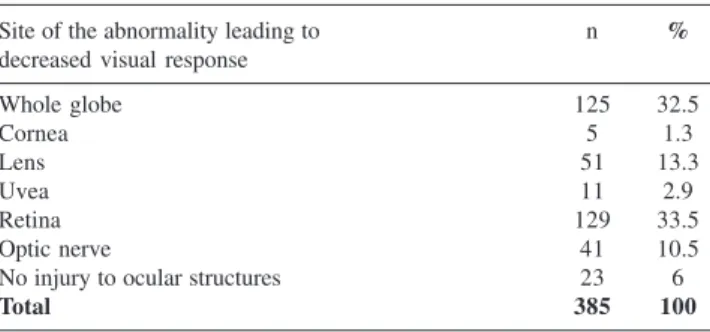
Concerning the location of the abnormality leading to decreased visual response, displayed in Table 3, the fol-lowing sites were observed: whole globe in 32.5% of the cases: cornea, 1.3%; lens, 13.3%; uvea, 2.9%; retina,
33.5%; optic nerve, 10.5%; and 6% had no changes in ocu-lar structures.
Optical aids were prescribed for 111 children (28.8%). For 69 children (17.9%), only optical aids for distant vi-sion had been prescribed, and for 12 children (3.1%), only optical aids for near vision. Both distant and near vision optical aids had been prescribed for 30 children (7.8%). Among the children above 6 years (n = 210), 52.9% re-ceived a prescription for optical aids.
The most widely used optical aids for distance were monocular and manual 2.8 X 26 mm (36.4%); 4.2 X 12 mm (29%); and 6 X 17 mm (26.8%) telescopic systems (Table 4).
The most widely used optical aids for near vision were 2 X magnifying bars (33.3%) and illuminated stand mag-nifiers with +28 diopters (19%) (Table 5).
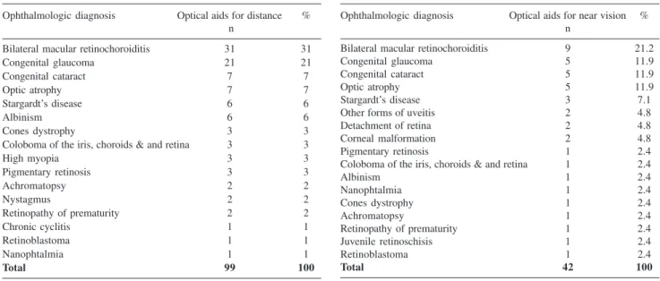
With respect to ophthalmologic diagnosis, optical aids were used more frequently in cases of bilateral macular retinochoroiditis, congenital glaucoma, congenital cataract, and optic atrophy (Tables 6 and 7).
Concerning visual acuity values, 18.2% of the aids were prescribed for children with mild low vision, 40.5% for children with moderate low vision, 34.3% for children with severe low vision, and 7% for children with profound low vision (Table 8).
DISCUSSION
In developing countries, available data on visual impair-ment,4,5,15,16 are scarce, and do not reflect the magnitude of
Table 1 - Distribution of the pediatric population by visual acuity and classes of visual impairment
Classes of visual impairment Visual acuity n %
Mild vision loss** 20/25 < VA d” 20/63
20/32, 20/40, 20/50, 20/63 68 17.7
1* 20/63 < VA d” 20/160
Moderate visual impairment** 20/80, 20/100,21/125, 20/160 137 35.5
2* 20/160 < VA d” 20/400
Severe visual impairment** 20/200, 20/250, 20/320, 20/400 100 26
3* 20/400 < VA d” 20/1000
Profound visual impairment** 20/500, 20/630, 20/800, 20/1000 33 8.6
4* 20/1000 < VA d” 20/2000 41 10.6
Near blindness** 20/1250, 20/1600, 20/2000 11 2.8
perception of figures 24 6.2
perception of light 6 1.6
5* no perception of light 6 1.6
Blindness**
Total 385 100
*World Health Organization (1993). International Statistical Classification of Diseases and Health-related Problems, 10th revision **International Council of Ophthalmology (2002). Visual Standards
Table 3 - Distribution of the pediatric population by site of the abnormality leading to decreased visual response
Site of the abnormality leading to n %
decreased visual response
Whole globe 125 32.5
Cornea 5 1.3
Lens 51 13.3
Uvea 11 2.9
Retina 129 33.5
Optic nerve 41 10.5
No injury to ocular structures 23 6
Total 385 100
Table 4 - Distribution of frequency of prescribed optical aids For far distance in the pediatric population with low vision
OPTICAL AID FOR DISTANCE n %
Monocular and manual telescopic system
2.5 X 23 mm 2 2
2.8 X 26 mm 36 36.4
4.2 X 12 mm 30 20.3
6 X 17 mm 25 25.3
8 X 21 mm 5 5
Spectacle-mounted binocular system 2.5 X 20 mm 1 1
Total 99 100
Table 5 - Distribution of frequency of prescribed optical aids for near distance in the pediatric population with low vision
OPTICAL AID FOR NEAR VISION n %
MOUNTED ON SPECTACLES
Addition of +4.00 ED 3 7
Spheric-prismatic lenses +6.00 ED + 8 PD base in 1 2.4 Spheric-prismatic lenses +8.00 ED = 10 PD base in 1 2.4
Addition of +10 ED monocular 2 4.8
+20 aspheric diopters (monocular) 1 2.4
+24 aspheric diopters (monocular) 2 4.8
HAND MAGNIFIERS
+12 aspheric diopters 4 9.5
+16 aspheric diopters 2 4.8
+20 aspheric diopters 2 4.8
STAND MAGNIFIER
Bar magnifier 2X magnification 14 33.3
+28 aspheric diopters with light 8 19
38 aspheric diopters with light 2 4.8
Total 42 100
Table 2 - Distribution of the pediatric population with decreased visual response by ophthalmologic diagnosis
n %
Congenital glaucoma 118 30.6
Primary 110 28.6
Secondary to mesoectodermic dysgenesis 8 2
Macular retinochoroiditisdue to congenital 64 16.6 toxoplasmosis
DISORDERS OF THE LENS
Congenital cataract 49 12.7
Treated Aphakia 31 8
Pseudophakia 6 1.6
Untreated 12 3.2
Ectopic lens 2 0.6
Marfan’s syndrome 1 0.3
Homocystinuria 1 0.3
Optic atrophy 39 10.1
Neonatal anoxia 7 1.8
Congenital infections 4 1
Postnatal infections - meningitis/ encephalitis 5 1.3
Leber’s optic neuropathy 4 1
Craniopharyngioma 3 0.8
Neurologic demyelinating disorders 6 1.6
Hydrocephalus 1 0.3
Bilateral optic neuritis 1 0.3
No defined etiology 8 2
Retinal and macular inherited disorders 45 11.7
Stargardt’s disease 12 3.1
Pigmentary retinosis 10 2.6
Albinism 10 2.6
Cones dystrophy 4 1
Achromatopsy 4 1
Leber’s congenital amaurosis 4 1
Juvenile retinoschisis 1 0.3
Ocular structures malformation 19 4.9 Coloboma of the iris, retina, and optic nerve 6 1.6
Microphtalmia 6 1.6
Optic nerve hypoplasia 2 0.51
Corneal disorders 4 1
Nanophtalmia 1 0.3
Retinopathy of prematurity 12 3.1 Congenital nystagmus 9 2.3 Detachment of retina 7 1.8
High myopia 4 1
Ocular toxoplasmosis 1 0.3
Undefined etiology 2 0.51
Brain-related visual disorder 8 2
Neonatal anoxia 3 0.8
Hydrocephalus 3 0.8
S. Dandy Walker 1 0.3
Cerebral vasculitis Juvenile rheumatoid arthritis 1 0.3 Other forms of uveitis 5 1.4
D. Vogt Koyanag-Harada 1 0.3
Chronic cyclitis 2 0.51
Juvenile rheumatoid arthritis 1 0.3
Undefined etiology 1 0.3
High myopia 6 1.6
Bilateral retinoblastoma 1 0.3 Bilateral leukoma (post-infection) 1 0.3
TOTAL 385 100
the problem of childhood visual impairment.4,5 Studies
Table 8 - Distribution of prescribed optical aids for far distance by class of visual impairment
Classes of visual impairment Visual acuity Optical aids for distant vision %
n
Mild vision loss** 20/25 < VA d•20/63 18 18.2
20/32, 20/40, 20/50, 20/63
1* 20/63 < VA d•20/160 40 40.5
Moderate visual impairment** 20/80, 20/100,21/125, 20/160
2* 20/63 < VA d•20/160 34 34.3
Severe visual impairment** 20/160 < VA d•20/40020/200, 20/250, 20/320, 20/400
3* 20/400 < VA d•20/1000 7 7
Profound visual impairment** 20/500, 20/630, 20/800, 20/1000
4* 20/1000 < VA d•20/2000 — —
Near blindness** 20/1250, 20/1600, 20/2000
perception of figures perception of light
5* no perception of light — —
Blindness**
Total 99 100
*World Health Organization (1993). International Statistical Classification of Diseases and Health-related Problems, 10th revision **International Council of Ophthalmology (2002). Visual Standards
Table 6 - Distribution of prescribed optical aids for far distance by ophthalmologic diagnosis
Ophthalmologic diagnosis Optical aids for distance % n
Bilateral macular retinochoroiditis 31 31
Congenital glaucoma 21 21
Congenital cataract 7 7
Optic atrophy 7 7
Stargardt’s disease 6 6
Albinism 6 6
Cones dystrophy 3 3
Coloboma of the iris, choroids & and retina 3 3
High myopia 3 3
Pigmentary retinosis 3 3
Achromatopsy 2 2
Nystagmus 2 2
Retinopathy of prematurity 2 2
Chronic cyclitis 1 1
Retinoblastoma 1 1
Nanophtalmia 1 1
Total 99 100
Table 7 - Distribution of prescribed optical aids for near distance by ophthalmologic diagnosis
Ophthalmologic diagnosis Optical aids for near vision % n
Bilateral macular retinochoroiditis 9 21.2
Congenital glaucoma 5 11.9
Congenital cataract 5 11.9
Optic atrophy 5 11.9
Stargardt’s disease 3 7.1
Other forms of uveitis 2 4.8
Detachment of retina 2 4.8
Corneal malformation 2 4.8
Pigmentary retinosis 1 2.4
Coloboma of the iris, choroids & and retina 1 2.4
Albinism 1 2.4
Nanophtalmia 1 2.4
Cones dystrophy 1 2.4
Achromatopsy 1 2.4
Retinopathy of prematurity 1 2.4
Juvenile retinoschisis 1 2.4
Retinoblastoma 1 2.4
Total 42 100
continental dimensions with diversified social, cultural, and economic characteristics, regional studies may show par-ticular features of each area and service. Therefore, the present study aims at contributing to the recognition of the profile of the pediatric population with decreased visual response.
The main causes of decreased visual responses were retinal injuries in 33.5% of the cases, whole globe abnor-malities in 32.5%, lens pathology in 13.3%, and optic nerve
injury in 10.5%. Data from developing countries indicate that corneal disorders account for 20% to 50% of the cases of blindness in childhood5,6,7,15; in the present study, we
found a prevalence of 1.3% of corneal disorders, the same as observed in industrialized countries (less than 2%).6 This
the Dominican Republic, and Peru, where they account for 20%.15,16
In the present study, the major cause of visual impair-ment was congenital glaucoma, due to the referral of pa-tients to the Congenital Glaucoma Service of the Ophthal-mologic Clinic. International data indicate that congenital glaucoma accounts for 6% of the cases of blindness in
childhood worldwide.6 Visual loss may be also secondary
to optic nerve injury, corneal opacities, cataract, and am-blyopia.1,18 The possibility of a better visual resolution with
the help of optical, nonoptical, and electronic aids should be considered as well in these cases.19
Macular retinochoroiditis due to congenital toxoplas-mosis was the second most frequent diagnosis (15.8%). Kara-José 20 and Carvalho 21 reported congenital
toxoplas-mosis as the major cause of visual impairment in the pediatric population seen at the Low Vision Department at the State University of Campinas. Tartarella 22 indicated
infections as the main etiology among the patients seen at the Early Visual Stimulation Department of Hospital São Paulo, 30% from toxoplasmosis-related ocular damage; and Haddad 23 observed this etiology in 11% of the cases seen
at the Brazilian Association for the Visually Impaired Peo-ple - Laramara.
Congenital cataract was found, in 12.7% of the cases in agreement with other local reports,21-24. Cataract accounts
for 5% to 20% of the cases of blindness in childhood,7,25
and children with untreated cataract represent 10% of those attending schools for the blind in developing countries.7 In
the present study, surgery was not indicated in 24.5% of the cataract cases, while 75.5% had been treated surgically. In 12.2% of these cases, an intraocular lens implantation was performed. Concerning the etiology of congenital cata-ract, one third is considered to be idiopathic, 50% are due to genetic mutations,15,18 and, in our context, rubella is an
evident cause.7,15 According to Foster (1992), rubella is
more relevant in countries where nutritional factors have been eradicated but where there is no effective immuniza-tion program for school-age girls.
Optic nerve atrophy was observed in 10.1% of the cases. Several reports have described the same cause in studies conducted in developed countries,5,19,26,27 where it appears
with increasing frequency.7 Faye (1984) found optic
atro-phy to be the major cause of visual impairment in patients aged 0 to 19 years seen at the New York Lighthouse Low Vision Service, from 1979 to 1983. It is difficult to isolate the cause of optic atrophy, particularly in developing coun-tries where medical records are incomplete. Several fac-tors might contribute, including heredity, exposure to toxic substances during pregnancy, hypoxia, trauma, and perina-tal infections.7 In this study, we found neonatal anoxia to
be the major cause (10.1%) of optic atrophy in the pediatric population studied.
Among inherited disorders of the retina, retinitis pigmentosa and Stargardt’s disease were most frequently observed. The progressive nature of several inherited disor-ders of the retina should be considered, and referral to visual rehabilitation services should be as early as possible.
Retinopathy of prematurity (ROP) was observed in 3.1% of the cases, and is considered a frequent cause of visual impairment in developing countries.7,16,18,27
Techno-logical advances have increased survival rates in very low birth weight preterm newborns, resulting in a higher inci-dence of ROP.7,15,16 In Latin American countries such as
Ar-gentina, Cuba, and Paraguay, ROP accounts for one third to one half of the cases of blindness or profound low vi-sion.15 In the United States, its frequency is low, between
8% to 19% of the cases of blindness, thanks to better neonatal intensive care.12 Catalano and Nelson 18 consider
that 90% of the cases of ROP resolve spontaneously, with minimal remaining damage and visual difficulties. A multicenter study1,26 revealed that 65.8% of the cases of
retinopathy of prematurity had no harmful effect on the visual function. Therefore, the cases observed in the present study correspond only to a part of the cases of ROP - ie, those with impaired visual response.
Referral to the Low Vision Service occurred similarly in all age groups. Early detection and attention to low vi-sion is essential for the baby’s global development, for the adaptation of optical aids in preschool and school-age chil-dren, and for family support, this being the major factor for the child’s global development.28 The school-age group
observed represented 52.9% of the study population. Visual difficulties may become more evident with school activi-ties; consequently, teachers and school staff have a critical role in the detection of visual problems, referral to special-ized medical care, and in the development of actions to in-tegrate the child into the educational system.
Moderate and severe visual impairment were the most frequent classes of visual loss and good results may be achieved when retinal image magnification aids are
indi-cated.14,28,29 Accordingly, we observed that 40.5% and
34.3% of optical aids for distance were prescribed to pa-tients with moderate and severe visual impairment, respec-tively.
Functional characteristics of the patient, the type of tasks he or she wants to perform, neuropsychomotor de-velopment, and individual needs should be considered in the choice of the low vision aids to be used.19,28,29 In the
Optical aids for distance were the most frequently used. In children, manual and monocular telescopic systems are
usually indicated.28 Daily tasks, such as reading
black-boards, may be performed with a visual acuity of 20/40.19,30
Therefore, telescopic systems of low magnification power, from 2 X to 4 X, may be effective when used for visual acuity up to 20/125.28 Telescopic systems of 2.8 X 20 mm,
4.2 X 12 mm, and 6 X 17 mm were the most frequently used (36.4%, 30.3%, and 25.3%, respectively).
The child’s accommodation allows for easy achieve-ment of retinal image magnification by decreasing the eye-object distance, so that optical aids for near vision tend to be used less frequently or to be of low dioptric power. In the present study, the most frequently used optical aid for near vision was the 2x magnifying bar. We should note that in some children with low vision, accommodation devel-ops more slowly,32 justifying the use of optical correction
or aids for near-vision image magnification.
Concerning ophthalmic diagnosis, optical aids were more often prescribed in cases of bilateral macular retino-choroiditis due to congenital toxoplasmosis, in which de-creased visual acuity and central and/or paracentral visual field defects, with variable location and density of scoto-mas, depending on the extent and deepness of macular in-volvement are present. In general, the peripheral field is preserved, allowing good results to be achieved with opti-cal aids for retinal image magnification.33
Family and educators should be oriented about visual function, about ocular disorders and their prognosis, and about the use of appropriate low vision aids. Cooperation between the ophthalmologist, the family, and the commu-nity is essential to ensure compliance to the orientations, thus contributing to a better effectiveness of the actions aimed at socially integrating the child with visual impair-ment.34
Based on the present study, we conclude the following:
• The main causes of visual impairment in childhood, in
this study, were congenital glaucoma and bilateral macular retinochoroiditis due to congenital toxoplasmo-sis.
• Retinal causes of visual impairment were more frequent.
• Moderate and severe visual impairment were the most
frequently observed classes of visual impairment.
• Among school-age children, 52.9% received a
prescrip-tion of optical aids.
• Regional studies should be conducted for a better
un-derstanding of the characteristics of the pediatric popu-lation with visual impairment.
• Prenatal factors (such as rubella and toxoplasmosis) and
perinatal factors (such as perinatal hypoxia and prema-turity) have an important role in determining visual de-ficiency in our setting. Actions towards primary, sec-ondary, and tertiary prevention should be emphasized to reduce avoidable causes of blindness and low vision.
RESUMO
Haddad MAO, Lobato FJC, Sampaio MW, Kara-José N. População infantil com deficiência visual: estudo de 385 casos. Clinics. 2006;61(3):239-46.
OBJETIVO: Analisar as características da população in-fantil atendida no Serviço de Visão Subnormal da Clínica Oftalmológica do Hospital das Clínicas da Faculdade de Medicina da Universidade de São Paulo.
MÉTODOS: Avaliação oftalmológica de 385 crianças e adolescentes, de abril de 1998 a dezembro de 2003, com idade média de 7 anos; 51,7% do sexo masculino e 48,3% do sexo feminino. Idade, diagnóstico oftalmológico, loca-lização anatômica da lesão ocular, acuidade visual e auxí-lios ópticos adaptados foram observados.
RESULTADOS: 45,4% tinham idade inferior a 6 anos e 54,6% tinham entre 6 e 16 anos de idade. 35,5% apresen-tavam baixa visão moderada, 26% baixa visão grave, 8,6% baixa visão profunda, 10,6% quase cegueira e 1,6% ceguei-ra. As principais causas da deficiência visual foram:
glaucoma congênito (30,6%), retinocoroidite macular por toxoplasmose congênita (16,7%), catarata congênita (12,8%), doenças hereditárias da retina e mácula (11,7%) e atrofia óptica (9,8%). Na população de crianças em ida-de escolar, 52,9% tiveram auxílios ópticos adaptados. Os auxílios ópticos para longe mais utilizados foram os siste-mas telescópicos de 2,8X 26 (34,4%), de 4,2 X 12 (30,3%) e de 6 X 17 (26,8%). O auxílio óptico para perto mais adap-tado foi a barra de ampliação de 2 X de aumento (33,3%).
CONCLUSÕES: Necessidade de prevenção primária (in-fecções congênitas), prevenção secundária (glaucoma con-gênito e retinopatia da prematuridade) e prevenção terciária (catarata congênita). A adaptação de auxílios ópticos nas crianças em idade escolar irá colaborar para o seu maior desempenho escolar e sua inclusão social.
REFERENCES
1. Teplin SU. Visual impairment in infants and young children. Inf Young Children. 1995;8:18-51.
2. Organização Mundial da Saúde. Classificação Internacional de Doenças e Problemas Relacionadas à Saúde - Décima Revisão. São Paulo:Edusp; 1993.
3. World Health Organization. Blindness and visual disability: part 3: other leading causes worldwide. Geneve, World Health Organization, 1997; pt 3 of 7, 3 p. (WHO fact sheet, n.144)
4. World Health Organization. Programme for the Prevention of Blindness - Management of low vision in children—Report of a WHO Consultation. Bangkok, 1992; 48 p. (WHO/PBL/93.27)
5. Gilbert C, Foster A. Childhood blindness in the context of vision 2020— the right to sight. Bull World Health Organ. 2001;79:227-32. 6. Gilbert C, Foster A. Blindness in children: control priorities and research
opportunities. Br J Ophthalmol. 2001;85:1025-7.
7. Foster A, Gilbert C. Epidemiology of childhood blindness. Eye. 1992;6:173-6.
8. Temporini ER, Kara-José N. Níveis de Prevenção de problemas oftalmológicos: propostas de investigação. Arq Bras Oftal. 1995;58:189-92.
9. Bonanomi, MTBC, Susanna Jr. R. Intravitreal triamcinolone acetonide as adjunctive treatment for neovascular glaucoma. Clinics. 2005;60:347-350.
10. Avakian A, Temporini ER, Kara-José N. Second eye cataract surgery: Perceptions of a population assisted at a university hospital. Clinics. 2005;60:401-406.
11. Oliveira R de SC de S, Temporini ER, Kara-José N, Carricondo PC, Kara-José AC. Perceptions of patients about cataract. Clinics. 2005;60:455-460.
12. Gilbert C, Foster A, Négrel AD, Thylefors B. Childhood blindness: a new form for recording causes of visual loss in children. Bulletin of World Health Organization. 1993;71:485-9.
13. Thylefors B. A simplified methodology for the assessment of blindness and its main causes. Wld Hlth Statist Quart. 1987;40:129-41. 14. Colenbrander A. Visual standards—aspects and ranges of vision loss.
International Council of Ophthalmology Report. Sydney, 2002. 33 p. 15. Muñoz B, West SK. Blindness and visual impairment in the Americas
and the Caribbean. Br J Ophthalmol. 2002;86:498-504.
16. Gilbert C, Canovas R, Canovas RK, Foster A. Causes of blindness and severe visual impairment in children in Chile. Dev Med Child Neurol. 1994;36:326-33.
17. Brito PR, Veitzman S. Causas de cegueira e baixa visão. Arq Bras Oftal. 2000;63:49-54.
18. Catalano RA , Nelson LB. Pediatric ophthalmology. Norwalk: Appleton & Lange; 1994.
19. Faye EE. Clinical low vision. 2nd ed. Boston: Little, Brown and Company; 1984.
20. Kara_José N, Carvalho KMM, Pereira VL, Venturini NHB, Gasparetto MEFR, Gushiken MT. Estudo retrospectivo dos primeiros 140 casos atendidos na Clínica de Visão Subnormal do Hospital de Clínicas da Unicamp. Arq Bras Oftalmol. 1998;51:65.
21. Carvalho KMM, Minguini N, Moreira Filho D, Kara-José N. Characteristics of a pediatric low-vision population. J Pediatr Ophthalmol Strabismus. 1996;35:162-5.
22. Tartarella MB, Nakano K, Castro CTM, Martins APM. Visão Subnormal em crianças. Arq Bras Oftalmol. 1991;54:221-4.
23. Haddad MAO, Sei M, Braga AP, Sampaio MW, Kara-José N. Causes of visual impairment in childhood and adolescence: a retrospective study of 1917 cases. In: Stuen C, Arditi A, Horowitz A, Lang MA, Rosenthal B, Seidman KR. Vision rehabilitation. Assessment, intervention and outcomes. New York: Swets & Zeitlinger; 2000. p. 371-5.
24. Kitadai SPS, Bonomo PP. Catarata Congênita: frequência etiológica. Arq Bras Oftal. 1994;57:404-6.
25. Tartarella MB, Kawakami LT, Scarpi MJ, Hayashi S, Bonomo PPO. Aspectos cirúrgicos em catarata congênita. Arq Bras Oftal. 1995;58:24-8.
26. Viisola M. Statistics on children and visual impairments. New York: Lighthouse International; 1999.
27. Blomé J, Tornqvist K. Visual impairment in Swedish children. Acta Ophthalmol Scand. 1997;75:681-7.
28. Haddad MAO, Sampaio MW, Kara-José N. Baixa visão na infância. Manual Básico para oftalmologistas. São Paulo: Laramara; 2001. 29. Sampaio MW, Haddad MAO, Kara-José N. Auxílios para baixa visão.
São Paulo: Laramara; 2001.
30. Lopez JMV. Apuntes Sobre Rehabilitacion Visual. Madrid: Organizacion Nacional de Ciegos Españoles; 1994.
31. Faye EE. El enfermo com deficit visual. ed. espanhola. Barcelona: Editorial Cientifico-Médica; 1972.
32. Lindsted E. Acommodation in the visually impaired child. In: WOO G. Low vision—principles and applications. Waterloo: Springer-Verlag; 1986. p. 425-35.
33. Haddad MAO, Braga AP, Sei M, et al. Low cost telescopic system: its effectiveness in cases of macular retinochoroiditis due to congenital toxoplasmosis. In: Stuen C, Arditi A, Horowitz A, Lang MA, Rosenthal B, Seidman KR. Vision rehabilitation. Assessment, intervention and outcomes. New York: Swets & Zeitlinger; 2000. p. 195-9.