From the Department of Gastroenterology1 and the Divisions of Emergency Medicine2 and Digestive Surgery3, Hospital das Clínicas, Faculty of Medicine, University of São Paulo – São Paulo/SP, Brazil.
E-mail: [email protected]
Received for publication on September 02, 2004. Accepted for publication on December 10, 2004.
LETTER TO THE EDITOR
CERVICAL NECROTIZING FASCIITIS DUE TO
BACTERIAL TONSILLITIS
Bruno Zilberstein3, Roberto de Cleva1, Renato Scarsi Testa2, Ubirajara Sene1, Rony Eshkenazy3, and Joaquim José Gama-Rodrigues3
ZILBERSTEIN B et al. Cervical necrotizing fasciitis due to bacterial tonsillitis. CLINICS 60(2):177-182, 2005.
Necrotizing fasciitis is a severe and potentially fatal soft tissue infection, but involvement of the head and neck is rare. We report on 4 cases of cervicalnecrotizing fasciitis arising from tonsillitis. One patient was diabetic and one had received steroids before disease development. One patient developed acute respiratory failure and died of septic shock. Three patients recovered, helped by early recognition, aggressive surgical intervention, appropriate broad-spectrum antibiotics, and supportive therapy. The common bacteria found in all abscess samples were Streptococcus sp., but mixed flora with anaerobic organisms was seen in all but 1 case.
Tonsillitis and peritonsillar abscess must be suspected as a cause of cervical necrotizing fasciitis and a successful result can be achieved with immediate aggressive treatment.
KEYWORDS: Necrotizing fasciitis. Tonsillitis. Peritonsillar abscess.
Necrotizing fasciitis is a highly aggressive infectious proc-ess characterized by spreading along the fasciae planes. It can rapidly involve overlying skin, subcutaneous tissues, muscle, and other adjacent soft tissues structures. The most frequently involved areas are the thorax, limbs, perineum, abdomen, and groin.1,2 Increased numbers of cases of
cervi-cal necrotizing fasciitis have been reported in recent years.3,4,5
Cervical necrotizing fasciitis is characterized by cutaneous necrosis, suppurative fasciitis, thrombosis of small blood ves-sels in the subcutaneous tissue, and extreme systemic toxic-ity. It is a severe condition, with a high death risk,5 and
prog-nosis is aggravated by spread of the infection through the fasciae, with development of mediastinitis and septic shock. Permanent mutilation and deformity of the face and sub-mandibular area is a frequent complication.
The most common causes of cervical necrotizing fasciitis are dental infection (dental abscess, gingivitis, pul-pits),1 blunt trauma,1 radiotherapy,6 and necrotizing fasciitis
of unknown origin.7 Tonsillitis and peritonsillar abscesses
are uncommon causes of necrotizing fasciitis. A literature review revealed only 13 cases with this etiology.8
Predis-posing factors include diabetes mellitus, steroid adminis-tration, arteriosclerosis, chronic renal failure, hypothy-roidism, obesity, alcoholism, cancer, cirrhosis, drug abuse, and a poor nutritional state, all involved with loss of host defenses.1,4 As it is a rare and unsuspected clinical
condi-tion, early diagnosis is not easy. We report on 4 cases of cervical necrotizing fasciitis arising from tonsillitis.
CASE REPORTS
Case 1
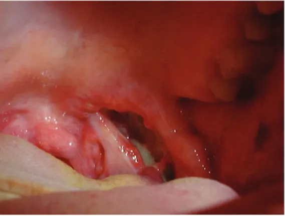
Figure 1 - Bacterial tonsillitis and abscess.
and extensive drainage of the neck. The platysma, superfi-cial trap muscles, omothyroids, and sternocleidomastoid
were removed. Subsequent culture revealed Streptococcus
viridans, Peptostreptococcus anaerobius, Peptostreptococcus micros, and Clostridium bifermentans. The patient was successfully treated with ampicillin and metronidazole. He gradually improved and was discharged from the hospital 18 days after admission.
Case 2
A 62-year-old man was admitted to the hospital for treat-ment of severe dermatomyositis. He received prednisone 80
mg/day. On the 12th hospital day he developed odynophagia
and dysphagia. The physical examination revealed bacte-rial tonsillitis, fever, and a palpable mass in the right sub-mandibular area. A CT scan showed diffuse enhancement of the superficial and deep right cervical fasciae; thicken-ing of the platysma, sternocleidomastoid, and strap mus-cles; and air and small fluid collections in multiple neck compartments. A right lateral cervicotomy with extensive
drainage was performed. Culture revealed S. viridans, and
Peptostreptococcus anaerobius. The patient was treated
with penicillin and metronidazole, but despite intensive care management, he developed mediastinitis and septic shock and died of multiple organ failure 8 days later.
Case 3
A 43-year-old man was admitted with tonsillitis and bi-lateral edema and erythema of the neck. He received cephalexin 2.0 gr. and sodium diclofenac for 3 days before admission, without improvement. A CT scan showed mul-tiple collections of air in the cervical region, without in-volvement of the upper mediastinum. The patient under-went bilateral cervical drainage through a midcervical in-cision. Culture revealed S. viridans and Clostridium sp. The patient was treated with penicillin and metronidazole and discharged 11 days after admission.
Case 4
A 40-year-old woman was admitted with tonsillitis, edema of the left neck, and trismus. The physical examina-tion revealed a palpable mass in the left submandibular area suggestive of a cervical abscess. A CT scan showed fluid collections with air in the left cervical region. A left ante-rolateral cervicotomy with drainage was performed. Culture revealed S. viridans and Corynebacterium sp. The patient was treated with penicillin and metronidazole and dis-charged after 23 days.
DISCUSSION
Cervical necrotizing fasciitis was defined by Monreland et al.9 and modified later to include infections beyond the
submandibular space as follows: (1) inflammation in the submandibular space, with little or no suppuration and with spread to the neck beyond the level of the hyoid bone; (2) involvement of more than one neck space, usually bilat-eral; (3) tissue necrosis with serosanguineous, putrid infil-tration; (4) involvement of connective tissue and fasciae and secondary in muscles and skin, but not of glandular struc-tures; and (5) contiguous—not lymphatic—spread.
Klabacha et al.10 proposed staging these inflammatory
processes as follows: type I—suppuration infection con-fined to the epidermis; type II—infection extending to the dermis; type III—infection extending to the superficial musculoaponeurotic layer of the face and superficial layer of the deep cervical fascia; type IV—infection extending to the musculature.
Normally, infection progresses rapidly and can involve the vascular structures causing small vessel thromboses. Necrotizing fasciitis of the head and neck usually results from polymorphic flora including numerous bacilli and cocci and occurs around the scalp and facial areas or pri-marily in the neck. In a series of 45 cases,3 anaerobes were
isolated in 32 cases (71%). Anaerobes only were isolated in 10 cases (22%), while a mixture of anaerobic and aero-bic organisms were found in 22 cases (49%). Aeroaero-bic bac-teria were only found in 3 patients (7%). Streptococcus sp.
were found in 8 cases. In a study of 20 cases,11
polymor-phic flora were also found, with aerobic gram positive cocci isolated in 18 cases and anaerobic flora in 11. Streptococ-cus sp. were found in 6. In this group,a mixed aerobic and anaerobic flora was identified, with a prevalence of Strep-tococcus sp.
Mortality for this condition have been reported to be
22% (10 of 45)3 and 50%12 but it has been decreasing in
recent years due to early diagnosis and advanced treatment in intensive care units. Even so, 1 patient in our 4 cases died (25%).
The most common clinical presentations are painful edema, erythema, warmth, tenderness, crepitation, and sub-mandibular abscess. Patients can develop mediastinitis and consequent septic shock.
Fasciitis is generally due to a dental or pharyngeal abscess (surgical or post-traumatic),3,11 radiotherapy,6 or unknown
ori-gin7). Tonsillar origin is rare; nevertheless, tonsillitis must be
fail-Table 1 - Review of all cases found in literature8.
Author/year N Cultures Associated diseases Complications Antibiotics Outcome
Wenig 1984 2 SH, FN, BM, EL, DM Thoracic and jugular P, Cl, G, C, CH Both died
PA, SS, SF, BF, CS vein thrombosis
Lalwani 1991 2 SH, MAF,BM - Mediastinal and thoracic C, M, P Both survived
Tovi 1991 1 E, PM, KP - Meningitis, mediastinitis, INA Died
and lung abscess
Scott 1994 1 SM, MAF - Mandible necrosis C, F, M, P Survived
Greinwald 1995 1 SE DM Thoracic AS, I Survived
Yii 1996 1 SM, MAF DM Thoracic and pharyngeal P, F, M, G Survived
perforation
Skitarelic 1999 1 SE, SH - - AS, G, I Survived
Djupesland 2000 3 SH, SM, BM B Mediastinal and thoracic P, M, T 1 died, 2 survived
Skitarelic 2003 1 SE - - P, AS, G, I Survived
TOTAL 1 3 5 died (24%),
12 survived Cultures: SH – Streptococcus hemolyticus, FN – Fusobacterium nucleatum, BM – Bacteroides melaninogenicus, EL – Eubacterium lentum, PA – Peptostreptococcus anaerobius, SS – Streptococcus sanguis, SF – Streptococcus faecalis, SV – Streptococcus viridans , BF –
Bacteroides fragilis, CS – Corynebacterium sp., MAF – Mixed anaerobic flora, E – Enterobacter, PM – Proteus mirabilis, KP – Klebsiella
pneumoniae, SE – Staphylococcus epidermidis, SM – Staphylococcus milleri, CL – Clostridium sp. Antibiotics: P – penicillin, G –
gentamicin, Cl – clindamycin, C – cefazolin, CH – chloramphenicol, M – metronidazole, AS – ampicillin/sulbactam, I – imipenem sodium, F – flucloxacillin, T – tobramycin, A – ampicillin, INA – Information not available. DM – diabetes mellitus, B – Bechterew’s disease, D – dermatomyositis
ure, old age, hypothyroidism, alcoholism, neoplasms, cirrho-sis, drug abuse, poor nutritional state, or use of corticosteroids.1,4,11 In our patients, 2 out 4 had
immunosup-pressive conditions due to diabetes and corticosteroid use. The divisions and subdivisions of the deep cervical fas-ciae determine the limits and the spread of the infectious process. Nevertheless, it is difficult to detect this infection before it has spread to the deep fasciae, when complications are more frequent.
Complications of cervical necrotizing fasciitis include airway obstruction, pneumonia, pulmonary abscess, septic shock, jugular venous thrombophlebitis, and mediastinitis. In order to exclude the cervical necrotizing fasciitis diag-nosis, x-ray of the affected area must reveal an absence of gas in the soft tissue. CT scan has been advocated for de-tecting gas, identifying the spread of infection in vascular sheaths, and detecting the extension of infection to remote areas (mediastinitis and pleural or pericardial effusions).13
Effective treatment and management of cervical necro-tizing fasciitis is based on early recognition, aggressive sur-gical intervention, use of broad-spectrum antibiotics, and supportive therapy. It is important to explore and drain all involved fascial planes. To carry out an adequate, wide, ex-tensive fasciotomy with exposure and exploration of all compartments, the surgeon must understand the complex anatomy of the cervical fasciae and deep neck spaces.14 This
was promptly performed in our patients with good results in 3 of 4 patients. In 1 patient, in spite of adequate surgical
treatment and appropriate antibiotic coverage, the infection spread rapidly to the mediastinum.
Other therapeutic considerations include management of hypocalcemia, anemia, and hypovolemia. Hypocalcemia results from the combination of ionic calcium with fatty ac-ids from fat necrosis, forming an insoluble soap. Intrave-nous calcium replacement may be required. Hemolysis of erythrocytes occurs from the actions of bacterial enzymes
and can result in anemia that may require transfusion.11
Fluid from extracellular edema usually occurs and requires crystalloid replacement.
In necrotizing fasciitis, the synergistic action of anaero-bic and facultative bacteria in a hypoxic environment pro-motes the production of destructive tissue enzymes and endotoxins that suppress host defenses, resulting in local and systemic dissemination of infection. Hyperbaric oxy-gen has been reported to be an effective adjuvant therapy to antibiotics and surgery in the treatment of necrotizing fasciitis of the extremities and trunk, witha mortality rate of 23% in the group receiving hyperbaric oxygen versus
66% in the group without hyperbaric oxygen.15 In another
study, the use of adjuvant hyperbaric oxygen therapy with an aggressive surgical approach and broad-spectrum intra-venous antibiotics in 12 patients with cervical necrotizing
fasciitis produced good results.15,16 Hyperbaric oxygen
RESUMO
ZILBERSTEIN B e col. Fasceite necrotizante cervical
secundária a amigdalite bacteriana. CLINICS 60(2):
177-182, 2005.
A fasceite necrotizante cervical é uma infecção grave de partes moles do pescoço. Trata-se de entidade rara, po-rém quando presente tem como principal origem um foco infeccioso odontogênico. São descritos 4 casos de fasceite necrotizante cervical a partir de tonsilites e abscesso peritonsilar, os quais, foram admitidos e tratados na unida-de unida-de terapia intensiva. Um dos pacientes era portador unida-de Diabetes Melittus não insulino- dependente e outro paci-ente havia recebido corticoterapia antes do desenvolvimen-to da infecção. Em um dos casos ocorreu mediastinite,
in-suficiência respiratória e o paciente evolui para o óbito em decorrência de choque séptico. Durante o tratamento, 3 pa-cientes evoluíram satisfatoriamente devido ao diagnostico precoce, tratamento cirúrgico agressivo e utilização de an-tibiótico terapia de largo espectro. A bactéria mais
comumente encontrada foi o Streptococcus sp, mas flora
mista com germes anaeróbios foi encontrada em 3 dos ca-sos descritos.
CONCLUSÕES: Deve-se suspeitar de tonsilite e absces-so peritonsilar como causa de fasceite necrotizante cervical para que tratamento agressivo e precoce seja realizado.
UNITERMOS: Fasceite necrotizante cervical.
Tonsilite. Abscesso peritonsilar.
Tonsillitis and peritonsillar abscesses are uncommon causes of cervical necrotizing fasciitis, with only 13 cases reported in literature8 (Table 1). We report on 4 patients with
cervical necrotizing fasciitis due to tonsillitis occurring within 8 months. One patient was diabetic and 2 were re-ceiving steroidal or nonsteroidal anti-inflammatory agents. One patient developed acute respiratory distress syndrome requiring mechanical ventilation and presented septic shock due to spread of infection to the mediastinum. Three
patients recovered because of early diagnosis, aggressive surgical intervention, use of appropriated broad-spectrum antibiotics, and supportive therapy.
REFERENCES
1 . Aimioni C, Cilione A R, Grandi E, Lombardi L, Merlo R, Pastore A. Cervical necrotizing fasciitis. Eur Arch. Otorhinolaryngol 1999;256:510-3.
2 . Reed JM, Anand VK. Odontogenic cervical necrotizing fasciitis with intrathoracic extension. Otolaryngology – Head and Neck Surgery 1992;107:596-600.
3 . Mathieu D, Neviere R, Teillon C, Chagnon JL. Cervical necrotizing fasciitis: clinical manifestations and management. Clin Infect Dis 1995;21:51-6.
4 . Kantu S, Harl-EL G. Cervical necrotizing fasciitis. Ann Otol Rhinol Laryngol 1997;106:965-70.
5 . Ferri E, Ianniello F, Salandin S. Necrotizing fasciitis of the neck in a patient with acute myeloid leukemia: clinical features, therapeutic strategy and review of the literature. Acta Otorhinolaryngol Ital 1998;18:116-22.
6 . Mortimore S, Thorp M. Cervical necrotizing fasciitis and radiotherapy: a report of two cases. J Laryngol Otol 1998;112:298-300.
7 . Gillis AR, Gillis TM. Necrotizing cervical fasciitis of unknown origin. J Otolaryngol 1992;21:171-3.
8 . Skitarelic N, Mladina M, Morovic M, Skitarelic N. Cervical necrotizing fasciitis: sources and outcomes. Infection 2003;31(1):39-44.
9 . Moreland LW, Corey J, Mackenzie R. Ludwig’s angina: case report and review of the literature. Arch Intern Med 1988;148:461-6.
10. Klabacha ME, Stankiewicz JA, Clift SE. Severe soft tissue infection of the face and neck: A classification. Laryngoscope 1982;92:1135-9.
11. Mohammedi I, Ceruse P, Duperret S, Vedrinne JM. Cervical necrotizing fasciitis: 10 years experience at a single institution. Intens Care Med 1999;25:829-34.
12. Krespi YP, Lawson W, Blaugund SM, Biller HF. Massive necrotizing fasciitis infections of the neck. Head Neck Surg 1981;3:475-81.
13. Becker M, Zbaren P, Hermans R, Becker CD, Marchal F, Kurt AM, et al. Necrotizing fasciitis of the head and neck: role of computed tomography in diagnosis and management. Radiology 1997;202:471-6.
14. Durazzo MD, Pinto FR, da Rocha Loures MS et al. Os espaços cervicais profundos e seu interesse nas infecções da região. Rev Ass Med Bras 1997;43:119-26.
15. Risman J, Zamboni AZ, Curtis A, Graham DR, Konrad HR, Ross DS. Hyperbaric oxygen therapy for necrotizing fasciitis reduces mortality and need for debridements. Surgery 1990;108: 847-50.