INTRODUCTION
Address to: Dra Janaína Luz Narciso-Schiavon. Depto Clínica Médica/HU Polydoro Ernani de São Thiago/UFSC. Rua Professora Maria Flora Pausewang s/no, 3º andar, Trindade, 88040-900 Florianópolis, SC, Brasil.
Phone: 55 48 3721-9014 e-mail: [email protected] Received 3 May 2013
Accepted 17 July 2013
Positive serology for viral hepatitis and donor
self-exclusion in Southern Brazil
Julia De Luca Maccarini
[1],
Carlos Alberto Kuntz Nazario
[1],
Jovino dos Santos Ferreira
[2],
William Queiroz Guimarães Wiegandt Ceglio
[2],
Rômulo Cavalcante Serpa
[2],
Vera Lúcia Paes Cavalcanti Ferreira
[2],
Leonardo de Lucca Schiavon
[1]and Janaína Luz Narciso-Schiavon
[1][1]. Núcleo de Estudos em Gastroenterologia e Hepatologia, Universidade Federal de Santa Catarina, Florianópolis, SC. [2]. Serviço de Hemoterapia, Hospital Universitário Polydoro Ernani de São Thiago, Florianópolis, SC.
ABSTRACT
Introduction: Despite the great advances in serological testing for transfusion-transmitted infections, the selection of blood donors by blood bank operators remains the only way to avoid transmission within the testing window period. Part of this selection is the self-exclusion form, on which the donors can exclude their blood from donation without any explanation. This study assessed the clinical and epidemiological characteristics related to positivity for viral hepatitis and to the use of the
confi dential self-exclusion (CSE) form. Methods: This transversal study analyzed the data collected from blood donors’ fi les in
a hospital in Southern Brazil. Univariate and multivariate analyses identifi ed the clinical and epidemiological variables related to
positive serologies of viral hepatitis and to whether the donor was self-excluded. Results: Of the 3,180 donors included in this study, 0.1% tested positive for HBsAg, 2.1% for anti-HBc, and 0.9% for anti-HCV. When the 93 donors with positive serologies for viral hepatitis were compared with those who were negative, a greater proportion of the positive serology group was found
to have had a history of blood transfusions (OR=4.908; 95%CI=1.628 - 14.799; p<0.01), had repeatedly donated (OR=2.147; 95%CI=1.236 - 3.729; p<0.01), and used the CSE form for self-exclusion (OR=7.139; 95%CI=2.045 - 24.923; p<0.01). No
variables were independently associated with self-exclusion. Conclusions: A history of blood transfusion, repeated donations, and self-exclusion are factors that should be considered during viral hepatitis screenings in blood banks.
Keywords: Hepatitis B. Hepatitis C. Blood donors.
Worldwide, there are approximately 400 million hepatitis
B virus (HBV) carriers and 170 million individuals chronically infected with the hepatitis C virus (HCV)1,2. The prevalences
of hepatitis B in south-central Brazil and the northern region of Brazil are 2 and 8%, respectively2. The prevalence of hepatitis C is 0.8% among all Brazilian blood donors (2.1% for northern donors and 0.7% for southern donors)3-5. Although
hepatitis B has a high risk of vertical, horizontal, and sexual transmission, parenteral transmission is the most common means of transmission for both hepatitis B and C1. The main risk factors
for parenteral transmission are intravenous drug use and blood and hemocomponent transfusions1.
In the State of Santa Catarina, in Southern Brazil, the
residual risk of HBV transmission by transfusion during the
testing window period is estimated to be 1 in every 15,400
donations, which is 16 times higher than that reported in the United States (1:250,000)6,7. For HCV, the residual risk is one in
every 127,300 donations, which is 12 times higher than the risk found in the United States7. In Australia, this risk is estimated
at approximately 1 in 633,000 transfusions for hepatitis B and 1 in 6,387,000 transfusions for hepatitis C8.
Post-transfusion hepatitis (PTH) was fi rst described in
1943. Studies evaluating the risk criteria for transmissible
infectious diseases in blood donors began in the 1960s and
1970s and compared voluntary and commercial donations9-11.
For a long time, a viral etiology was suspected for PTH; in 1965, the Australia antigen was discovered and was later called the hepatitis B surface antigen (HBsAg)12-15. After the
discovery of the hepatitis A virus and its exclusion as the PTH cause, it became clear that a large portion of PTH was caused
by other unknown viruses; such infections were called non-A non-B hepatitis (NANB hepatitis)16-18. In the 1970s and 1980s,
extensive research was conducted to establish the etiologic agent of NANB hepatitis, leading to the discovery of the hepatitis C virus in 198919,20. Since then, recent studies have established that
80-90% of NANB hepatitis is caused by HCV12,19-21.
Safety in blood donation is a major concern at blood transfusion centers22. Efforts to guarantee safety focus on
METHODS RESULTS
risk of transfusion transmissible infections23. Despite the
development of assays to detect infectious diseases, transmission by transfusion still occurs for reasons such as the testing window period, the convalescent phase of an HBV infection, or a chronic HBV infection with a low HBsAg7,24-26. Additionally, new pathogens transmitted by risky behavior that are not yet tested in blood donors may appear26.
The selection of donors is still the only way to prevent the transmission of infected blood donated during the immunological testing window8,27,28. One of the steps in this selection is the confi dential self-exclusion (CSE) form on
which the blood donors can exclude their donated blood without
providing any justifi cation28,29. Donors who self-exclude could
do so because they think that they have risk factors or because they are donating blood to obtain their serological test results29.
The fi rst CSE system was developed in the United States in
1984, and since then, similar systems have been used in several countries22. In Brazil, the use of CSE has been mandatory with
blood donations since 200330,31. Few studies have evaluated the
effectiveness of CSE in preventing the transmission of infectious diseases by blood donation26,28, so this study aimed to evaluate the clinical and epidemiological characteristics associated with a positive serology for hepatitis B and C and with the use of the CSE form.
Subjects
This cross-sectional study reviewed the records of blood donations at the Blood Bank of the University Hospital of the
Federal University of Santa Catarina (HU/UFSC). The subjects
were adults who met the selection criteria of the blood bank and who donated blood between January 2009 and December 2010.
Individuals who were seropositive for human immunodefi ciency virus (HIV), had insuffi cient records of clinical and laboratory
data, and made repeated donations were excluded from the study
(only the fi rst donation was included).
Methods
Epidemiological, laboratory, and other clinical variables were obtained from histories taken during routine donations at the blood bank. Patients were evaluated for a positive viral hepatitis serology. Those individuals who showed a positive or indeterminate HBsAg, a reactive or indeterminate
antibody to hepatitis B core antigen (anti-HBc), or a reactive or
indeterminate anti-HCV were considered positive.
The clinical and epidemiological variables assessed
in the study were the following: age (in years), gender, ethnicity (Caucasian or not), marital status (married, in a stable relationship, single, separated, divorced, or widowed), education (in years), having a homosexual relationship, body mass index (defi ned as weight/height²), being a healthcare
professional, history of previous surgery, history of receiving
blood transfusions, sexual promiscuity (two or more partners in the last year), previous use of illicit drugs, tattoos, reason for
donation (spontaneous: unselfi sh donation, made with a caring attitude; or directed: donation bound or tied to a patient), fi rst
donation or not, donation with the intention of obtaining the
serology results, and CSE status (suggestion of the donor after the interview that their blood should be discarded).
Statistical analysis
The continuous variables were analyzed using Student's t-test or the Mann-Whitney test, when appropriate. The
categorical variables were compared using the chi-square (χ2) or Fisher's exact test. A p-value < 0.05 indicated statistical signifi cance. A comparative analysis of the data was performed
according to the serological positivity for viral hepatitis. A logistic regression analysis was used to identify the variables that were independently associated with the presence of positive serologic tests for viral hepatitis. An analysis comparing the data with the CSE decision was also performed. All of the tests were performed by the statistical program Statistical Package for the
Social Sciences, Version 17.0 (SPSS Inc., Chicago, IL, USA).
Ethical considerations
This study complied with the ethical recommendations of the Helsinki Declaration of 2000 and was approved by our institutional review board under the number 1900.
Patient characteristics
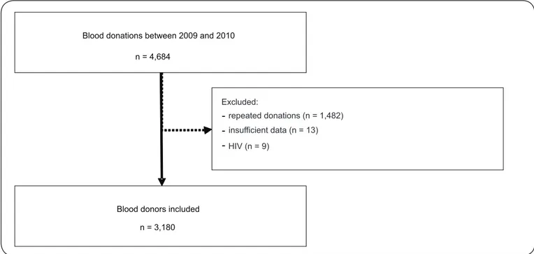
During 2009 and 2010, 4,684 blood donations were collected
at the Blood Bank of the HU/UFSC. Of these donations, 1,482 were excluded because they were donated by the same donor
(repeated donations), 13 were excluded due to insuffi cient data, and 9 were excluded due to reactive HIV serology. Therefore,
the study included 3,180 individual donors (Figure 1). Of the donors with a positive serology for viral hepatitis,
two had a positive or indeterminate HBsAg test (both were also reactive for anti-HBc). Sixty-three donors were reactive for
anti-HBc alone, and 28 were anti-HCV reactive or indeterminate
(2 of these 28 donors were also reactive for anti-HBc), totaling
93 individuals with a positive serology for viral hepatitis. Among the donors who presented as reactive for anti-HBc alone, 43 were also tested for anti-HBs, with 24 found to be reactive, 18 found to be negative, and 1 with an indeterminate result (Figure 2).
The demographic characteristics of the 3,180 blood donors included in the study were as follows: the mean age was 27.2 ± 9.7 years, 59.4% were men, 96.2% were Caucasian, 20.8% were married or were in a stable relationship, 97.2% had undergone more than 8 years of education, and 23.4% were either overweight or obese. The epidemiological characteristics of the donors were as follows: 9.9% were health professionals,
21.6% had undergone previous surgery, 0.9% had undergone
blood transfusion, 0.3% had previously used illicit drugs, and 8.1% had a tattoo. With regard to the reasons for donating
Blood donations between 2009 and 2010
n = 4,684
Blood donors included
n = 3,180
Excluded:
-
repeated donations (n = 1,482)-
insufficient data (n = 13)-
HIV (n = 9)FIGURE 1 - Flow diagram of the potential candidates for inclusion in the study, reasons for exclusion, and subjects enrolled. HIV: Human immunodefi ciency virus.
Tested for anti-HBS n = 43 Isolated anti-HBc+
n = 63
Anti-HCV+
n = 28
Anti- HBc+ n = 2 HBsAg+
n = 2
Blood donors with positive serology
for viral hepatitis
n = 93
An- HBs+ n = 24
A nti -HBs negative n = 18
A nti -HBs indeterminate n = 1
Anti- HBc+ n = 2
donation), 55.3% donated for the fi rst time, and 0.1% donated to
obtain the results of the blood exams. Among all the individuals included in the study, 0.7% chose self-exclusion of their blood.
Comparative analysis of individuals with positive serology
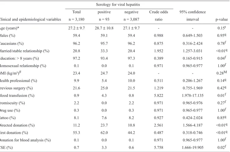
Compared with individuals with negative serology for viral hepatitis, those with positive serology had higher rates
of being in a stable relationship or marriage (33.3% vs 20.4%, p < 0.01), having a past history of blood transfusion (4.3% vs 0.8%, p < 0.01), giving a directed donation (23.7% vs 10.8%, p < 0.01), giving their fi rst donation (62% vs 44.2%, p < 0.01), and choosing to self-exclude (3.3% vs 0.6%, p = 0.02)
(Table 1). Furthermore, donors with positive serology had less frequently attended the complete compulsory education program
(93.4% vs 97.3%, p = 0.04).
The multivariate analysis showed that the following variables were independently associated with positive serology for viral
hepatitis: history of previous blood transfusions (adjusted OR=4.908, 95%CI=1.628-14.799, p < 0.01), directed donation (adjusted OR=2.147, 95%CI=1.236-3.729, p < 0.01), and choosing to self-exclude (adjusted OR=7.139, 95%CI=2.045-24.923, p < 0.01).
TABLE 1 - Distribution of the clinical and epidemiological variables of 3,180 blood donors at the University Hospital of the Federal University of Santa Catarina between 2009 and 2010, according to their viral hepatitis serology results.
Serology for viral hepatitis
Total positive negative Crude odds 95% confi dence
Clinical and epidemiological variables n = 3,180 n = 93 n = 3,087 ratio interval p-value
Age (years)* 27.2 ± 9.7 28.7 ± 10.8 27.1 ± 9.7 - - 0.15t
Males (%) 59.4 59.1 59.4 0.988 0.649-1.503 0.95q
Caucasians (%) 96.2 95.7 96.2 0.875 0.316-2.424 0.78f
Married/stable relationship (%) 20.8 33.3 20.4 1.952 1.257-3.031 <0.01q
Education: > 8 years (%) 97.2 93.4 97.3 0.389 0.165-0.915 0.04f
Homosexual relationship (%) 0.1 0.0 0.1 0.971 0.965-0.977 1.00f
BMI (kg/m²)# 23.4 24.7 24.0 - - 0.28M
Health professional (%) 9.9 5.4 10.0 0.511 0.206-1.267 0.14q
Previous surgery (%) 21.6 25.0 21.5 1.219 0.755-1.969 0.42q
Blood transfusion (%) 0.9 4.3 0.8 5.822 1.978-17.135 0.01f
Promiscuity (%) 2.2 0.0 2.2 0.971 0.965-0.976 0.27f
Drug use (%) 0.3 0.0 0.3 0.971 0.965-0.977 1.00f
Tattoo (%) 8.1 7.6 8.2 0.927 0.424-2.024 0.85q
Directed donation (%) 11.2 23.7 10.8 2.561 1.566-4.187 <0.01q
First donation (%) 55.3 62.0 44.2 0.487 0.318-0.746 <0.01q
Donation for blood analysis (%) 0.1 0.0 0.1 0.971 0.965-0.977 1.00f
CSE (%) 0.7 3.3 0.6 5.758 1.666-19.905 0.02f
BMI: body mass index; CSE: confi dential self-exclusion; *Mean ± standard deviation; #Median; tStudent’s t-test; mMann-Whitney test; qchi-squared test; fFisher’s exact test.
Comparative analysis of individuals with confi dential self-exclusion
We did not observe any signifi cant differences between the
donors who did or did not choose CSE of the donated blood with
regard to age (p = 0.63), gender (p = 0.51), ethnicity (p = 0.56), marital status (p = 1.00), education (p = 1.00), sexual orientation (p = 1.00), body mass index (p = 0.13), being a health professional (p = 1.00), history of previous surgery (p = 0.28), history of previous blood transfusions (p = 1.00), sexual promiscuity (p = 1.00), illicit drug use (p = 1.00), tattoos (p = 1.00), directed donation (p = 0.16), fi rst donation (p = 0.79), or donation to obtain blood test results (p = 1.00) (Table 2).
DISCUSSION
Research on blood donors has shown a predominance of
male donors, with a prevalence of 63.9-71.3%32-35. With respect to ethnicity, 96.2% of blood donors in the present study were
prevalence of single (79.2%) donors and donors who completed high (97.2%) school was greater than that found in previous studies (34.4-49.0% and 54.7-63.7%, respectively)33,34, possibly refl ecting the university population in this state.
The mean age in the present study was 27.2 ± 9.7 years. When the donors were distributed by age group, we found that the largest number of donations were from individuals
between 20 and 24 years of age (35.2% of donors). This
result is inconsistent with the results of the Epidemiological
Profi le of Brazilian Blood Donor study performed in 2004,
which indicated a higher frequency of donations from
donors between 30 and 39 years of age (28.3% in Brazil and 27% in Southern Brazil)34. In similar studies, the mean age of the donors was found to be between 36.5 ± 11.0 and 45.0 + 10.9 years32,33. This difference can be explained by the
location of the blood bank at a university hospital and by the several blood donation campaigns promoted in every course,
especially during the fi rst year of university study. Sharma
et al.37 reported that when the donor group is composed of
mostly student volunteers, a lower proportion of donors with positive serology is observed37. Other studies addressing the
epidemiological characteristics of blood donors showed that the presence of serological markers increases with the age of the donor33,38.
TABLE 2 - Comparative analysis of the clinical and epidemiological characteristics of 3,180 blood donors at the University Hospital of the
Federal University of Santa Catarina according to their confi dential self-exclusion status.
Self-exclusion
yes no
Clinical and epidemiological variables n = 21 n = 3,159 p-value‡
Age (years)* 26.1 ± 9.6 27.2 ± 9.7 0.63
Males (%) 52.4 59.5 0.50
Caucasians (%) 95.2 96.2 0.56
Married/stable relationship (%) 19 20.8 1.00
Education: > 8 years (%) 100 97.2 1.00
Homosexual relationship (%) 0.0 0.1 1.00
BMI (kg/m²)# 22.8 23.4 0.13
Health professional (%) 9.5 9.9 1.00
Previous surgery (%) 9.5 21.6 0.28
Blood transfusion (%) 0.0 0.9 1.00
Promiscuity (%) 0.0 2.2 1.00
Illicit drug use (%) 0.0 0.3 1.00
Tattoo (%) 4.8 8.2 1.00
Directed donation (%) 0.0 11.2 0.16
First donation (%) 47.6 44.8 0.79
Donation for analysis (%) 0.0 0.1 1.00
BMI: body mass index; *Mean ± standard deviation; #Median; ‡Student’s t-, Mann-Whitney, χ2,or Fisher’s exact test, when appropriate for
group comparisons.
The prevalence of viral hepatitis markers in this study
(HBsAg: 0.1%, anti-HBc: 2.1%, anti-HCV: 0.9%) was lower
compared with that in countries of high endemicity and similar to levels observed in areas with a low prevalence of these
infections. In a study in Pakistan, 4.7% of the donors were
found to be HBsAg-positive39. In Vietnam, the prevalences
were 11.4 and 51.7% for HBsAg and anti-HBc, respectively40.
These countries are located in South and Southeast Asia, regions with a high prevalence of hepatitis B41. In the United
States, the prevalence of HBsAg in blood donors is 0.2%42. In
Europe, the distribution of individuals with HBsAg positivity is
heterogeneous and varies according to region; Northern Europe has the lowest prevalence (less than 0.1%), Western Europe has
a prevalence of 0.1-0.5%, and Southern and Eastern Europe have higher rates of 2.4%43.
Worldwide, Egypt has the highest number of individuals
with hepatitis C infection, with 16.8% of their donors being
anti-HCV reactive44. However, the prevalence of hepatitis C
infection in the general population of developed countries varies from 1-2%45. In the United States, for example, approximately
0.5% of volunteer blood donors are anti-HCV reactive45.
The prevalence of positive serology for viral hepatitis found in this study is similar to those reported previously for Brazil
anti-HCV: 0.34-1.14%)46-48. The prevalence of hepatitis B markers in Brazil varies greatly by region. The literature shows that Southern Brazil is an area of low endemicity, but the Western State of Santa Catarina has high endemicity. Data
from 2001 showed an HBsAg prevalence of 0.6% in the blood
donors in Santa Catarina, 1.5% in the Western region, 0.3% in
the Southern region, and 0.6% in the capital, Florianópolis48.
The anti-HBc rates also vary by location, with rates of 5.4% state-wide, 12.7% in the Western region, 2.9% in the Southern region, and 3.5% in Florianópolis48. Although the presence of
anti-HBc antibodies in individuals who are HBsAg-negative
may represent a previous infection (anti-HBs reactive), there are
reports of the transmission of hepatitis B by blood transfusion from individuals in this group. For this reason, in places of intermediate prevalence, such as Brazil, anti-HBc-reactive individuals are treated as carriers, and the donated blood bags are discarded. A prevalence of 3% positivity for anti-HCV is estimatedin the world population1. However, in Brazil, there is
a prevalence of 0.8% of anti-HCV reactivity in blood donors1.
Data indicate a prevalence of 0.3% of infection in the entire
State of Santa Catarina (0.2% in the west, 0.4% in the south, and 0.5% in the capital)48.
Although blood transfusion is a recognized risk factor for the transmission of hepatitis B and C, PTH is currently virtually eradicated and is limited to a residual risk estimated only by mathematical models7. However, there is evidence suggesting
that the global epidemic of HCV infection during the second half of the 20th century was initiated and maintained by the increasing
use of parenteral therapies and blood transfusions49. The main
factors contributing to the reduction of post-transfusion hepatitis were the exclusion of commercial donations and the introduction in blood blanks of a serological screening program that was made progressively more sensitive11. After the implementation
of mandatory HBsAg tests for blood donors in the United States in 1972, there was a 25% reduction in the rate of PTH in that country11. In Brazil, serological tests for HBV did not become
mandatory until 197850. Eighteen percent of multi-transfused
patients were infected with HCV in the period before the
introduction of mandatory screening for anti-HCV in 1993;
afterward, the infection rate was reduced to 1.4%5,51.
Paid donations have been banned in Brazil for the past 33 years, although there are still some rewards provided to donors, such as free meals after donation33,52. The experiences
of countries that stopped making remunerations to donors have
shown that altruistic donations are insuffi cient to maintain
a positive balance in the blood banks52. Linked donations,
including donations among friends and relatives of patients who need transfusions52, are needed. These types of donations
are performed by a wide spectrum of donors, ranging from family and friends to individuals who present themselves as such but who are actually receiving payment from the patient’s family53. Several studies have demonstrated a relationship
between directed donation and higher rates of positive serology for infections transmitted by blood transfusions37,54-56. Dorsey et al. observed that this result could be explained by a greater
number of fi rst-time donators54.
In this study, a self-exclusion prevalence of 0.7% was found,
which means that 22 bags of blood were discarded between 2009 and 2010 due to self-exclusion. This result is consistent with the
fi ndings of another study conducted in Southern Brazil, where
0.5% of the donations were discarded due to self-exclusion33.
However, studies performed in Uberaba, State of Minas Gerais and Londrina, State of Paraná, showed higher self-exclusion
rates (2.7 and 3.2%, respectively)57,58. Although no clinical or
epidemiological variables were related to CSE in this study,
previous studies have shown that males and fi rst-time donors
respond yes on the CSE form more frequently28,57-60. However, the greater the number of repeated donations, the fewer times the CSE form was used to self-exclude the donor’s blood57,60.
Although a relationship between CSE and positive serology for viral hepatitis has been previously demontrated22,57,61, the ability of this practice to identify individuals in the testing window period was not previously addressed. The sensitivity and positive predictive values of CSE are quite low22,58,61-63, leading to the disposal of many blood bags from potentially healthy donors. The main factors related to problems with the
CSE form are the diffi culty of understanding the question26,28,57,58, the low socioeconomic status of the donors58,59, and the
ineffective explanations by health professionals58. Another
aspect to be considered is the varied format and content of the form, which are different for each blood bank57,58. It was
shown that blood banks that use forms with plain text and color
pictures have a lower frequency of affi rmative responses to the
self-exclusion choice, leading to a reduction in the disposal of low-risk blood bags and to an increase in the sensitivity of the procedure. Kean et al.64 observed a reduction of self-exclusion from 0.7% to 0.3% when the donor was properly and timely orientated to the CSE form64. Sumnig et al. demonstrated that the self-exclusion rate decreased from 0.7% to 0.5% after a change in the format of the question26.
Even with a high sensitivity and a signifi cant positive
predictive value, Martins et al.57 found three cases of positive serology after self-exclusion in less than 6 months (two cases of hepatitis C and one of HIV)57. Other longitudinal studies should
be conducted to address the real value of CSE. Educational measures, changes in the format and content of self-exclusion forms, and campaigns aimed to increase donors’ motivation to re-donate rather than only captivate new donors are possible actions that may decrease the rate of self-exclusion and avoid the loss of safe donations.
Considering the literature and the fi ndings of this study, it
ACKNOWLEDGMENTS
REFERENCES
The authors declare that there is no confl ict of interest.
CONFLICT OF INTEREST
This paper is presented as partial fulfi llment of the requirements
for the Medical Doctor (MD) degree from the Federal University of Santa Catarina.
1. Violantea MD, Nunes-Nateras R. Epidemiology of Hepatitis Virus B and
C. Arch Med Res 2007; 38:606-611.
2. Te HS, Jensen DM. Epidemiology of Hepatitis B and C Viruses: A Global
Overview. Clin Liver Dis 2010; 14:1-21.
3. Alleyne GA. Ensuring safe blood in the Americas. Rev Panam Salud
Publica 2003; 13:65-67.
4. Ropero AM, Danovaro-Holliday MC, Andrus JK. Progress in vaccination
against hepatitis B in the Americas. J Clin Virol 2005; 34 (suppl II):14-19.
5. Sociedade Brasileira de Hepatologia. Relatório do Grupo de Estudo da
Sociedade Brasileira de Hepatologia. Epidemiologia da Infecção pelo
Vírus da Hepatite C no Brasil. Gastroenterol Endosc Dig 1999; 18:53-58.
6. Dodd RY, Notari EP, Stramer SL. Current prevalence and incidence of infectious disease markers and estimated window-period risk in
the American Red Cross blood donor population. Transfusion 2002;
42:975-979.
7. Maresch C, Schluter PJ, Wilson AD, Sleigh A. Residual infectious disease risk in screened blood transfusion from a high-prevalence population:
Santa Catarina, Brazil. Transfusion 2008; 48:273-281.
8. Polizzotto MN, Wood EM, Ingham H, Keller AJ, Australian Red Cross Blood Service Donor and Product Safety Team. Reducing the risk of transfusion-transmissible viral infection through blood donor selection: the
Australian experience 2000 through 2006. Transfusion 2008; 48:55-63.
9. Beeson PB. Jaundice occurring one to four months after transfusion of
blood or plasma. Report of seven cases. JAMA 1943; 121:1332-1334.
10. Grady GF, Chalmers TC. Risk of post-transfusion viral hepatitis. N Engl
J Med 1964; 271:337-342.
11. Walsh JH, Purcell RH, Morrow AG, Chanock RM, Schmidt PJ.
Posttransfusion hepatitis after open-heart operations. Incidenceafter
the administration of blood from commercial and volunteer donor
populations. JAMA 1970; 211:261-265.
12. Tobler LH, Busch MP. History of posttransfusion hepatitis. Clinical
Chemistry 1997; 43:1487-1493.
13. Blumberg BS, Alter HJ, Visnich S. A “new” antigen in leukemia sera.
JAMA 1965; 191:541-546.
14. Gocke DJ, Greenberg HB, Kavey NB. Hepatitis antigen. Detection of
infectious blood donors. Lancet 1969; 2:248-249.
15. Gocke DJ, Greenberg HB, Kavey NB. Correlation of Australia antigen
with posttransfusion hepatitis. JAMA 1970; 77:877-879.
16. Feinstone SM, Kapikian AZ, Purcell RH. Hepatitis A. Detection by immune electron microscopy of a virus-like antigen associated with acute
illness. Science 1973; 182:1026-1028.
17. Stevens CE, Silbert JA, Miller DR, Dienstag JL, Purcell RH, Szmuness W. Serologic evidence of hepatitis A and B virus infections in thalassemia
patients: a retrospective study. Transfusion 1978; 18:356-360.
18. Dienstag JL, Purcell HR, Alter HJ, Feinstone SM, Wong DC, Holland PV.
Non-A, non-B post-transfusion hepatitis. Lancet 1977; 1:560-562.
19. Ezzell C. Candidate cause identifi ed of non-A, non-B hepatitis [News]. Nature 1988; 333:195.
20. Choo QL, Kuo G, Weiner AJ, Overby LR, Bradley DW, Houghton M. Isolation of a cDNA clone derived from a blood-borne non-A, non-B viral hepatitis genome. Science 1989; 244:359-361.
21. Alter HJ. To C or not to C: these are the questions. Blood 1995; 85:1681-1695.
22. Kasraian L, Tavasoli A. Positivity of HIV, hepatitis B and hepatitis C in patients enrolled in a confi dential self-exclusion system of blood donation: a cross-sectional analytical study. Sao Paulo Med J 2010; 128:320-323 23. Eder AF, Menitove JE. Blood donations´ past, present, and future:
TRANSFUSION´S golden anniversary. Transfusion 2010; 50:1870-1877. 24. Lee WM. Hepatitis B virus infection. N Engl J Med 1997;
337:1733-1745.
25. Su PJ, Chen TC, Cheng HR, Li L, Lin KS, Kao JH, et al. The clinical signifi cance of occult hepatitis B transfusion in Taiwan - a look-back study. Transfus Med 2010; 1:33-41.
26. Sümnig A, Konerding U, Kohlmann T, Greinacher A. Factors infl uencing confi dential unit exclusions in blood donors. Vox Sanguinis 2010; 98: e231-e240.
27. Soldan K, Ramsay ME, Robinson A, Hall AJ. Estimation of the risk of hepatitis B virus, hepatitis C virus and human immunodefi ciency virus infectious donations entering the blood supply in England, 1993-2001. Vox Sanguinis 2003; 84:274-286.
28. Castro V. The role of confi dential unit exclusion on blood safety. Rev Bras Hematol Hemoter 2009; 31:213-214.
29. Sloand EM, Pitt E, Klein HG. Safety of the blood supply. JAMA 1995; 274:1368-1373.
30. Ministério da Saúde. Resolution - RDC Nº 153 - [Technical Roules to Hemotherapy Procedures]. Brasília: Diário Ofi cial da União; 2004. 31. Ministério da Saúde - Resolution - RDC Nº 343 – [Technical Roules to
Hemotherapy Procedures]. Brasília: Diário Ofi cial da União; 2003. 32. Baha W, Foullous A, Dersi N, They-They TP, Alaoui K, Nourichafi N, et al.
Prevalence and risk factors of hepatitis B and C virus infections among the general population and blood donors in Morocco. BMC Public Health 2013; 13:50.
33. Silveira L, Schiavon LL, Silva KP, Lopes TB, Zaccaron MR, Narciso-Schiavon JL. Clinical and epidemiological profi le of blood donors with positive serology for viral hepatitis in southern Brazil. Rev Soc Bras Med Trop 2011; 44:269-273.
34. Agência Nacional de Saúde (Anvisa). Brazilian Blood Donor Profi le. [Internet]; Brasília: Anvisa 2004 [Cited 2012 August 02]. Available at: http://www.anvisa.gov.br/hotsite/doador_sangue/pdsbfi les/pdf/Tabdoadores/ Brasil_d.pdf/.
35. Almeida-Neto C, Murphy EL. Prevalence of serologic markers for hepatitis B and C viruses in Brazilian blood donors and incidence and residual risk of transfusion transmission of hepatitis C virus. Transfusion 2013; 53:827-834.
36. Instituto Brasileiro de Geografi a e Estatística (IBGE). Brazilian Demographic Census [Internet]; Brasília: IBGE; 2010. [Cited 2012 August 08]. Available at: http://www.ibge.gov.br/home/estatistica/ populacao/censo2010/default.shtm/.
37. Sharma RR, Cheema R, Vajpayee M, Rao U, Kumar S, Marwaha N, et al. Prevalence of markers of transfusion transmissible diseases in voluntary and replacement donors. Natl Medic J India 2004; 17:19-21.
38. Chávez JH, Campana SG, Hass P. An overview of hepatitis B in Brazil and in the state of Santa Catarina. Rev Panam Salud Publica 2003; 14:91-96. 39. Mujeeb AS, Nanan D, Sabir S, Altaf A, Kadir M. Hepatits B and C
infection in fi rst-time blood-donors in Karachi - a possible subgroup for sentinel surveillance. East Mediterr Health J 2006; 12:735-741. 40. Viet L, Lan NT, Ty PX, Bjorkvoll B, Hoel H, Gutteberg T, et al. Prevalence
41. Candotti D, Allain JP. Transfusion-transmitted hepatitis B virus infection. J Hepatol 2009; 51:789-809.
42. Glynn SA, Kleinman SH, Schreiber GB, Busch MP, Wright DJ, Smith JW, et al. Trends in incidence and prevalence of major
transfusion-transmissible viral infections in US blood donors, 1991 to 1996. Retrovirus Epidemiology Donor Study (REDS). JAMA 2000; 284:229-235.
43. Goudeau A. Epidemiology and eradication strategy for hepatitis
B in Europe. The European Regional Study Group. Vaccine 1990; 8:S113-S116.
44. Awadalla HI, Ragab MH, Nassar NA, Osman MAH. Risk factors of hepatits C infection among egyptian blood donors. Cent Eur J Public
Health 2011; 19:217-221.
45. Di Bisceglie AM. Hepatitis C. Lancet 1998; 351:351-355.
46. Andrade AF, Oliveira-Silva M, Silva SGC, Motta IJF. Seroprevalence of
hepatitis B and C virus markers among blood donors in Rio de Janeiro,
Brazil, 1998-2005. Mem Inst Oswaldo Cruz 2006; 101:673-676.
47. Vasconcellos HCFF, Yoshida CFT, Vanderborght BOM, Schatzmayr HG. Hepatitis B and C prevalence among blood donors in south region of
Brazil. Mem Inst Oswaldo Cruz 1994; 89:503-507.
48. Rosini N, Mousse D, Spada C, Treitinger A. Seroprevalence of HBsAg, Anti-HBc and Anti-HCV in Southern Brazil, 1999-2001. Braz J Infec Dis
2003; 7:262-267.
49. Prati D. Transmission of hepatitis C virus by blood transfusions and other
medical procedures: A global review. J Hepatol 2006; 45:607-616.
50. Ministério da Saúde. State of São Paulo. Decree No. 12479 of 18 October
1978 [Internet]. Published in the Government Secretariat, on 18/10/1978;
1978. [Cited 2012 August 12]. Available at http://www.anvisa.gov.br/ legis/decretos/12479_78.htm/.
51. Ministério da Saúde. Ordinance No. 1376, of November 19, 1993
[Internet]. Published in the Diário Ofi cial da União on 02/12/1993;
1993. [Cited 2012 August 12]. Available at http://www.planalto.gov.br/
ccivil_03/decreto-lei/del1376.htm/.
52. Guerra CCC. Fim da doação remunerada de sangue no Brasil faz 25 anos.
Rev Bras Hematol Hemoter 2005; 27:1-3.
53. World Health Organization (WHO) [Internet]. WHO Global Database on
Blood Safety, 2004-2005. Geneva: WHO; 2008. [Cited 2012 August 12].
Available at: http://www.who.int/bloodsafety/global_database/en/. 54. Dorsey KA, Moritz ED, Steele WR, Eder AF, Stramer SL. A comparison
of human immunodefi ciency virus, hepatitis virus, hepatitis B virus, and
human T-lymphotropic virus marker rates for directed versus volunteer blood donations to the American Red Cross during 2005 to 2010.
Transfusion 2013; 53:1250-1256.
55. Pereira A, Sanz C, Tassies D, Ramirez B. Do patient-related blood donors
represent a threat to the safety of the blood supply? Haematologica 2002;
87:427-433.
56. Sultan F, Mehmood T, Mahmood MT. Infectious pathogens in volunteer and replacement blood donors in Pakistan: a ten-year experience. Int J Infect Dis 2007; 11:407-412.
57. Martins RJ, Martins RA, Moraes-Souza H, Barbosa VF, Pereira GA,
Eustáquio JMJ. Self-exclusion profi les of blood donors of the Regional Blood Bank in Uberaba, Brazil (HRU) in the period of 1996 to 2006. Rev Bras Hematol Hemoter 2009; 31:222-227.
58. Vogler IH, Saito M, Spinoza AA, Silva MC, Munhoz E, Reiche EMV.
Effectiveness of confi dential unit exclusion in screening blood donors of
the regional blood bank in Londrina, Paraná State. Rev Bras Hematol
Hemoter 2011; 33:347-352.
59. Farhadi E, Gharehbaghian A, Karimi G, Samiee S, Tavasolli F, Salimi Y.
Effi cacy of the Confi dential Unit Exclusion Option in Blood Donors in Tehran, Iran, Determined by Using the Nucleic Acid Testing Method in 2008-2009. Hepat Mon 2011; 11:907-912.
60. Thaikruea L, Nantachit N, Leetrakool N, Fongsatitkul L, Sompan P, Heaton A, et al. Assessment of a self-deferral form for screening blood-donors, Chiang Mai University Hospital, Thailand. Southeast Asian J
Trop Med Public Health 2008; 39:906-912.
61. Korelitz JJ, Williams AE, Busch MP, Zuck TF, Ownby HE, Matijas LJ, et al. Demographic characteristics and prevalence of serologic markers
among donors who use the confi dential unit exclusion process: the Retrovirus Epidemiology Donor Study. Transfusion 1994; 34:870-876. 62. Zou S, Notari EPt, Musavi F, Dodd RY. Current impact of the confi dential
unit exclusion option. Transfusion 2004;44:651-657.
63. Food and Drug Administration [Internet]. Department of Health &
Human Services. Revised recom mendations for the prevention of human
immunodefi ciency virus (HIV) transmission by blood and blood products;
1992. [Cited 2012 September 01]. Available at: http://www.fda.gov/downloads/
BiologicsBloodVaccines/GuidanceComplianceRegulatoryInformation/
OtherRecommendationsforManufacturers/MemorandumtoBloodEstablishments/
UCM062834.pdf/.
64. Kean CA, Hsueh Y, Querin JJ, Keating LJ, Allensworth DD. A study of