406 406
Bacal et al
Histoplasmosis after heart transplantation
Arq Bras Cardiol 2001; 76: 406-8.
Instituto do Coração of Hospital das Clínicas - FMUSP
Mailing address: Fernando Bacal – InCor - Av. Dr. Enéas C. Aguiar, 44 - 05403-000 - São Paulo, SP, Brazil
Fernando Bacal, Ana Cristina Magalhães Andrade, Bruno Cupertino Migueletto, Edimar Alcides Bocchi, Noedir A. G. Stolf, Alfredo Inácio Fiorelli, Tânia Mara V. Strabelli, Luis Alberto Benvenuti,
Carlos Manuel Brandão, Giovanni Bellotti, José Antônio Franchini Ramires
São Paulo, SP - Brazil
Histoplasmosis as a Late Infectious Complication Following
Heart Transplantation in a Patient with Chagas’ Disease
Case Report
Infectious complications following heart transplan-tation are an important cause of morbidity and mortality. Generally, bacterial infections are predominant; however, fungal infections can be responsible for up to 25% of in-fectious events.
We report the case of a patient who presented with histoplasmosis as an infectious complication five years after heart transplantation due to a chagasic cardiopathy. This association has rarely been reported in the interna-tional literature.
Infectious events constitute an important cause of mor-bidity and mortality following heart transplantation, being responsible for 17% to 40% of deaths 1-3.
Usually, a predominance of bacterial infections exists, with peak incidence in the first month. The lungs are the main site of attack, and lung infections the
predo-minant type of hospital infections. After the 6th month
posttransplantation, with the reduction in immune-de-pression intensity, the predominant out of hospital infec-tions are similar to those observed in immune-competent persons.
In the literature, reported fungal infections in patients who undergo heart transplantation ranges between 10% and 25% 4, with the peak incidence in the first and second
months, although some infections may occur after the 6th
month. Deep mycosis is usually associated with greater mortality 3.
The occurrence of histoplasmosis in patients who un-dergo heart transplantation is rare, with few reports publi-shed in the literature. Our purpose is to report such a unique association.
Case Report
The patient is a 37-year-old Caucasian male transplan-ted five years ago due to a chagasic cardiopathy. He is un-der ambulatory follow-up at the Congestive Heart Failure and Heart Transplantation Unit of the Instituto do Coração (InCor), and is taking 200mg/day of cyclosporin, 50mg/day of azathioprine, 5mg/day of prednisone, and 90mg/day of diltiazem. Approximately one month earlier, the patient expe-rienced sporadic, uncontrolled fever, associated with inten-se holocranium headache, diffuinten-se myalgia, and a decline in general status. Laboratory examinations conducted during the
patient’s first appointment showed: leukocytes 3,400/mm3
(18% band forms and 58% segmented) and low platelets (126,000/mm3). Empirical therapy was initiated with 500mg of
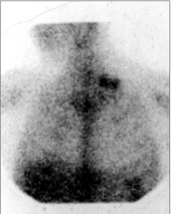
ciprofloxacin every 12 h, for 10 days; azathioprine was dis-continued and ambulatory re-evaluation was scheduled. After the use of the antimicrobial, the patient reported an improvement in fever; however, he mentioned that he had experienced the onset of coughing with white sputum, a loss of 4kg of weight in one month, and a vesicular eruption on the lower lip. During a routine follow-up examination of the transplanted patient, a mild diffuse hyperconcentration in the pulmonary fields and a focal hypercaptation area of a moderate degree on the left supraclavicular region was re-vealed in the cardiac scintigraphy with gallium 67, where the presence of lymphatic ganglion was investigated. The stu-dy for an active cardiac inflammatory process was negative (figure 1).
Because the patient remained febrile, with low leuko-cytes (2,700/mm3) and platelets (113,000/mm3) and with
le-sions on the lower lip, we chose to admit the patient to in-vestigate the cause of the fever.
The physical examination on admission revealed the following: the patient was hydrated, and eupneic; his blood pressure was 130/90mmHg, and his heart rate was 120bpm. Examination of the oral cavity revealed gum hypertrophy, in addition to the presence of hematic cicatricial lesions on the
Arq Bras Cardiol 2001; 76: 406-8.
Bacal et al Histoplasmosis after heart transplantation
407 407
lower lip. No alterations were noted at the neurological exa-mination. The abdomen had mild splenomegaly. Cervical palpation showed ganglion in an anterior chain of approxi-mately 1.5cm, elastic and attached to deep planes. During hospitalization, the patient experienced two febrile peaks (38.4°C and 38.8°C).
Five pairs of blood cultures were collected, which did not reveal bacterial growth. It is important to emphasize that the cultures were followed for only five days according to the usual protocol, and were not examined for fungal growth.
The thoracic radiography was normal. No bacterial growth was discovered in the urine culture. Thoracic tomo-graphy was performed and did not reveal alterations in the pulmonary parenchyma. Ultrasonography of the left supra-clavicular region revealed three solid nodules, hypoechoge-nic and confluent, measuring up to 2.2cm (Fig. 2).
A biopsy of the cervical lymphonodus was performed, revealing a sinusoidal histiocytic reaction, with the nume-rous rounded fungi of uniform size present, measuring 2.2
µm in diameter and histoplasma sp. morphology (Fig. 3). The histochemistry study of bacteria and BAAR were negative. Histological slides of the lymphonodus were submitted to immunehistochemistry investigation with polyclonal and monoclonal antibodies for T. cruzi, with negative results. The indirect xenodiagnosis was negative. A diagnosis of histoplasmosis was then made, and therapy with
ampho-tericin B was started. Ten days after the onset of treatment, the patient showed significant improvement in his general condition and appetite.
Discussion
The signs of infection after heart transplantation are ex-tremely variable according to the criteria adopted; however, infections are responsible for up to 40% of the deaths. Bac-terial infections occur most frequently, especially in the first month after the transplantation. The frequency of fungal in-fections is also variable, although a reduction in the number
Fig. 1 - Scintigraphy with gallium revealing a diffuse hyperconcentration of the tracer in the pulmonary fields and a moderate degree focal hypercollection area on the left supraventricular region. An abnormal radioligand concentration in cardiac area projection was not observed.
Fig. 2 - Ultrasonography of the left supraventricular region revealing the presence of confluent adenomegaly.
408 408
Bacal et al
Histoplasmosis after heart transplantation
Arq Bras Cardiol 2001; 76: 406-8.
and severity of these events has been reported 4.
Histoplas-mosis in patients who undergo heart transplantation is rare. Uip et al.4 reported in a series of 100 patients, that 47 had
fungal infections, with just one single episode of histoplas-mosis.
In the present patient, the differential diagnosis was performed to eliminate reactivation of Chagas’ disease and lymphoproliferative disease.
Investigation of Chagas’ disease reactivation is of fun-damental importance, because it can produce graft dysfunc-tion and, due to its systemic character, leads to death 5-7. It
can simulate rejection and must be differentiated from other infectious diseases. It may evolve asymptomatically and produce nonspecific symptoms, such as a decline in general condition, anorexia, anemia, icterus, long-term fever, liver in-volvement, central nervous system impairment, with heada-ches and localized signals. T. cruzi may invade the bone marrow and cause cytopenias. Cutaneous assault is also characteristic, with the presence of hard, painful subcuta-neous nodes and the likely presence of phlogistic signs. For the diagnosis of reactivation of T. cruzi infection, the parasite must be demonstrated. Xenodiagnosis may also be efficiently used to demonstrate the parasite, where the indirect method is preferred 8. In the present case, both
indirect xenodiagnosis and immunehistochemistry investi-gation of T. cruzi in the lymphonodus were negative, exclu-ding the possibility of Chagas’ disease reactivation.
A higher incidence of neoplasias occurs in patients who undergo heart transplantation, especially cutaneous and lymphoproliferative neoplasias; the diagnosis is made by a biopsy of the tumoral mass, detected by the clinical exa-mination or imaging methods. In the present case, despite the presence of lymphonodemegaly in the cervical chain, the anatomo-pathological examination of the lymphonodus ruled out the possibility of lymphoma.
Histoplasmosis occurs by inhalation of fungi conidi-um, which reach the alveolus and undergo phagocytosis, initiating a process of multiplication inside the alveolar macrophages. Through the lymphatic passage, the parasi-tes reach regional lymphonodus where they produce a new
inflammatory focus, thus forming the primary pulmonary and ganglionic complex. In this phase, the hematogenous dissemination of the fungi may occur determining the for-mation of new focuses. After a variable period of 10-18 days, activation of cellular immunity determines an intense inflammatory reaction, with formation of granuloma with clot necrosis, fibrotic encapsulation and, eventually, calcifica-tion. This primary infection, generally regressive, usually occurs in immune-competent patients 9.
In immune-compromised patients, the primary infec-tion or the re-infecinfec-tion may assume a progressive character, with variable severity from patient to patient, without forma-tion of granuloma, solely with a histiocytic reacforma-tion, as in the present case.
In Brazil, numerous inquiries with histoplasmin have demonstrated expressive levels of positiveness in the stu-dy population, with a higher prevalence occurring in the Southeast region of the country 9,10. Clinical manifestations
of histoplasmosis are variable, from light symptomatology, confused with flu symptoms in the regressive forms, to ex-tremely severe situations in disseminated progressive forms. Our patient probably had the chronic disseminated form, which is more prevalent in immune-compromised sub-jects, with concomitant oropharynx or larynx lesions in up to 70% of the cases. Sometimes, lips, gums, tongue, pharynx, or larynx lesions constitute the only manifestation of the di-sease, which is accompanied by low and intermittent fever, asthenia, and weight loss.
The definitive diagnosis is obtained in the laboratory by means of mycological, histological, or immune techniques, where the culture is individually the most sensitive diagnostic tool. In the present case, however, the diagnosis was made exclusively by the anatomo-pathological examination. The response to treatment and the prognosis are generally good, but depend fundamentally on the patient’s immune condition 11,12.
We conclude that in the evaluation of heart transplant patients who are febrile, a thorough clinical investigation must be performed, aiming at an early diagnosis and imme-diate onset of treatment, avoiding the expressive morbidity and mortality of infectious events in these patients.
1. Miller LW, Naftel DC, Bourge RC, et al and the Cardiac Transplant Research Da-tabase Group. Infection after heart transplantation: A multiinstitutional study. J Heart Lung Transplant 1994; 13: 381-92.
2. Uip DE, Amato Neto VA, Strabelli TMV, et al. Infecções em 100 pacientes subme-tidos a Transplante Cardíaco. Arq Bras Cardiol 1995;64: 537-40.
3. Camargo LFA, Uip D. Infecções em pacientes submetidos a transplante cardíaco. Rev Soc Cardiol Estado de São Paulo 1995; 6: 679-85.
4. Uip DE, Neto VA, Strabelli TMV, Bocchi E, et al. Infecções fúngicas em 100 paci-entes submetidos a transplante cardíaco. Arq Bras Cardiol 1996; 6: 675-7. 5. Bocchi EA, Bellotti G, Mocelin A, et al. Heart Transplantation for Chronic
Cha-gas Heart Disease. Ann Thorac Surg 1996; 61: 1727-33.
6. Stolf NAG, Higushi ML, Bocchi EA, et al. Heart Transplantation in patients with Chagas disease cardiomyopathy. J Heart Transplant 1987; 6: 307-12.
References
7. Bocchi EA, Bellotti G, Uip DE, et al. Long term follow up after heart transplanta-tion in Chagas’ disease. Transplantatransplanta-tion Proc 1993; 25: 1329-30. 8. Bronfeu E, Rocha FSA, Machado GBN, et al. Isolamento de amostras do
Trypano-soma cruzy por xenodiagnóstico e hemocultura de pacientes na fase crônica da Doença de Chagas. Mem Inst Osvaldo Cruz 1989; 84: 237-40.
9. Wheat JL, et al. Disseminated histoplasmosis in the acquired immune deficiency syndrome: clinical findings, diagnosis and treatment, and review of the literatu-re. Medicine 1990; 69: 361.
10. Goodwin RA, et al. Histoplasmosis in normal hosts. Medicine 1981; 60: 231. 11. Londero , Atewanke, B. Histoplasmose capsulata. J Bras Medicina 1988;
55: 94-109.