BASIC RESEARCH
Department of Physiology, Universiti Kebangsaan, Malaysia Medical Center - Kuala Lumpur/Malaysia.
Email: [email protected]
Received for publication on March 05, 2010 First review completed on March 22, 2010 Accepted for publication on April 12, 2010
PIPER SARMENTOSUM
INCREASES NITRIC OXIDE PRODUCTION IN OXIDATIVE
STRESS: A STUDY ON HUMAN UMBILICAL VEIN ENDOTHELIAL CELLS
Azizah Ugusman, Zaiton Zakaria, Chua Kien Hui, Nor Anita Megat Mohd Nordin
doi: 10.1590/S1807-59322010000700010
Ugusman A, Zakaria Z, Hui CK, Megat Mohd Nordin NA. Piper sarmentosum increases nitric oxide production in oxidative stress: a study on human umbilical vein endothelial cells. Clinics. 2010;65(7):709-14.
OBJECTIVE: Nitric oxide produced by endothelial nitric oxide synthase (eNOS) possesses multiple anti-atherosclerotic properties. Hence, enhanced expression of eNOS and increased Nitric oxide levels may protect against the development of atherosclerosis.
Piper sarmentosum is a tropical plant with antioxidant and anti-inlammatory activities. This study aimed to investigate the effects of Piper sarmentosum on the eNOS and Nitric oxide pathway in cultured human umbilical vein endothelial cells (HUVECs).
METHODS: HUVECs were divided into four groups:control, treatment with180 μM hydrogen peroxide (H2O2), treatment with 150 μg/mL aqueous extract of Piper sarmentosum, and concomitant treatment with aqueous extract of PS and H2O2 for 24 hours. Subsequently, HUVECs were harvested and eNOS mRNA expression was determined using qPCR. The eNOS protein level was measured using ELISA, and the eNOS activity and Nitric oxide level were determined by the Griess reaction.
RESULTS: Human umbilical vein endothelial cells treated with aqueous extract of Piper sarmentosum showed a marked induc-tion of Nitric oxide. Treatment with PS also resulted in increased eNOS mRNA expression, eNOS protein level and eNOS activity in HUVECs.
CONCLUSION: Aqueous extract of Piper sarmentosum may improve endothelial function by promoting NO production in HUVECs.
KEYWORDS: Piper sarmentosum, Nitric oxide, Endothelial nitric oxide synthase, Oxidative stress, Human umbilical vein en-dothelial cells.
INTRODUCTION
Nitric oxide (NO) in the endothelium is synthesized by endothelial nitric oxide synthase (eNOS) via the conversion of L-arginine to L-citrulline. The reaction requires the presence of the following cofactors: nicotinamide adenine dinucleotide phosphate (NADPH), calcium/ calmodulin (CaM), lavin adenine dinucleotide (FAD), lavin mononucleotide (FMN) and tetrahydrobiopterin (BH
4). 1,2
Nitric oxide has been recognized as a major anti-atherogenic factor because of its vasoprotective activity.3
Nitric oxide induces vasorelaxation by activating soluble guanylate cyclase; thus, NO plays an important role in
regulating the vascular tone. Nitric oxide has also been shown to inhibit oxidation of low-density lipoprotein (LDL) and antagonize platelet aggregation by inhibiting platelet activation.3 Moreover, NO inhibits nuclear
factor-κ B-dependent expression of adhesion molecules that mediate recruitment of leukocytes to the endothelium in the early phase of atherosclerosis. Nitric oxide has been shown to suppress abnormal proliferation of vascular smooth muscle cells, which contribute to the narrowing of atherosclerotic vessel walls.3 Based on these anti-atherosclerotic properties,
the enhancement of endothelial NO production may play an important role in the prophylaxis or treatment of cardiovascular diseases.4
Oxidative stress plays an important role in the pathogenesis of atherosclerosis and cardiovascular diseases by promoting endothelial dysfunction, inlammation and lipid/lipoprotein peroxidation as well as by reducing
NO bioavailability.5 Loss of normal NO production
dysfunction.6 Endothelial dysfunction is characterized
by a reduction in the bioavailability of NO, which is followed by increased levels of endothelium-derived
vasoconstrictors such as endothelin-1.7 This imbalance
leads to impairment of endothelium-derived relaxation (EDR). Arteries may thereby be predisposed to increased vascular tone and vasospasm. Impairment of EDR was found in atherosclerotic vessels even before vascular
structural changes had ensued.8
Piper sarmentosum (PS) is a creeping terrestrial herbaceous plant that belongs to the Piperaceae family. It is commonly found in the tropical and subtropical regions
of the world, such as the Asian region.The leaves and roots
of this plant have been used for the treatment of toothache, fungoid dermatitis on the feet, cough, asthma and pleurisy.
9 Chloroform extracts of Piper sarmentosum have the
ability to act as an anti-malarial agent against Plasmodium
falciparum and Plasmodium berghei.10 The aqueous extract
of the entire plant has been reported to have hypoglycemic effects in experimental rats.11 Furthermore, the ethanolic
extract of PS exerted anti-carcinogenic effects through an intrinsic apoptosis pathway in HepG2 cells.13 The
aqueous extract of PS also exhibited anti-nociceptive and anti-inlammatory activities in vivo.14Recent research has
demonstrated that various extracts prepared from PS leaves have antioxidant and anti-tuberculous activities.12To date,
however, there is no direct evidence linking PS to the eNOS system. Based on the properties of PS extracts mentioned above, the present study was designed to investigate the effects of PS on the eNOS system and NO synthesis in human umbilical vein endothelial cells (HUVECs). The results of the present study may help in the prevention and treatment of atherosclerosis, which is linked to various cardiovascular diseases.
MATERIALS AND METHODS
Preparation of aqueous extract of Piper sarmentosum
Leaves of PS were collected in Sungai Buloh, Malaysia, and identified by a plant taxonomist from the Forest Research Institute of Malaysia (voucher specimen: FRI 45870). The leaves were washed with tap water, cut into small pieces, sun-dried and ground into powder form. A
10% aqueous extract of Piper sarmentosum (AEPS) was
prepared by soaking 100 g of the powdered leaves in 900 mL of puriied water followed by incubation in a high-speed mixer at 80 °C for 3 hours. After cooling, the extract was iltered using mesh and further concentrated. The aqueous extract was then freeze-dried to powdered form and kept at 4 °C until the experiments.
Cell culture and treatment protocols
Human umbilical vein endothelial cells were obtained from umbilical cord veins using 0.1 % type I collagenase (Gibco-Invitrogen Corp., Grand Island, N.Y.) digestion. Cells were grown in medium-200 (Cascade Biologics, USA) supplemented with LSGS (low-serum growth supplement;
Cascade Biologics, USA) at 37 0C in a humidified
atmosphere of 5% CO2 and 95% air. Human umbilical vein
endothelial cells were identiied by the typical endothelial cell cobblestone morphology and positive expression of vonWillebrand factor and CD31 in immunocytochemistry. The culture medium was changed every other day until the cells reached conluence. Human umbilical vein endothelial cells from passage 3 at 80% conluency were used for the experiments. The cells were divided into four groups:control
(CTRL), treatment with180 μM hydrogen peroxide (H2O2)
to induce oxidative stress; treatment with 150 μg/mL AEPS, and concomitant treatment with 150 μg/mL AEPS and 180 μM H2O2. All treatments were given for 24 hours.
Quantitative reverse transcription polymerase chain re-action (qPCR) for analysis of eNOS mRNA expression
After treatment for 24 hours, total ribonucleic acid (RNA) from HUVECs was extracted using TRI Reagent (Molecular Research Center, Cincinnati, USA) according to a previously published protocol.15 Polyacryl carrier
(http://frodo:wi.mit.edu/cgi-bin/primer3/primer3-www.cgi) was used to design the primers from the NIH GenBank database. The following sequences were used as primers for eNOS [GenBank: NM_000603] and GAPDH [GenBank: BC020308]: CTCCAGCCCCGGTACTACTC (forward) and TTAGCCACGTGGAGCAGACT (reverse), and TCCCTGAGCTGAACGGGAAG (forward) and GGAGGAGTGGGTGTCGCTGT (reverse), respectively. The qPCR reaction was performed in a BioRad iCycler (Bio-Rad, USA) with 1 μl of cDNA, 5 μM of each forward and reverse primer and 12.5 μl of IQ SYBR Green Supermix (Bio-Rad, USA). The reaction proile consisted of 40 cycles using the following parameters: 95 C (10 seconds) and 61 C (30 seconds). The reaction kinetics of each primer set and protocol were veriied with the melting proile, and product size was further conirmed with 2% agarose gel electrophoresis stained with ethidium bromide (Sigma,
St Louis, USA). The threshold cycle (CT) value was
determined, and the relative mRNA expression of eNOS was calculated with the following equation: 2∆∆CT, where
∆∆CT =
CT GAPDH - CT eNOS.
Enzyme-linked immunosorbent assay (ELISA) for eNOS protein analyses
The eNOS protein level of the cultured HUVECs was determined using the Quantikine human eNOS ELISA kit (R&D Systems). Human umbilical vein endothelial cells were washed twice with phosphate-buffered saline (PBS), manually scraped from the culture lask and lysed with 400 μL of lysis buffer. The assay was performed using 100 μL of the cell lysate. The cell lysate was pipetted into the 96-well plate so that any eNOS that was present would be bound to the immobilized antibody in the plate. After washing away any unbound substances, eNOS conjugate was added to the wells. This was followed by the addition of substrate solution and stop solution. The optical density of each well was determined at 450 nm using an ELISA microplate reader.
Determination of eNOS Activity
The eNOS activity was determined using a commercially available kit (Nitric Oxide Synthase Colorimetric Assay, Calbiochem, USA). The principle of this assay was based on the measurement of nitrite produced by eNOS in the sample in a timed reaction. Human umbilical vein endothelial cells were scraped from the culture lask, homogenized in PBS and centrifuged at 10,000 g for 20 minutes. Then, the cell lysate in the supernatant solution was iltered through a 0.45 μm ilter prior to ultracentrifugation at 100,000 g for
15 minutes. A total of 40 μL ofthe cell lysate was diluted
with 20 μL ofassay buffer. Next, the samples were mixed with NADPH, nitrate reductase, cofactor preparation solution and lactate dehydrogenase (LDH). Total nitrite was measured at 540 nm with Griess reagents (sulfanilamide
and naphthalene–ethylenediamine dihydrochloride). The
concentration of nitrite in the sample wascalculated using a standard curve. The eNOS activity was expressed as nmol of nitrite/min per mL of sample.
Determination of endothelial nitric oxide production
Production of NO by HUVECs was measured via its stable oxidation product, nitrite, using a commercial kit (BIOXYTECH Nitric Oxide Colorimetric Assay, OXIS
Research, USA). Briely,50 μL ofthe culture medium was
diluted with 35 μL of assay buffer and mixed with10 μL of nitrate reductase and 10 μL NADH. After 20 minutes of incubation to convert nitrateto nitrite, total nitrite was measured at 540 nm with Griess reagents (sulfanilamide and
naphthalene–ethylenediamine dihydrochloride).
Statistical analysis
Data were tested for normality using the Kolmogorov-Smirnov test, and all variables were normally distributed. Data were expressed as mean ± SEM. Statistical analyses
between two groups were performed withStudent’spaired
t-test using SPSS version 16.0 software. Values of p < 0.05 were considered statistically signiicant.
RESULTS
eNOS mRNA expression in HUVECs
Treatment of HUVECs with AEPS significantly enhanced eNOS mRNA expression by 2.3-fold (4.405 ± 0.892 x 10⁻³) compared with the control group (1.915 ±
0.428 x 10⁻³) (Figure 1). In the oxidative stress-induced
group, HUVECs treated with H2O2 also showed a
significantly higher (2.2-fold) level of eNOS mRNA expression (4.280 ± 0.760 x 10⁻³) compared with the control
group. However, concomitant treatment of HUVECs with both AEPS and H2O2 did not result in a signiicant increase
in eNOS mRNA expression (2.95 ± 0.697 x 10⁻³) compared
to the control group.
eNOS protein level in HUVECs
The aqueous extract of PS signiicantly increased the
eNOS protein level by 1.4-fold (1.706 ± 0.154 x 10³ pg/mL)
mL) (Figure 2), which was consistent with the changes in
eNOS mRNA. Treatment of HUVECs with H2O2 alone also
resulted in a signiicantly higher (1.3-fold) level of eNOS protein (1.669 ± 0.137 x 10³ pg/mL) compared with the
control group. Concomitant treatment of HUVECs with both AEPS and H2O2 did not result in a signiicant increase
in eNOS protein level (1.549 ± 0.096 x 10³ pg/mL) compared
with the control group.
eNOS activity in HUVECs
Treatment of HUVECs with AEPS significantly
promoted eNOS enzyme activity (5.237 ± 0.55x10⁻² nmoles/
mL/min) compared with the control group (4.393 ± 0.74 x 10⁻² nmoles/mL/min) (Figure 3). In the oxidative
stress-induced group, HUVECs treated with H2O2 also showed a
signiicantly higher level of eNOS enzyme activity (5.863 ± 0.57 x 10⁻² nmoles/mL/min) compared with the control
group. Concomitant treatment of HUVECs with both AEPS and H2O2 did not result in a signiicant increase in eNOS
enzyme activity (5.228 ± 1.473 x 10⁻² nmoles/mL/min)
compared with the control group.
Nitric oxide production in HUVECs
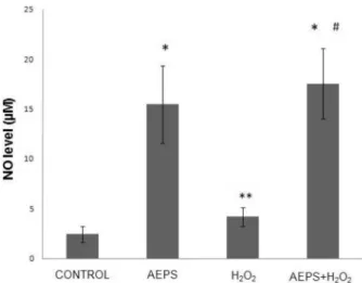
The aqueous extract of PS signiicantly promoted NO production in HUVECs by 6.3-fold (15.446 ± 3.879 μM) compared with the control group (2.454 ± 0.799 μM) (Figure 4). In the oxidative stress-induced group, HUVECs treated with H2O2 also showed a signiicantly higher (1.7-fold increase) level of NO production (4.175 ± 0.966 μM) compared with the control group. The greatest increase in NO production (17.536 ± 3.55 μM) was observed in
HUVECs treated with both AEPS and H2O2; the level of NO
production was signiicantly higher than that in the control and H2O2 groups.
DISCUSSION
Oxidative stress can contribute to the development and progression of atherosclerosis by promoting endothelial dysfunction, inlammation and lipid peroxidation, as well Figure 1 - eNOS mRNA expression in HUVECs.
Figure 2 - eNOS protein level in HUVECs.
Figure 3 - eNOS activity in HUVECs.
as by reducing NO bioavailability.16 Based on the results of
a previous study, we used 180 μM H2O2 to induce oxidative stress in HUVECs.17 This concentration of H
2O2 increased
eNOS mRNA expression, eNOS protein level and eNOS activity (Figure 1, 2, 3). In another study, incubation of bovine aortic endothelial cells with 150 μM H2O2 for 24 hours also caused an increase in eNOS mRNA, eNOS
protein and eNOS enzyme activity.18
The NO level was higher in the H2O2-treated group compared to the control group. This may have been due to induction of NO production by H2O2 as part of the self-protective mechanism of the cells. The dose of H2O2 used in this study was not lethal to HUVECs; therefore, the cells were still able to increase their endogenous NO production after an H2O2 challenge. However, H2O2 also caused oxidative destruction of the synthesized NO, which explains why the increase in NO in the H2O2-treated group was not as high as in the other groups (i.e., the AEPS and the combined AEPS and H2O2 groups) (Figure 4).
In this study, the responses to H2O2 are in agreement with earlier reports.5 The expression of eNOS in endothelial cells
is regulated by NO through a negative feedback mechanism at both the transcriptional and translational levels. Hydrogen peroxide-derived upregulation of eNOS was mediated by diminished NO availability and a consequent reduction in the negative feedback regulatory action of NO on eNOS expression. This represents a compensatory protective mechanism of the cells to a reduction in NO availability induced by acute exposure to H2O2.5 Hydrogen peroxide has
been shown to increase eNOS activity by inducing changes in the phosphorylation status of the enzyme. This response represents an attempt by the endothelial cells to maintain NO bioactivity under conditions of increased oxidative stress.19
It is well recognized that NO produced by eNOS plays a protective role against the development of atherosclerosis and endothelial dysfuntion.20,21 The results of this study
show that AEPS(150 μg/mL) signiicantly increased NO
production in HUVECs (Figure 4). AEPS also induced increases in eNOS mRNA, protein and activity (Figure 1, 2, 3). The higher amount of eNOS protein caused a higher level of eNOS activity. This resulted in an increase in NO production by HUVECs. The results of the present study suggest that AEPS may improve endothelial function by augmenting NO production in human endothelial cells. Thus, AEPS could reduce the risk of atherosclerosis.
Antioxidants are known to enhance the biological actions of NO by protecting NO against oxidative destruction by
reactive oxygen species.21 AEPS has been shown to exhibit
antioxidant properties.12 Thus, AEPS can directly protect
NO from oxidative destruction by H2O2 (Figure 4). The aqueous extract of PS also promoted NO production from HUVECs by increasing eNOS protein synthesis and enzyme activity (Figure 2, 3). Therefore, both protection of NO from oxidative destruction and enhancement of eNOS activity by AEPS caused an increase in NO level.
We observed the largest increase in NO production in the group that received AEPS and H2O2. Since AEPS can directly protect NO from oxidative destruction by H2O2, NO is available in the cells at a higher level. A higher level of NO in the cells reduced the mRNA expression and protein
synthesis of eNOS via a negative feedback mechanism.5
Previous phytochemical screening of PS revealed the presence of a variety of natural products, such as amides,
polyphenols and flavonoids.12 Myricetin, apigenin and
quercetin are examples of flavonoids identified in PS
leaves.22 Quercetin improved endothelial dysfunction by
increasing NO synthesis in HUVECs. The increase in NO synthesis was attributed to an enhancement of eNOS activity via increased calcium concentration.23 Quercetin has also
been reported to exert endothelium-dependent vasodilatation of porcine aortic rings.24 Therefore, in the present study,
the lavonoids appear to be the active constituents of AEPS responsible for enhancing NO production in HUVECs.
STUDY LIMITATIONS
This experiment was an in vitro study that investigated some fundamental biomolecular and cellular activities. The data suggest that AEPS could reduce the risk of atherosclerosis by increasing the bioavailability of NO to defend against oxidative stress. A clinical placebo-controlled study may be needed before employing AEPS as an effective supplement. If this study were performed in a clinical setting, we could attempt to determine dosage, pharmacokinetics and pharmacodynamics of AEPS.
CONCLUSION
REFERENCES
1. Govers R, Rabelink T. Cellular regulation of endothelial nitric oxide synthase. Am Physiological Soc. 2001;280:193-206.
2. Förstermann U. Janus-faced role of endothelial NO synthase in vascular disease: uncoupling of oxygen reduction from NO synthesis and its pharmacological reversal. J Biol Chem.2006;387:1521-33.
3. Naseem K. The role of nitric oxide in cardiovascular diseases. Mol Aspects Med.2005;26:33-65.
4. Ignarro L, Napoli C. Novel features of nitric oxide, endothelial nitric oxide synthase, and atherosclerosis. Current Atherosclero Reps.
2004;6:281-7.
5. Zhen J, Lu H, Wang X, Vaziri N, Zhou X. Upregulation of endothelial and inducible nitric oxide synthase expression by reactive oxygen species. Amer J Hypertension. 2008;21:28-34.
6. Park S, Jung W, Moon S, Ko C, Cho K, Kim Y, et al. Chunghyuldan activates NOS mRNA expression and suppresses VCAM-1 mRNA expression in human endothelial cells. Can J Physiol Pharmacol. 2005;83:1101-8.
7. Bonetti P, Lerman L, Lerman A. Endothelial Dysfunction A Marker of Atherosclerotic Risk. Am Heart Assoc. 2003;23:168-75.
8. Kawashima S. Malfunction of Vascular Control in Lifestyle-Related Diseases: Endothelial Nitric Oxide (NO) Synthase/NO System in Atherosclerosis. J Pharmacol Sci. 2004;96:411-9.
9. Rukachaisirikul T, Siriwattanakit P, Sukcharoenphol K, Wongvein C, Ruttanaweang P, Wongwattanavuch P, et al. Chemical constituents and bioactivity of Piper sarmentosum. J Ethnopharmacol. 2004;93:173-6.
10. Najib Nik A Rahman N, Furuta T, Kojima S, Takane K, Ali Mohd M. Antimalarial activity of extracts of Malaysian medicinal plants. J Ethnopharmacol. 1999;64:249-54.
11. Peungvicha P, Thirawarapan S, Temsiririrkkul R, Watanabe H, Kumar Prasain J, Kadota S. Hypoglycemic effect of the water extract of Piper sarmentosum in rats. J Ethnopharmacol. 1998;60:27-32.
12. Hussain K, Ismail Z, Sadikun A, Ibrahim P. Antioxidant, anti-TB activities, phenolic and amide contents of standardised extracts of Piper sarmentosum Roxb. Nat Pro Res. 2009;23:238-49.
13. Hisham Z, Haryani W, Zaidah Z, Fauzi S, Sahidan S, Rohaya M. Intrinsic anticarcinogenic effects of Piper sarmentosum ethanolic extract on a human hepatoma cell line. Cancer Cell Int. 2009;9:6.
14. Zakaria Z, Patahuddin H, Mohamad A, Israf D, Sulaiman M. In vivo anti-nociceptive and anti-inlammatory activities of the aqueous extract of the leaves of Piper sarmentosum. J Ethnopharmacol. 2010;128:42-8.
15. Chua K, Aminuddin B, Fuzina N, Ruszymah B. Insulin-transferrin-selenium prevent human chondrocyte dedifferentiation and promote the formation of high quality tissue engineered human hyaline cartilage. Eur Cell Mater. 2005;9:58-67.
16. Kuzkaya N, Weissmann N, Harrison D, Dikalov S. Interactions of peroxynitrite, tetrahydrobiopterin, ascorbic acid, and thiols: implications for uncoupling endothelial nitric-oxide synthase. J Biol Chem. 2003;278:22546-54.
17. Haizah AH, Zaiton Z, Zulkhairi A, Mohd Ilham A, Nor Anita MMN, Zaleha AM.Piper sarmentosum as an antioxidant on oxidative stress in human umbilical vein endothelial cells induced by hydrogen peroxide. J Zhejiang Univ-Sc B. 2010;in press.
18. Drummond G, Cai H, Davis M, Ramasamy S, Harrison D. Transcriptional and posttranscriptional regulation of endothelial nitric oxide synthase expression by hydrogen peroxide. Circ Res. 2000;86:347-54.
19. Thomas S, Chen K, Keaney J. Hydrogen peroxide activates endothelial nitric-oxide synthase through coordinated phosphorylation and dephosphorylation via a phosphoinositide 3-kinase-dependent signaling pathway. J Biol Chem. 2002;277:6017-24.
20. Landim M, Casella Filho A, Chagas A. Asymmetric dimethylarginine (ADMA) and endothelial dysfunction: implications for atherogenesis. Clinics. 2009;64:471-8.
21. Ignarro L, Byrns R, Sumi D, de Nigris F, Napoli C. Pomegranate juice protects nitric oxide against oxidative destruction and enhances the biological actions of nitric oxide. Nitric Oxide. 2006;15:93-102.
22. Miean K, Mohamed S. Flavonoid (myricetin, quercetin, kaempferol, luteolin, and apigenin) content of edible tropical plants. J Agric Food Chem. 2001;49:3106-12.
23. Kuhlmann C, Schaefer C, Kosok C, Abdallah Y, Walther S, Lüdders D, et al. Quercetin-Induced Induction of the NO/cGMP Pathway Depends on Ca 2-Activated K Channel-Induced Hyperpolarization-Mediated Ca 2+-Entry into Cultured Human Endothelial Cells. Planta Med. 2005;71:520-4.