Original Article
Artigo Original
Camila Dantas Martins1 Renata Maria Moreira Moraes Furlan2 Andréa Rodrigues Motta3 Maria Cândida Ferrarez Bouzada Viana4
Descritores
Aleitamento Materno Eletromiograia Comportamento de Sucção Músculos Faciais Sistema Estomatognático
Keywords
Breast-feeding Electromyography Sucking Behavior Facial Muscles Stomatognathic System
Correspondence address: Camila Dantas Martins
Rua São Lázaro, 1301/404, Bairro Sagrada Família, Belo Horizonte (MG), Brasil, CEP: 31035-580.
E-mail: [email protected] Received: 01/27/2015
Study carried out at the Speech Language Pathology and Audiology Ambulatory, Universidade Federal de Minas Gerais – UFMG – Belo Horizonte (MG), Brazil.
(1) Graduate Program in Health of the Child and Adolescent, Universidade Federal de Minas Gerais – UFMG – Belo Horizonte (MG), Brazil.
(2) Graduate Program in Structure Engineering, Universidade Federal de Minas Gerais – UFMG – Belo Horizonte (MG), Brazil.
(3) Speech Language Pathology and Audiology Department, Universidade Federal de Minas Gerais – UFMG – Belo Horizonte (MG), Brazil.
Electromyography of muscles involved
in feeding premature infants
Avaliação eletromiográica dos músculos envolvidos
na alimentação de recém-nascidos prematuros
ABSTRACT
Purpose: To measure and compare the electrical activity of masseter, temporal, and suprahyoid muscles in premature newborn infants during breast-feeding and cup-feeding. Methods: This cross-sectional observational study was carried out by the electromyographic assessment of 36 preterm infants, 53% of whom were male, with mean gestational age of 32 weeks and birth weight of 1,719 g, fed via oral route, by full breast-feeding and supplementation of diet, through cup with expressed breast milk, until 15 days after hospital discharge. Children with neurological disorders, genetic syndromes, oral-motor, and/or congenital malformations were excluded. The different methods of feeding and the variables gestational age at birth, corrected gestational age, chronological age, birth weight and size, head circumference, and Apgar scores at 1 and 5 minutes were analyzed and compared by appropriate statistical analysis. Results: No difference was observed between breast-feeding and cup-feeding in the analysis of the temporal and masseter muscles. However, higher activity of suprahyoid musculature was observed during cup-feeding (p=0.001). The other variables were not correlated with the electrical activity of the muscles during the different feeding methods. Conclusion: There may be a balance between the activity of the temporal and masseter muscles during breast-feeding and cup-feeding. There was higher activity of suprahyoid musculature during cup-feeding. This can be explained by the greater range of tongue movement, as premature infants usually perform tongue protrusion to get the milk from the cup.
RESUMO
INTRODUCTION
Breast feeding, due to its nutritional and immunological aspects, is the best source of food for the newborn (NB). Breast-feeding (BF) is considered the most appropriate Breast-feeding method for the development of the structures and functions of the sto-matognathic system(1), in addition to being important for
stab-lishing a healthy mother–baby interaction(2). The preterm neonate
(PTNN), considered the risk, usually neurologically disorga-nized, may have absence of protection and/or feeding relexes, thus, they may have dificulties in BF, being necessary the use of alternative feeding methods(3,4). The safe feeding through oral
route (OR) requires the maturation of the structures and func-tions of the stomatognathic system, and this maturity is related to the gestational age (GA) of the infant(5). The successful feeding
through OR is important for the neurobehavioral development of the PTNN and for their cognitive functions in the future(6).
When the BF is not possible to be onset, whether by the absence of the mother at the moment of feeding, mammary alterations, maternal disease, among others, the cup is one of the methods recommended by the Ministry of Health to initiate the transition from the tube to the OR(7). The United Nations
Children’s Fund and the World Health Organization have invested in the implementation of the Child Friendly Hospital Initiative, which approaches the offer of breast milk milked through the cup when it is not possible to breast-feed(8-10). In a study carried
out with the objective of comparing the feeding through glass and the BF, it was veriied that some tongue and jaw movements during the use of the cup are similar to those necessary to the successful BF and that the use of this tool reduces the time of use of probes and consequently the time of hospitalization(11).
Besides, studies have indicated that the use of the cup for BF supplementation is associated with the higher prevalence of BF at the moment of the hospital discharge(12).
In clinical practice, no standardized method has been veri-ied to be used in the transition for OR and/or supplementation of the diet in the Neonatal Progressive Care Nursery (NPCN). In a systematic review in which the use of the cup as an alterna-tive feeding method for PTNN was evaluated, no consensus was found among the researchers in regard to the supplementation of the diet in this population through the use of these tools(13).
It is believed that the most appropriate method is the one that provides the safest and most eficient feeding way, with oro-facial muscle activity most similar to the one used during BF.
An important tool to investigate the muscle activity of NBs and infants is the surface electromyography (sEMG)(10,14,15).
The sEMG refers to an objective method that evaluates the alterations in electrical potential from the skeletal muscle con-traction; it is characterized by being a noninvasive and radi-ation-free test(16). Its use in studies to determine the action of
the oral muscles in the numerous feeding ways is recent, and few studies are available in the literature about the theme(10,17).
The studies carried out show similarities between the electri-cal muscle activity in BF and during cup-feeding(10,14,17) and the
reduced activity of the masseter muscle in the bottle-feeding than in the cup-feeding(17-20).
A study carried out with PTNN(14) evaluated the electrical
activity of the masseter suprahyoid muscles during the use of the cup, translactation, and BF. No difference was observed in the activity of the muscles studied between the different kinds of feeding. Comparing the masseter and the suprahy-oid muscles, the author concluded that there is a balance between the activity of these muscles during translactation and BF. In the use of the cup, another mechanism can be observed, the masseter muscle being more active than the suprahyoid muscles, suggesting that the translactation is a more physiological transition method to the OR. The author pointed out the need of a greater discussion on the feeding methods used in the transition of feeding among PTNNs and their inluence on the muscles involved during BF, and, in further publications, proposed a protocol for the evaluation of the electrical activity of the masseter and the suprahyoid muscles in PTNN during feeding(15).
As mentioned earlier, it becomes relevant to carry out new studies, which use objective tests, aiming at evaluating the activity of the muscles involved in the feeding processes of children, especially among preterms, once that these analyses are scarce and inconclusive. Thus, this study had the objective of measuring and comparing the electrical activity of the tem-poral, masseter, and suprahyoid muscles through an sEMG in preterms during BF and cup-feeding.
METHODS
It is an observational cross-sectional study, involving 36 PTNN, 53% males and 47% females, with GA at birth ranging from 27 to 36 weeks, and average weight at birth of 1,719 g, who were born in the Hospital das Clínicas of the Universidade Federal de Minas Gerais, in Belo Horizonte, and remained hos-pitalized in the NPCN. The parents/guardians for the children, at the moment of hospital discharge, were invited to take part in the study. The research was carried out at the Speech Language and Audiology Laboratory of the institution, up until 15 days after the discharge, from August 2012 to July 2013.
The sample calculation was made by using the Stata soft-ware, version 10.0. The premises adopted were conidence interval of 95%, error margin of 5%, and a total of 36 children. Patients who, at the time of birth, presented weight equal to or less than 2,500 g, GA equal to or less than 37 weeks, with up to 15 days after hospital discharge at the time of the test, who were under mixed BF, with supplementation of the cup, were included in this study. Those who had genetic syndromes and craniofacial malformations, and who used bottles and/or artii-cial silicone nipple between the date of hospital discharge and the test, besides those who were not able to go under sEMG for any reason, were excluded.
suprahyoid muscles, respectively. The suprahyoid muscle encompasses the digastric, stylohyoid, mylohyoid, and genio-hyoid. The remaining channels were disabled.
We used disklike bipolar surface capture electrodes, pedi-atric, pre-jelliied, disposable, by Hall. As a data collection material, the ground electrode by Medi-Trace, procedure gloves, disposable coffee cups, and gauze and alcohol 70%, for the hygiene of the child’s skin, were used. The test was carried out in a sound treated room, without electrical or electromagnetic interference, according to the recommen-dations of the sEMG for a noninvasive assessment of mus-cles (SENIAM)(21).
The test consisted of the placement of a bipolar electrode on the surface of the skin, in the region of each of the researched muscles (temporal, masseter, and suprahyoid), unilaterally to the left, being this the side of the face the NB was not in con-tact with the maternal breast at the moment of the BF, reducing, thus, the interferences. The muscles evaluated were identiied through touch. The electrodes were positioned over the muscles being studied, and the ground electrode, placed in the frontal region of the child(10). The mothers were oriented to attend to
the test between 2 and 3 hours before BF.
Initially, with the objective of removing the excessive oil and making the contact of the electrodes easier, the skin of the child was cleaned with gauze soaked in alcohol 70%(21).
Thereafter, the muscles were identiied, and the ground and surface electrodes were properly positioned. The electromyo-graphic records occurred always in the same order, so that there was a standardization: BF and, later on, offering of milk from the cup. Each trial lasted 60 seconds. The mother was oriented to milk enough milk, before the beginning of the test, to complete the total volume of the cup (60 mL), avoiding the lack of milk during the data collection. During BF, the mother was found in state of alert, sitting down, with their feet on the ground. The body of the child remained fac-ing the front and close to the front of the mother, head and spine aligned with the chin close to the breast(22). In
cup-feeding the NB, in state of alert, remained sitting down on the researcher’s lap. The edge of the cup was touched to the lips of the child and it was waited until they would drink the milk(7). The positioning to the maternal breast and the
offering of diet through cup was carried out by the same researcher. The stabilizing of child was waited and then the recordings began. The time spent during the electromyo-graphic evaluation, including the procedures that preceded it, lasted approximately 40 minutes.
The acquisition of the signal was performed with the sample rate of 1,000 Hz. Then, the signal was filtered (20–500 Hz), rectiied, and sent to a microcomputer where it was processed by a speciic software for the acquisition and processing of the data (Software AqDados, version 5.05; Lynx Tecnologia Eletrônica LTDA®). The variable analyzed in this research was the value of the root mean square (RMS) of the electromyographic signal, calculated by the software, RMS being the square root of the squares of the signal throughout the whole measurement(16).
The mean values of RMS for the temporal, masseter, and suprahyoid muscles were compared between the feeding methods (breast and cup) and between the gender and Apgar variables at the irst and ifth minutes of life for both evalu-ated feeding methods. For the comparison of the Apgar at the irst minute, the individuals were grouped into the following categories: Apgar from 1 to 3, from 4 to 7, and from 8 to 10. For the comparison of Apgar at the ifth minute, the individu-als were grouped into the following categories: Apgar from 4 to 7 and from 8 to 10.
In the statistical analysis, measures of central and disper-sion trends were used. For the groups with normal distribution, as a hypothesis test, we used the paired t-test and analysis of variance for the comparison of the groups. We used the lin-ear correlation coeficient of Splin-earman with the objective of measuring the degree of association or mutual linear relation between the two variables. The value of p<0.05 was adopted as signiicance level in all analyses.
The research was approved by the ethics committee of the Universidade Federal de Minas Gerais under the number CAAE – 0597.0.203.000-11. All parents and legal guardians of the children signed the informed consent.
RESULTS
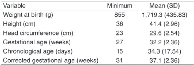
In Table 1, the indings related to the continuous variables analyzed in this study are described.
In Table 2, the means of the electrical activity of the tem-poral, masseter, and suprahyoid muscles in PTNN during the BF and the cup-feeding are presented. When comparing both feeding methods and the muscle groups evaluated, we obtained a difference only for the suprahyoid muscles (p=0.001), notic-ing greater activity of these muscles durnotic-ing cup-feednotic-ing.
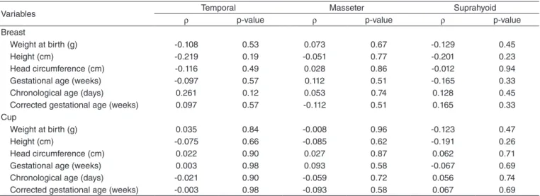
When comparing the mean of electrical activity of the temporal, masseter, and suprahyoid muscles, during BF and cup-feeding, between the gender variables, Apgar in the irst and ifth minutes of life (Table 3), we observed no difference. The values of the Spearman correlation coeficient for the variables weight at birth, height, head circumference, GA at birth, chronological age, and corrected GA presented low val-ues and did not indicate association between these variables and the electrical activity of the temporal, masseter, and suprahyoid muscles in both the feeding methods being studied (Table 4).
Table 1. Descriptive analysis refers to the continuous variables: weight at birth, height, head circumference, gestational age at birth, chronological age, and corrected gestational age
Variable Minimum Mean (SD)
Weight at birth (g) 855 1,719.3 (435.83)
Height (cm) 36 41.4 (2.96)
Head circumference (cm) 23 29.6 (2.54)
Gestational age (weeks) 27 32.2 (2.36)
Chronological age (days) 15 34.3 (17.54)
Corrected gestational age (weeks) 31 37.1 (2.36)
DISCUSSION
No signiicant differences were found in this study between the electrical activity of the temporal and masseter muscles when comparing BF to cup-feeding. In relation to the suprahy-oid muscles, a higher activity was veriied during cup-feeding, this being a signiicant data. Such inding may be justiied by the higher range of movement of the tongue during cup-feed-ing when compared to suction. Durcup-feed-ing cup-feedcup-feed-ing, usually, between the PTNN the protrusion of the tongue is observed to obtain milk. Later on, this movement is replaced by the movement of absorption(23).
In a research through which the activity of the masseter, temporal, and buccinator muscles during BF, bottle-feed-ing, and cup-feeding was measured, using the sEMG, the author concluded there was similarity between the mus-cle activity of the group in BF and during cup-feeding(10),
which agrees to the findings of this study for the masseter and temporal muscles.
A study that analyzed the electrical activity of the mas-seter and suprahyoid muscles, during cup-feeding, translac-tation, and BF, no difference was observed in the activity of the muscles studied between the different types of feeding. These indings are different from the ones found in this study, once the electrical activity of the suprahyoid muscles was more active with cup-feeding. The author also compared the electrical activity between the masseter and suprahyoid mus-cles during the different feeding methods and found a signii-cant difference between the activities of the masseter and the suprahyoid muscles during cup-feeding, with higher activ-ity of the masseter muscle. The author concluded that there is a balance between the activities of the masseter and the suprahyoid muscles during feeding by translactation and in BF. In cup-feeding, there seems to be another mechanism of activity of these muscles, considering that the masseter
Table 2. Comparison between the different feeding methods (breast and cup) and the mean of electrical activity of the temporal, masseter, and suprahyoid muscles (in µV)
Variable Breast Cup p-value
Mean (SD) Mean (SD)
Temporal 0.0088 (0.0068) 0.0510 (0.1291) 0.058
Masseter 0.0088 (0.0014) 0.0189 (0.0440) 0.173
Suprahyoid 0.0114 (0.0088) 0.0475 (0.0597) 0.001*
*t-Test
Caption: SD = standard deviation
Table 3. Comparison between the variables of gender, Apgar values in the first and fifthminutes of life, in relation to the mean of the electrical activity of the temporal, masseter, and suprahyoid muscles (µV) for both the evaluated feeding methods
Variables Temporal Masseter Suprahyoid
p-value Breast
Gender* 0.962 0.788 0.516
Apgar 1st minute** 1 to 3
0.709 0.482 0.898
4 to 7 8 to 10 Apgar 5th minute**
4 to 7
0.960 0.488 0.720
8 to 10 Cup
Gender* 0.936 0.342 0.096
Apgar 1st minute** 1 to 3
0.5466 0.9217 0.6313
4 to 7 8 to 10 Apgar 5th minute**
4 to 7
0.646 0.727 0.482
8 to 10
*t-Test; ** ANOVA test
Table 4. Spearman correlation between the epidemiological and physical variables and the mean electrical activity of the temporal, masseter, and suprahyoid muscles (µV) for both the feeding methods evaluated
Variables Temporal Masseter Suprahyoid
ρ p-value ρ p-value ρ p-value
Breast
Weight at birth (g) -0.108 0.53 0.073 0.67 -0.129 0.45
Height (cm) -0.219 0.19 -0.051 0.77 -0.201 0.23
Head circumference (cm) -0.116 0.49 0.028 0.86 -0.012 0.94
Gestational age (weeks) -0.097 0.57 0.112 0.51 -0.165 0.33
Chronological age (days) 0.261 0.12 0.053 0.74 0.128 0.45
Corrected gestational age (weeks) 0.097 0.57 -0.112 0.51 0.165 0.33
Cup
Weight at birth (g) 0.035 0.84 -0.008 0.96 -0.123 0.47
Height (cm) -0.075 0.66 -0.085 0.62 -0.191 0.26
Head circumference (cm) 0.022 0.90 0.027 0.87 0.062 0.71
Gestational age (weeks) 0.003 0.98 0.093 0.58 -0.067 0.69
Chronological age (days) -0.021 0.90 -0.059 0.72 0.056 0.74
Corrected gestational age (weeks) -0.003 0.98 -0.093 0.58 0.067 0.69
muscles are more active than the suprahyoid ones, which suggests that translactation seems to be a more physiologi-cal method when compared to BF(14).
When comparing the electrical activity of the evalu-ated muscles in both the studied feeding methods and the score of Apgar in the first and fifth minutes of life, no sig-nificant relations were observed. These data, however, dif-fer from the findings of a study analyzing the factors that could influence the BF among PTNN. The authors verified that these factors include the maternal diseases, obstetric complications, and the score of Apgar in the first minute of life(24). The Apgar score in the first minute may be related
to the maturation of the structures and functions of the sto-matognathic system, interfering, therefore, on BF success. The fact that no correlation was found between these two variables may be related to the reduced number of PTNN with Apgar score below 4.
The sample variables GA at birth, corrected age, chrono-logical age, weight at birth, and height and head circumference at birth do not present correlation with the electrical activity of the muscles studied during the different feeding methods. The articles measuring the electrical activity of the muscles involved in the feeding of NBs do not investigate this relation. A relevant aspect of the research refers to the fact that both feeding methods were evaluated in the same population, to reduce the confounding variables.
In the institution where this study was conducted, the method of choice in the transition from OR is the suction of the maternal breast. When BF is not possible, the cup is a preconized method as it regards a friendly hospital of the child. This way, the use of a bottle was not evaluated in this study. In a research that evaluated the electrical activity of the masseter muscle of infants born at term during BF and bottle-feeding, the authors veriied low activation of the mas-seter muscle during bottle-feeding when compared to BF and emphasized that such fact may compromise the devel-opment of the masticatory function(19). The same way, other
authors(20), when studying the electrical activity of the
tem-poral, masseter, orbicularis oris, and suprahyoid muscles in babies born at term in exclusive BF, bottle-feeding, and mixed feeding, veriied that the masseter muscle presented reduced electrical activity in bottle-feeding when compared to BF. Another inding of the literature refers to the hyperfunction of the buccinator muscle in bottle-feeding, verifying in a research that evaluated the temporal, masseter, and buccina-tor muscles during the BF, cup-feeding, and bottle-feeding of infants born at term(10).
Because it is an objective, non-invasive, fast and radiation-free method(16), the sEMG was the instrument chosen in this
study to evaluate the differences between the electrical activity of the temporal, masseter and suprahyoid muscles during BF and cup-feeding. In the literature researched, it was observed that the studies using the sEMG in the feeding process of PTNN are scarce(17).
In this study, there was no normalization because for its cal-culation a voluntary maximum contraction of a given muscle is necessary(21) and this contraction is impossible in this kind
of population. The limitations of this study include not having considered the hospitalization time, the time at which the NB was fed by probe, the time at which they were fed by OR, and the time since the hospital discharge to the test.
Other studies using the sEMG are required as it is an objec-tive test, with larger samples when compared to other feeding methods, especially between the PTNN, so that the most physi-ological method determined in the transition into OR and/or dietary supplementation for this population, ensuring thus the success of BF.
CONCLUSION
The results indicate there is a balance between the activ-ity of the temporal and masseter muscles during BF and cup-feeding. The suprahyoid muscles presented themselves as more active in cup-feeding, which may be justiied by the wider range of movement of the tongue, as usually the PTNN perform the protrusion of the tongue to obtain milk during the feeding with this tool.
*CDM was responsible for the design of the study, for the collection, tabulation, and analysis of the data, as well as for the elaboration of the manuscript; RMMF helped in the design of the study, and in the collection and tabulation of the data; MCFBV and ARM were responsible for the project, design of the study, and overall guidance of the execution stages.
REFERENCES
1. Carvalho GD. Amamentação e o sistema estomatognático. In: Carvalho MR, Tamez RN. Amamentação: bases cientíicas para a prática proissional. Rio de Janeiro: Guanabara-Koogan, 2002. p. 37-49.
2. Reyna BA, PicklerRH. Mother-infant synchrony. J Obstet Gynecol Neonatal Nurs. 2009;38(4):470-7.
3. Hernandez AM. Atuação fonoaudiológica em neonatologia: uma proposta de intervenção. In: Andrade CRF.Fonoaudiologia em berçário normal e de risco. São Paulo: Lovise, 1996. p. 43-95.
4. Neiva FCB. Neonatologia: Papel do fonoaudiólogo no berçário. São José dos Campos: Pulso, 2004. p. 225-234.
5. Amaizu N, Shulman RJ, Schanler RJ, Lau C. Maturation of oral feeding skills in preterm infants. Acta Paediatr. 2008;97(1):61-7.
6. Pickler RH, McGrath JM, Reyna BA, McCain N, Lewis M, Cone S, et al. A model of neurodevelopmental risk and protection for preterm infants. J Perinat Neonat Nurs. 2010;24(4):356-65.
7. Brasil. Ministério da Saúde. Secretaria de Atenção à Saúde. Departamento de Ações Programáticas Estratégicas. Atenção humanizada ao recém-nascido de baixo peso: método canguru. 3. ed. Brasília: Editora do Ministério da Saúde, 2013.
8. Lang S. Aleitamento do lactente: cuidados especiais. São Paulo: Editora Santos, 1999.
9. Dowling DA, Meier PP, Dii JM, Blatz M, Martin RJ. Cup feeding for preterm infants: mechanics and safety. J Hum Lact. 2002;18(1):13-20. 10. Gomes CF, Trezza EMC, Murade ECM, Padovani CR. Surface
11. Kuehl J. Cup feeding the newborn: What you should know. J Perinat Neonatal Nurs. 1997;11(2):56-60.
12. Pedras CTPA, Pinto EALC, Mezzacappa MA. Uso do copo e da mamadeira e o aleitamento materno em recém-nascidos prematuros e a termo: uma revisão sistemática. Rev Bras Saúde Matern Infant. 2008;8(2):163-9.
13. López CP, Silva RG. Métodos de alimentação alternativa para recém-nascidos prematuros. Rev Paul Pediatr. 2012;30(2):278-82.
14. Raposo RD. Atividade dos músculos masséter e supra-hioideos em recém-nascidos pré-termo durante o uso do copinho, da translactação e na amamentação. [Tese]. Recife: Universidade Federal de Pernambuco; 2012. 15. Raposo RD, Silva HJ. Proposta de um protocolo de avaliação da atividade elétrica dos músculos masseter e supra-hióideos em recém-nascidos pré-termo durante a alimentação. Rev CEFAC. 2013;15(4):803-14. 16. Rahal A, Pierotti S. Eletromiograia e cefalometria na Fonoaudiologia.
In: Ferreira LP, Bei-Lopes DM, Limongi SCO (organizadores). São Paulo: Roca; 2004. p. 237-53.
17. Gomes CF, Thomson Z, Cardoso JR. Utilization of surface electromyography during the feeding of term and preterm infants: a literature rewiew. Dev Med Child Neurol. 2009;51:936-42.
18. Inoue N, Sakashita R, Kamegai T. Reduction of masseter muscle activity in bottle-fed babies. Early Hum Dev. 1995;18:185-93.
19. Sakashita R, Kamegai T, Inoue N. Masseter muscle activity in bottle feeding with the chewing type bottle teat: evidence from electromyographs. Early Hum Dev. 1996;45:83-92.
20. Tamura Y, Horikawa Y, Yoshida S. Co-ordination of tongue movements and peri-oral muscle activities during nutritive sucking. Dev Med Child Neurol. 1996;38:503-10.
21. Hermens HJ, Freriks B, Disselhorst-Klug C, Rau G. Development of recommendations for SEMG sensors and sensor placement procedures. J Electromyogr Kinesiol. 2000;10:361-74.
22. WHO [Internet]. Promovendo o aleitamento materno [cited 2013 Jan 12]. Available from: www.bvsms.saude.gov.br/bvs/publicacoes/album_ seriado_aleitamento_maternpdf
23. Lang S, Lawrence CJ, Orme RL. Cup feeding: an alternative method of infant feeding. Arch Dis Child. 1994;71:365-9.