Reliability of Symptoms Suggestive of Angina in Patients with
Chronic Obstructive Pulmonary Disease
Yilmaz Gunes
1, Mustafa Tuncer
1, Unal Guntekin
1, Hasan Ali Gumrukcuoglu
1, Serkan Akdag
1, Bulent Ozbay
2, Bunyamin
Sertogullarindan
2Departamento de Cardiologia 1, Departamento de Doenças Torácicas2, Faculdade de Medicina, Universidade Yuzuncu Yil, Van - Turquia
Summary
Background: Due to overlapping symptoms and inadequate exercise capacity, noninvasive diagnosis of coronary artery disease (CAD) may be under- or overestimated in patients with chronic obstructive pulmonary disease (COPD).
Objective: To assess outcomes of coronary angiography in COPD patients depending on baseline clinical characteristics.
Metods: Medical records of 157 patients with COPD and 157 patients without COPD matched for baseline clinical characteristics who had undergone diagnostic coronary angiography for the first time were reviewed retrospectively.
Results: The frequency of significant CAD was significantly lower in COPD patients than in the control group (52.8% vs. 80.2%, p<0.001). Frequencies of CAD risk factors (older age, hypertension, diabetes, smoking history) were significantly more frequent among COPD patients having significant CAD. Among patients reporting stable angina pectoris, significant CAD was detected in 32.7% of COPD patients and 71.0% of non-COPD patients (p<0.001). However, among the patients with a diagnosis of unstable angina pectoris, significant CAD was detected in 87.5% of COPD patients and 90.2% of non-COPD patients (p=0.755).
Conclusion: Diagnosis of CAD in COPD patients by symptomatology may be difficult. However, clinical diagnosis of CAD in the setting of unstable angina is accurate in most of the COPD patients. Therefore, further noninvasive diagnostic methods or careful follow up may be more appropriate for COPD patients reporting stable angina pectoris. (Arq Bras Cardiol 2009;92(5):334-338)
Key Words: Coronary artery disease; pulmonary disease, chronic obstructive; angina pectoris.
Mailing address: Yilmaz Gunes •
Yuzuncu Yil University, Faculty of Medicine, Cardiology Department, Van - Turkey
E-mail: [email protected]
Manuscript received May 1, 2008; revised manuscript received June 6, 2008; accepted July 4, 2008.
Introduction
Coronary artery disease (CAD) is not rare in patients with chronic obstructive pulmonary disease (COPD) because both diseases share common risk factors, especially smoking, increased age and decreased physical activity1. Chronic bronchitis has also been identified as an independent predictor of the occurrence of CAD2.Individuals with diagnosed COPD undergoing treatment are at increased risk for hospitalizations and deaths due to cardiovascular diseases3. Symptoms related with myocardial ischemia, such as chest constriction, chest pain and dyspnea on exertion are also symptoms reported by COPD patients. Additionally, symptoms are triggered by exertion, stress or exposure to cold and alleviated by rest in both conditions. Patients presenting with symptoms suggestive of angina pectoris are usually referred for cardiac evaluation. However, noninvasive assessment of coronary disease in COPD is difficultbecause patients with COPD are often ventilatory-limited atexercise. Exercise tests may not be performed or
may not give satisfactory results and pharmacological stress testing may be associated with bronchospasm4,5. Therefore, noninvasive diagnosis of CAD may be under- or overestimated in patients with COPD. Furthermore, due to common risk factors and similar symptoms, coronary angiography may be performed more frequently in those patients.
In this study, we retrospectively assessed characteristics of COPD patients who had undergone diagnostic coronary angiography and compared the symptoms and clinical variables of those patients with a non-COPD patient group.
Methods
Institutional Review Board of hospital.
COPD was defined accordingto guidelines (Global Initiative for Chronic Obstructive LungDisease, GOLD), with a post-bronchodilator FEV1/FVC ratioof less than 70%6. COPD severity was graded as mild (FEV1>80% predicted), moderate (FEV1= 50 to 80% predicted) and severe (FEV1= 30 to 50% predicted). None of the patients presented very severe COPD (FEV1≤ 30% predicted). According to GOLD6, a clinical diagnosis of COPD should be considered in any patient who has dyspnea, chronic cough or sputum production, and/or a history of exposure to risk factors for the disease. The diagnosis should be confirmed by spirometry.
In this regard, the control group consisted of patients in whom COPD was excluded based on the patient’s history and physical examination and in case of suspected disease, by spirometry.
Coronary angiography was performed after clinical evaluation by at least one cardiologist. The reasons for angiography were unstable angina, wall motion abnormalities on echocardiography, typical angina, angina-like symptoms and suspicious electrocardiographic (ECG) findings for coronary ischemia and positive treadmill test. Unstable angina was defined according to Braunwald classification7 and to the presence of dynamic ECG changes. Medical records and coronary angiograms were reviewed by two observers blinded to patients’ data. In case of conflict a third expert was consulted.
Significant coronary artery disease was defined as stenosis of at least 70 percent of the diameter of at least one major epicardial artery or presence of evident coronary slow flow. The number of vessels with significant stenosis was counted and classified into 0-, 1-, 2- and 3-vessel disease. Left main coronary lesion with ≥50% reduction of luminal diameter was considered to be 2-vessel disease.
Echocardiographic examination was available for 136 patients with COPD and 143 patients in the control group. Transthoracic examination was performed according to current recommendations8.Pulmonary artery systolic pressure (PASP) was calculated from tricuspid insufficiency flow in the parasternal short axis and apical 4-chamber view, and the highest tricuspid regurgitation velocity was taken into account.
Electrocardiographic parameters could not be overviewed since an important percentage of the ECG recordings were faded.
Statistical analysis
Quantitative variables are expressed as mean ± standard deviation (SD), and qualitative variables as numbers and percentages. Differences between independent groups were assessed by t-tests for quantitative data and Chi-square test for qualitative variables. Pearson’s correlation analysis was used to assess the correlation between variables. Multivariate regression analysis was used to analyze the value of different baseline characteristics as independent predictors of CAD in COPD patients. All analyses were performed in the SPSS program for Windows, version 10.0. A two-tailed p value of <0.05 was considered statistically significant.
Results
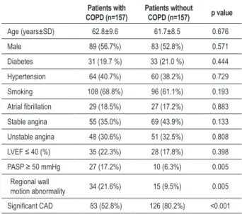
Clinical characteristics of the study population are listed in Table 1. Reasons for coronary angiography were; stable angina in 55 (35.0 %) patients, unstable angina in 48 (30.6%) patients, regional wall motion abnormality on echocardiography in 34 (21.6 %) patients, positive exercise test in 9 (5.7%) patients, suspicious ECG findings for ischemia in 8 (5.1%) patients assessed before elective surgery, malign arrhythmia in 2 (1.3 %) patients and complete atrioventricular block in 1 (0.6 %) patient. Regional wall motion abnormalities and PASP≥ 50 mmHg were significantly higher in the COPD group. Treadmill exercise test was performed by only 14 COPD patients. Of the 48 COPD patients evaluated as having unstable angina pectoris, 15 (31.2%) had just new-onset chest pain, 4 (8.3%) had progressive increase in chest pain, 14 (29.1%) had chest pain associated with ST depression, 11 (22.9%) had chest pain and negative T wave, 4 (8.3%) had chest pain and left bundle branch block (LBBB). Six (12.5%) patients with unstable angina were found to have normal coronary arteries; 3 had only chest pain, 2 had negative T wave and one had LBBB. Although 71.0% of the non-COPD patients describing stable angina had significant CAD, only 32.7% of the COPD patients describing stable angina had significant CAD (Table 2).
Coronary artery disease risk factors were significantly more frequent in COPD patients with significant CAD (older age, more frequent diabetes, hypertension, smoking history and lower LVEF) compared to COPD patients with normal coronary arteries (Table 3). However, there were no significant differences between risk factors in CAD patients with and without COPD (Table 2). Multivariate regression analysis including age, gender, diabetes, smoking, hypertension and severity of COPD revealed that only age (beta=0.247, p=0.02) and severity of COPD (beta=0.435, p<0.001) were independent predictors of significant CAD in COPD patients.
Table 1 - Clinical characteristics of the study population
Patients with COPD (n=157)
Patients without
COPD (n=157) p value
Age (years±SD) 62.8±9.6 61.7±8.5 0.676
Male 89 (56.7%) 83 (52.8%) 0.571
Diabetes 31 (19.7 %) 33 (21.0 %) 0.444
Hypertension 64 (40.7%) 60 (38.2%) 0.729
Smoking 108 (68.8%) 96 (61.1%) 0.193
Atrial ibrillation 29 (18.5%) 27 (17.2%) 0.883
Stable angina 55 (35.0%) 69 (43.9%) 0.133
Unstable angina 48 (30.6%) 51 (32.5%) 0.808
LVEF ≤ 40 (%) 35 (22.3%) 28 (17.8%) 0.398
PASP ≥ 50 mmHg 27 (17.2%) 10 (6.3%) 0.005
Regional wall
motion abnormality 34 (21.6%) 15 (9.5%) 0.005
Table 2 - Comparison of ratio of detection of signiicant CAD depending on clinical characteristics between patients with and without COPD
Ratio of CAD in COPD patients
Ratio of CAD in non-COPD patients
p value
Age (years±SD) 64.8±8.9 65.6±9.8 0.452
Male 58/89 (65.1%) 62/83 (74.7%) 0.188
Diabetes 25/31 (80.6 %) 27/33 (81.8 %) 1.0
Hypertension 45/64 (70.3%) 48/60 (80.0%) 0.299
Smoking 65/108 (60.2%) 66/96 (68.7%) 0.242
Stable angina 18/55 (32.7%) 49/69 (71.0%) <0.001
Unstable angina 42/48 (87.5%) 46/51 (90.2%) 0.755
LVEF ≤ 40% 28/35 (80.0%) 21/28 (75.0%) 0.763
Regional wall
motion abnormality 18/34 (52.9%) 12/15 (80.0%) 0.113 CAD: Coronary artery disease, COPD: Chronic obstructive pulmonary disease, LVEF: Left ventricular ejection fraction.
Table 3 - Comparison of clinical characteristics of COPD patients
with and without signiicant CAD.
Patients with normal coronary
arteries (n=74)
Patients with
signiicant CAD
(n=83)
p value
Age (years±SD) 58.3±9.6 65.0±8.9 <0.001
Male 34 (45.9%) 55 (66.2%) 0.024
COPD duration 85.6 + 32.5 68.1 + 30.6 0.002
Diabetes 9 (12.1%) 22 (26.5%) 0.010
Hypertension 23 (31.1%) 41 (49.4%) 0.023
Smoking 41 (55.4%) 67 (80.7%) 0.001
Atrial ibrillation 13 (17.5%) 16 (19.3%) 0.839
Stable angina 37 (50.0%) 18 (21.7%) <0.001
Unstable angina 6 (8.1%) 42 (50.6%) <0.001
LVEF ≤ 40% 7 (9.4%) 28 (33.7%) <0.001 Regional wall
motion abnormality 16 (21.6%) 18 (21.7%) 1.0 COPD: chronic obstructive pulmonary disease, CAD: Coronary artery disease, SD; Standard deviation, LVEF; Left ventricular ejection fraction. Severity of COPD was mild in 21 (13.4%), moderate in
93 (59.2%) and severe in 43 (27.4%) patients. Twenty-nine (18.5%) COPD patients had 3-vessel disease, 30 (19.1%) had 2-vessel and 24 (15.3%) had one-vessel disease. There was a significant correlation between COPD severity and presence of CAD (r=0.674, p=0.006) and number of vessels involved (r=0.675, p<0.001).
Discussion
There is a high coexistence between COPD and CAD because both diseases share similar risk factors9-12. In a recent large multi-center clinical trial in COPD patients the cause of death was cardiovascular in 26% of the cases13.
In a small study, coronary angiography was performed in 60 COPD patients over 40 years of age and ischemic heart disease was diagnosed in 53.3% of the examinees14. Similarly, in our study, significant CAD was detected in 52.8 % of the COPD patients undergoing coronary angiography. Patients with more severe COPD were reported to be more likely to have coexisting CAD and more likely to be hospitalized due to or die from cardiovascular causes3. In accordance, we found that the severity of COPD significantly correlated with the extent of CAD and the severity of COPD was an independent predictor for the presence of CAD.
Patients presenting with symptoms suggestive of angina pectoris are usually referred for cardiac evaluation. The most widely used test to obtain objective evidence of myocardial ischemia is the treadmill exercise testing with an average sensitivity and specificity of 75%15. Patients with an inconclusive test result and patients who are incapable of performing a satisfactory treadmill exercise test have to be further assessed by specific noninvasive diagnostic tests, such as Thallium-201 perfusion scintigraphy or pharmacological stress echocardiography to identify those patients with a high probability of disease, whereupon coronary angiography
is reasonable and cost-effective16. Due to lack of further noninvasive tests apart from the treadmill exercise in the region of our center and because of common risk factors, angina-like symptoms and inadequate exercise capacity, coronary angiography was performed in the majority of COPD the patients with possible associated ischemic heart disease.
Among patients with chest pain suggestive of angina pectoris who underwent coronary angiography, 10-30% were found to have normal or near-normal coronary arteries at the angiography and no evidence of coronary vasospasm17. In the present study, 67.3% of the COPD patients describing stable angina pectoris had normal or near-normal coronary arteries, while 90.2% of the COPD patients presenting with unstable angina pectoris, according to symptoms or the ECG, had significant CAD. Therefore, more attention should be paid to the differentiation of angina-like symptoms in COPD patients in the absence of suspicion of acute coronary syndrome. Further noninvasive diagnostic methods such as scintigraphy or stress echocardiography or careful follow-up may be more appropriate for COPD patients describing stable angina.
secondary to overload of right ventricle may result in the misinterpretation of wall motion on echocardiography19. In our study, 47.1% of the COPD patients documented as having regional wall motion abnormality on the echocardiography had normal or near-normal coronary arteries.
A possible explanation for the description of angina in COPD patients having normal coronary arteries in our study may be the presence of endothelial dysfunction. Long-term cigarette smoking is associated with impaired endothelium-dependent coronary vasodilatation, regardless of the presence or absence of atherosclerotic wall thickening20. Chronic bronchitis itself has also been identified as an independent predictor of the occurrence of CAD. Studies of the coronary circulationsuggest that during the natural history of atherosclerosis,impairment of receptor-mediated activation of the endotheliumis the first event21. In resected lung specimens studied in vitro, it has been shown that endothelial function is abnormal in COPD. The endothelial function of the renal circulation has also been shown to be abnormal in patients with COPD. This abnormality may also occur in other systemic vascular territories and may explain the coexistence of coronary heart disease and COPD in a different manner22.
Limitations
The present study has the disadvantages of retrospective studies. There may be several limitations associated with the
use of medical records. There may be diagnostic errors and not all data may be recorded, including COPD diagnosis and clinical characteristics. The number of study patients may be low to generalize our results.
Conclusions
In our retrospective study, we observed that diagnosis of CAD in COPD patients by symptomatology alone is difficult. However, the clinical diagnosis of CAD in the setting of unstable angina is accurate in most of the COPD patients. Therefore, further noninvasive diagnostic methods or careful follow-up may be more appropriate for COPD patients describing stable angina pectoris.
Potential Conflict of Interest
No potential conflict of interest relevant to this article was reported.
Sources of Funding
There were no external funding sources for this study.
Study Association
This study is not associated with any post-graduation program.
References
1. Falk JA, Kadiev S, Criner GJ, Scharf SM, Minai OA, Diaz P. Cardiac disease in chronic obstructive pulmonary disease. Proc Am Thorac Soc. 2008; 5: 543-8.
2. Haider AW, Larson MG, O’Donnell CJ, Evans JC, Wilson PW, Levy D. The association of chronic cough with the risk of myocardial infarction: the Framingham Heart Study. Am J Med. 1999; 106: 279-84.
3. Curkendall SM, DeLuise C, Jones JK, Lanes S, Stang MR, Goehring E Jr, et al. Cardiovascular disease in patients with chronic obstructive pulmonary disease, Saskatchewan Canada cardiovascular disease in COPD patients. Ann Epidemiol. 2006; 16: 63-70.
4. Balan KK, Critchley M. Is the dyspnea during adenosine cardiac stress test caused by bronchospasm? Am Heart J. 2001; 142: 142-5.
5. Thurnheer R, Laube I, Kaufmann PA, Stumpe KD, Stammberger U, Bloch KE, et al. Practicability and safety of dipyridamole cardiac imaging in patients with severe chronic obstructive pulmonary disease. Eur J Nucl Med. 1999; 26: 812-7.
6. Rabe KF, Hurd S, Anzueto A, Barnes PJ, Buist SA, Calverley P, et al. Global Initiative for Chronic Obstructive Lung Disease. Global strategy for the diagnosis, management, and prevention of chronic obstructive pulmonary disease: GOLD executive summary. Am J Respir Crit Care Med. 2007;176 (6): 532-55.
7. Braunwald E. Unstable angina: a classification. Circulation. 1989; 80: 410-4.
8. Lang RM, Bierig M, Devereux RB, Flachskampf FA, Foster E, Pellikka PA, et al. Recommendations for chamber quantification: a report from the American Society of Echocardiography’s Guidelines and Standards Committee and the Chamber Quantification Writing Group, developed in conjunction with the European Association of Echocardiography, a branch of the European Society of Cardiology. J Am Soc Echocardiogr. 2005; 18: 1440-63.
9. Camilli AE, Robbins DR, Lebowitz MD. Death certificate reporting of confirmed airways obstructive disease. Am J Epidemiol. 1991; 133: 795-800.
10. Hansell AL, Walk JA, Soriano JB. What do chronic obstructive pulmonary disease patients die from?: a multiple cause coding analysis. Eur Respir J. 2003; 22: 809-14.
11. Mapel DW, Dedrick DW, Davis K. Trends and cardiovascular co-morbidities of COPD patients in the Veterans Administration Medical System, 1991-1999. COPD. 2005; 2: 35-41.
12. Banasiak W, Pociupany R, Wilkins A, Ponikowski P. Characteristics of patients with coronary artery disease managed on an outpatient basis in the population of Poland. Results of the multicentre RECENT trial. Kardiol Pol. 2007; 65: 132-40.
13. McGarvey LP, John M, Anderson JA, Zvarich M, Wise RA. Chronic obstructive pulmonary disease. Ascertainment of cause-specific mortality in COPD: operations of the TORCH Clinical Endpoint Committee. Thorax. 2007; 62: 411-5.
14. Pavlov NR, Chereiskaia NK, Fedorova SI. Early diagnosis of ischemic heart disease in patients with chronic obstructive pulmonary diseases. Ter Arkh. 1999; 71: 52-6.
15. Grech ED. ABC of interventional cardiology: pathophysiology and investigation of coronary artery disease. BMJ. 2003; 326: 1027-30.
16. Hackshaw BT. Excluding heart disease in the patient with chest pain. Am J Med. 1992; 92: 46-51.
17. Crea F, Lanza GA. Angina pectoris and normal coronary arteries: cardiac syndrome X. Heart. 2004; 90: 457-63.
21: 473-7.
19. Ryan T, Petrovic O, Dillon JC, Feigenbaum H, Conley MJ, Armstrong WF. An echocardiographic index for separation of right ventricular volume and pressure overload. J Am Coll Cardiol. 1985; 5: 918-27.
20. Zeiher AM, Schachinger V, Minners J. Long-term cigarette smoking impairs endothelium-dependent coronary artery vasodilator function. Circulation.
1995; 92: 1094-100.
21. Zeiher AM, Drexler H, Saurbier B, Just H. Endothelium-mediated coronary blood flow modulation in humans: effects of age, atherosclerosis, hypercholesterolemia, and hypertension. J Clin Invest. 1993; 92: 652-62.