Origin of the Sinoatrial and Atrioventricular Nodal Arteries in South
Indians: an Angiographic Study
Lakshmi Ramanathan
1, Prakash Shetty
2, Soubhagya R. Nayak
1, Ashwin Krishnamurthy
1, Ganesh K. Chettiar
1, Annamalai
Chockalingam
3Departamento de Anatomia - Centro de Ciências Básicas, Kasturba Medical College, Bejai, Mangalore, Karnataka1; Departamento de Anatomia - Father Muller Medical College, Father Muller Road, Kankanady, Mangalore, Índia2; Departamento de Fisiologia - Centro de Ciências Básicas, Kasturba Medical College, Bejai, Mangalore, Karnataka3 - India
Summary
Background: To study the arterial supply of the conducting system and its correlation with the dominance of the coronary arteries in the South Indian population.
Objective: To determine angiographically the origins of the sinoatrial nodal artery (SAna) and atrioventricular nodal artery (AVna) in Indians.
Methods: The study included 300 consecutive patients (114 females, 186 males; mean age, 55 years) living in the southern coastal region of India, who underwent coronary angiography either for the symptoms of chest pain, angina pectoris or positive Treadmill Test. The angiograms contained both coronary arteries (right and left) in the right and left anterior oblique position. The origin of SAna and AVna from the coronary arteries was observed and correlated with the arterial dominance.
Results: The SA (sinoatrial) node was supplied by the right coronary artery (RCA) in 53% of the cases, by the circumflex (Cx) branch of left coronary artery (LCA) in 42.66%, and by both coronary arteries in 4.33% of cases. The AV (atrioventricular) node was also more often supplied by the RCA (72.33% of cases) than by the Cx branch of the LCA (27.66%), and surprisingly in none of the cases was this node supplied by both coronary arteries.
Conclusion: The results of the present study may help cardiac surgeons, particularly in surgeries related to certain valvular disorders, due to the proximity of the nodal branches to the valve complex. (Arq Bras Cardiol 2009;92(5):314-319)
Key words: Coronary vessels; sinoatrial node; atrioventricular node; angiography.
Mailing address: Soubhagya R. Nayak •
Department of Anatomy, Centre for Basic Sciences, Kasturba Medical Col-lege, Bejai, Mangalore, 575004, Karnataka - India
E-mail: [email protected]
Manuscript received January 02, 2008; revised manuscript received March 14, 2008; accepted March 14, 2008.
Introduction
Anterior atrial branches of the RCA are arranged in anterior, lateral and posterior groups to supply the corresponding surfaces of the right atrium. One important atrial branch is the SA nodal artery which is usually the second branch of the RCA and supplies the SA node in 65% of the cases; in the remaining 35%, the SA node is supplied by a branch of the Cx artery1. According to
April, SAna also arises from Kugel’s artery (when an atrial branch of the circumflex communicates with the similar branch of the right coronary artery, such an anastomotic branch is called Kugel’s artery)2. The arterial supply of the AV node arose in 80% of the
hearts from the RCA in the posterior atrioventricular sulcus at a point just beyond the origin of the posterior interventricular artery. In 20% of the hearts the AV node arose from the terminal part of the Cx at the crux of the heart [1]. The study on the arterial supply of the SA node will benefit clinical knowledge andsurgical
procedures as well as methods of cardiac examinationand treatment. Proper precautions should be taken to preserve the main arterial supply to the SA node during surgery3.
Sokolov and Varegin4, Krupa5, DiDio et al6, Sow et al7
and Onciu et al8 studied the origin of SAna. Krupa9, Sow
et al10, Arid et al11 studied the origin of AVna. Hutchinson1,
Vieweg et al12, Futami et al13 studied the origin of both SAna
and AVna. We studied the origin of both SAna and AVna and also the coronary dominance with the help of coronary arteriography.
The aim of the present study was to identify the origin of SAna and AVna from different coronary arteries and to determine the correlation of both arteries above with the artery of dominance. In this study, angiographic analysis of the coronary arteries was the preferred method of study as it is most commonly used in today’s clinical setting and such a study would be of more clinical significance than studies done on cadaver hearts.
Methods
costal region of India, who underwent coronary angiography either for the symptoms of chest pain, angina pectoris or positive Treadmill Test at Kasturba Medical College, Mangalore, India. The coronary angiograms selected were recorded on CDs. These were then studied using Cardiac SoftView (a software program to read coronary angiogram). The angiograms contained both coronary arteries (right and left) in various views, but to visualize the AVna and the SAna from either of the two coronary arteries, the preferred projections that were used here were the left anterior oblique view of the right coronary artery and the right anterior oblique view of the left coronary artery. The SAna when arising from the RCA can be recognized as its second branch after the conus artery that runs dorsally and extends over the upper portion of the atrial septum to terminate in the region of the ostium of the superior vena cava after arching laterally in front of the later. When the SAna is supplied by the circumflex branch of the LCA it passes to the right over the left auricle and reaches the SA node after encircling the superior vena cava.
The AVna is usually seen as a branch of the dominant artery. The artery arises from the crux, and is said to be a branch of the first posterior interventricular artery. It is consistent with the inverted U turn that is seen at the crux in the case of the RCA and a smooth curve seen at the crux in the case of the circumflex branch of the left coronary artery.
The following parameters were chosen for the study:
(i) Origin of the posterior interventricular artery to determine the coronary dominance.
(ii) The origin of SAna and AVna from the coronary arteries.
(iii) The origin of the nodal arteries were then correlated with dominance
These parameters were then tabulated and analyzed.
Results
In the present study, 161 (53.66%) of our 300 patients had a right dominant coronary arterial system (posterior descending artery originating from the right coronary artery), 67 (22.33%) had a left dominant coronary arterial system (posterior descending artery originating from the left circumflex), and 72 (24%) showed codominance. The SA node was supplied by the RCA [Fig. 1] more frequently (53% of cases) than by the Cx branch of LCA [Fig. 2] (42.66%), and in 4.33% of the cases this node was supplied by both coronary arteries. The AV node was supplied by the RCA (72.33% of the cases) [Fig. 3] more frequently than by the Cx branch of the LCA (27.66%) [Fig. 4], and surprisingly in none of the cases was this node supplied by both coronary arteries [Table 1]. It was found that (i) in 35% of the cases both the SA and AV nodal arteries originated from the RCA, (ii) in 11% of the cases both the SA and AV nodal arteries originated from the Cx branch of LCA, and (iii) in 54% of the cases both the SA and AV nodal arteries originated from different coronary arteries [Table 2]. The AV nodal artery was a branch of the dominant artery in 71.66% of the cases [Table 3] in comparison to the SA nodal artery which was a branch of the dominant artery only in 36% of the cases [Table 4].The source of arterial supply of the SA
node and AV node, as found by various authors and in the present study, is shown in Table 5.
Discussion
Hutchinson found that in 50% of the hearts, both the SA and AV nodes were supplied by branches of the right coronary artery. In 7%, both were supplied by branches from the left coronary artery1.
The SAna of the coronary arteries is anatomically regarded as a significant artery since it is used as a landmark to identify
Table 1 - Arteries giving rise to the S.A nodal and A.V nodal branches
Coronary artery
/branches No of SAna arising from
No of AVna arising from
RCA 159 (53%) 217 (72.33%)
Cx 128 (42.66%) 83 (27.66%)
RCA-Cx 13 (4.33%)
-Total 300 (100%) 300 (100%)
S.A - sinoatrial, A.V - atrioventricular, SAna - sinoatrial nodal artery, AVna
- atrioventricular nodal artery, RCA - right coronary artery, Cx - circumlex
artery,
Table 2 - Arterial supply to the S.A node and A.V node
Node Arterial supply No of cases
S.A & A.V
RCA 105 (35%)
Cx 33 (11%)
RCA-Cx 162 (54%)
S.A - Sinoatrial, A.V - Atrioventricular, RCA - right coronary artery, Cx -
circumlex artery.
Table 3 - Relation between A.V nodal supply and dominance
AV nodal supply in the case of From RCA From Cx
Right dominance 156 4
Left dominance 7 59
Co dominance 54 20
AV - arterioventricular, AVna - arterioventricular nodal artery
Table 4 - Relation between S.A nodal supply and dominance
SAna in the case of From RCA From Cx From RCA-Cx
Right dominance 81 75 8
Left dominance 43 25
-Co dominance 35 28 5
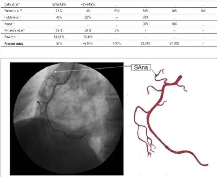
Figure 1 -Right coronary artery in the left anterior oblique position showing the sinoatrial nodal branch. SAna, sinoatrial nodal artery.
Table 5 - Arterial supply of the SA node & AV node as reported by various authors
Authors / Reference No.. Arterial supply of the SA node by (%) Arterial supply of the AV node by (%)
RCA LCA RCA & LCA RCA LCA RCA & LCA
Berdaj et. al 14 66% 34% _ _ _ _
Berdaj et. al 15 _ _ _ 73% 27% _
Didio et. al 6 58%+4.9% 42%+4.9% _ _ _ _
Futami et al 13 73 % 3% 23% 80% 10% 10%
Hutchinson 1 47% 27% -- 80% _ _
Krupa 9 _ _ _ 90% 10% _
Kyriakidis et al19 59 % 38 % 3% - -
-Sow et al 7 64.45 % 24.44% - - -
-Present study 53% 42.66% 4.33% 72.33% 27.66%
-the SA node, in addition to its clinical significance. Berdajs et al14 reported that the SAna crosses the superior posterior
border of the interatrial septum in 54% of the cases. From these data they concluded that the risk for intraoperative damage to the sinus node artery during the superior transseptal approach to the mitral valve is high14. Berdajs et al15 studied the AVna by
coronary angiogram; they found that the SAna originated from the RCA in 73% of the examined cases, and from the LCA in 27% of the cases. The left AVna was closely related to the mitral valve attachment, especially at the area of the left proximal part of the posterior leaflet. By comparing their data with the available literature, they reported that there is a high risk of intraoperative damage to the left AVna during manipulation of
the mitral valve annulus fibrosus15. In the present study, only
in 27.66% of the cases did the AVna originate from the LCA. In addition to being an excellent guide to the location of the sinoatrial node, the topic investigated demands further study due to its application in cardiac surgery3.
Nerantzis et al16 observed that the SAna played a major
Figure 2 -Left coronary artery in the right anterior oblique position showing the sinoatrial nodal branch from the circumlex artery; SAna - sinoatrial nodal artery; Cxa - circumlex artery.
Figure 3 - Right coronary artery in left anterior oblique position showing the atrioventricular nodal branch; AVna - atrioventricular nodal artery.
A study by Bokeriya et al17 on the variations of the SAna
and AVna showed 14 different variations of the SAna with regard to its connections with the anterior atrial sulcus and orifices of the superior vena cava, and they also identified 6 variants of the AVna with regard to its connections with the interventricular septum. The importance of these variations
was stressed because any surgery on these arteries could damage the surrounding structures17.
Krupa5 published a study on SAna in human hearts, where
base of the superior vena cava5. The origin of the SAna from
the proximal portion or trunk of the LCA, was less frequent (12%±3.2) than from the Cx (30%±4.5) and neither sex nor race influenced the origin of Sana6.
Sow et al7 studied the SA node artery by
injection-dissection and found that it was a solitary artery in 88.89% of cases and double in 11.11%, and the artery of origin was not influenced by coronary dominance. However, in the present study the AVna was a branch of the dominant artery in 71.66% of the cases. When it came from the RCA, the course of SAna varied greatly in relation to the site of origin – from either the atrial artery or from one of its collateral branches. When it came from the Cx its course was relatively uniform. These findings are especially important during certain stages of cardiac surgeries like atriotomies, surgical repair of valvular disorders and congenital malformations, which expose the SAna7. The
origin of the SAna from both coronary arteries in our study (4.33%) was much less frequent than in the study conducted by Futami et al13, but the frequency of RCA origin of the
AVna found by Futami et al13 was quite similar to that of
the present study (72.33%). Not even in a single occasion did the AVna originate from both coronary arteries in the present study, in comparison to the 10% of cases found by Futami et al13.
Sokolov and Varegin4 investigated 60 heart preparations
to observe the variation in the SAna. They reported that large atrial branches of RCA or LCA usually supply the SA node and showed some variations in the site of origin from the RCA and LCA. The SAna may arise from the right
Figure 4 - Left coronary artery in right anterior oblique position showing the atrioventricular artery from the circumlex artery; AVna - atrioventricular nodal artery; Cxa - circumlex artery.
intermediate or right posterior atrial branches. In the LCA variant it can arise from the anterior left, posterior left or intermediate left atrial branches4. Abuin and Nieponice18
studied the origin of AVna in 20 hearts. They found that in 40% of these hearts Kugel’s artery was supplying the AV node. The right descending superior artery supplied the AV node in 70% of the cases. These findings may be of major significance both in cardiology and in cardiovascular surgery18. Arid et al11 aimed at studying the anatomical
characteristics of the major arterial source of the AV node of 40 hearts by various anatomical and radiological techniques but they could only obtain results from 23 of them. They concluded that the most common atrial source of the AV node (21/23) was the RCA, but numerous variations in origin and topography were found11.
Kyriakidis et al19 mentioned that the knowledge of the
References
1. Hutchison MCE. A study on the atrial arteries in man. J Anat. 1978; 25: 39-54.
2. April WE. Clinical anatomy. 3rd ed. Baltimore: Williams & Wilkins; 1997. p. 275-7.
3. Yalcin B, Kirici Y, Ozan H. The sinus node artery: anatomic investigations based on injection-corrosion of 60 sheep hearts. Interac Cardiovasc Thorac Surg. 2004; 3: 249-53.
4. Sokolov VV, Varegin MP. Anatomy of the sinoatrial node and the supply of its vascularization in man. Arkh Anat Gistol Embriol. 1990; 98: 5-12.
5. Krupa U. The sinuatrial nodal artery in the human heart. Folia Morphol (Warsz). 1993; 52: 29-37.
6. DiDio LJ, Lopes AC, Caetano AC, Prates JC. Variations of the origin of the artery of the sinoatrial node in normal human hearts. Surg Radiol Anat. 1995; 17: 19-26.
7. Sow ML, Ndoye JM, Lô EA. The artery of the sinoatrial node: anatomic considerations based on 45 injection-dissections of the heart. Surg Radiol Anat. 1996; 18: 103-9.
8. Onciu M, Tuta LA, Baz R, Leonte T. Specifics of the blood supply of the sinoatrial node. Rev Med Chir Soc Med Nat Iasi. 2006; 110: 667-73.
9. Krupa U. The atrioventricular nodal artery in the human heart. Folia Morphol (Warsz). 1993; 52: G1-9.
10. Sow ML, Ndoye JM, Lô EA. The artery of the atrioventricular node: an anatomic study based on 38 injection-dissections. Surg Radiol Anat. 1996; 18: 183-7.
11. Arid JM, Armstrong O, Rogez JM, Robert R, Lardoux MC, Leborgne J. Arterial vascularisation of the atrioventricular node. Surg Radiol Anat. 2000; 22: 93-6.
12. Vieweg WV, Alpert JS, Hagan AD. Origin of the sinoatrial node and atrioventricular node arteries in right, mixed, and left inferior emphasis systems. Cathet Cardiovas Diagn. 1975; 1: 361-73.
13. Futami C, Tanuma K, Tanuma Y, Saito T. The arterial blood supply of the conducting system in normal human hearts. Surg Radiol Anat. 2003; 25: 42-9.
14. Berdajs D, Patonay L, Turina MI. The clinical anatomy of the sinus node artery. Ann Thorac Surg. 2003; 76:732-5.
15. Berdajs D, Kunzli A, Shurr U, Zund G, Turina MI, Genonni M. Clinical anatomy of the atrioventricular node artery. J Heart Valve Dis. 2006; 15: 225-9.
16. Nerantizis CE, Toutouzas P, Avgoustakis, D. The importance of the sinus node artery in the blood supply of the atrial myocardium: an anatomical study of 360 cases. Acta Cardiol. 1983; 38: 35-47.
17. Bokeriya LA, Mikhallin SL, Rerishvill AS. Anatomical variants of sinoatrial and atrioventricular node arteries. Cor Vasa. 1984; 26: 220-8.
18. Abuin G, Nieponice A. New findings on the origin of the blood supply to the atrioventricular node: clinical and surgical significance. Text Heart Inst J. 1998; 25: 113-7.
19. Kyriakidis M, Vyssoulis G, Barbetseas J, Toutouzas P. A clinical angiographic study of the arterial blood supply to the sinus node. Chest. 1988; 94: 1054-7.
Conclusion
In our study, the origin of the SAna was mainly from the RCA (53%) and LCA (42.66%); only in 4.33% cases was the SAna supplied by both coronary arteries. The AVna originated either from the RCA (72.33%) or from the Cx branch of LCA (27.66%). However, more angiographic studies of coronary arteries in different races should be performed to provide conclusive data on the origin of the SAna and AVna. The results of the present study may help cardiac surgeons, particularly during surgical correction of certain valvular disorders and congenital malformations of the coronary arteries.
Potential Conflict of Interest
No potential conflict of interest relevant to this article was reported.
Sources of Funding
There were no external funding sources for this study.
Study Association