Effect of Methyl Jasmonate on antioxidative
enzyme activities and on the contents of ROS
and H
2
O
2
in
Ricinus communis
leaves
alexandra Martins dos santos soares
1,2, thiago freitas de souza
1, tânia Jacinto
3,
olga lima tavares Machado
1*
1 Laboratório de Química e Função de Proteínas e Peptídeos, Centro de Biociências e Biotecnologia, Universidade
Estadual do Norte Fluminense Darcy Ribeiro, Campo dos Goytacazes-RJ, Brazil.
2 Present address: Laboratório de Bioquímica,Centro de Ciências Agrárias e Ambientais, Universidade Federal do
Maranhão (CCAA-UFMA), BR 222, Km 4, Chapadinha – MA. CEP 65500-000, Brazil
3 Laboratório Biotecnologia, Centro de Biociências e Biotecnologia, Universidade Estadual do Norte Fluminense
Darcy Ribeiro, Campo dos Goytacazes-RJ, Brazil.
* Corresponding author: Laboratório de Química e Função de Proteínas e Peptídeos
Centro de Biociências e Biotecnologia, Universidade Estadual do Norte Fluminense Darcy Ribeiro, Av. Alberto Lamego, 2000, Parque Califórnia, Campos dos Goytacazes-RJ, 28013-602; Tel 27261464; Fax 55-22-27261520 ; e-mail: [email protected]
Received: 18 January 2010; Accepted: 13 September 2010
abstract
Jasmonates are a class of plant hormones that mediate various aspectsin gene and metabolic regulation, defense, stress responses, reproduction and, possibly, communication. Oxidative stress stimulates synthesis of antioxidant metabolites and enhances antioxidant enzyme activities that could protect plant tissues. The aim of this study was to verify the effects of methyl jasmonate (JAME) treatment on the reactive oxygen species (ROS) and on the activities of H2O2 scavenging enzymes, such as superoxide
dismutase (SOD; EC 1.15.1.1), catalase (CAT; EC 1.11.1.6), ascorbate peroxidase (APX EC; 1.11.1.1), and guaiacol peroxidase (GPX; EC 1.11.1.7) in Ricinus communis leaves. The activity of CAT and GPX was transient while SOD activity decreased and APX increased after treatment with JAME. In addition, JAME exposure induced ROS accumulation.
Key words: Methyl jasmonate, oxidative stress, antioxidant enzymatic defenses, catalase, superoxide dismutase, peroxidase,
Ricinus comunnis, signal transduction
abbreviations:Prx, peroxidase (EC 1.11.1.7); APX, ascorbate peroxidase (EC; 1.11.1.1); SOD, superoxide dismutase (EC 1.15.1.1); CAT, catalase (EC 1.11.1.6); AJ, jasmonic acid; JAME, methyl jasmonate; ROS, reactive oxygen species.
introDUction
Plants, similar to animals, face many problems: a need
for regulation of metabolic processes and reproduction and
efficientdefense against enemies. They respond to a variety of
Jasmonic Acid (JA) and its methyl ester (JAME) are linoleic acid-derived cycle-pentanone based compounds and key molecules of the octadecanoid signaling pathway. Jasmonates are one of the simplest non-traditional plant hormones with diverse roles and functions, including a potential role in plant defense as part of the complex signaling pathways JAME does not only regulate a variety of plant-developmental responses, but is also induced by pathogen attack or wounding, which often leads to the generation of reactive oxygen species (ROS), including hydrogen peroxide (H2O2), superoxide anions (O2-) and hydroxyl free radicals (•OH) (Faurie et al., 2009; Parra-Lobato et al., 2009). ROS have the potential to interact with many cellular components, triggering stresses in plant cell culture, leading to membrane damage and, as a result, there is an immediate cellular response to trigger plant defense signals. Plants possess antioxidant defense systems, consisting of enzymatic and non-enzymatic components, which normally maintain ROS balance within the cell. For instance, they use a diverse array of enzymes, as well as low molecular weight antioxidants such as ascorbate and glutathione (GSH) to scavenge different types of ROS (Mittler, 2002; Jubany-Mari et al., 2010).
Anti-oxidative enzymes include superoxide dismutase (SOD), which catalyzes the disproportion of superoxide radicals to hydrogen peroxide and dioxygen; catalase (CAT) and peroxidase (Prx), which remove H2O2. SOD are
metalloenzymes with three known classes, depending on the active site metal cofactor (Mn, Fe, or Cu/Zn), and which play a key role in protection against oxidative stress. Catalases are tetrameric homoproteins that exist as multiple isozymes encoded by nuclear genes. They are located mostly in peroxisomes and glyoxisomes.
Unlike peroxidase, catalase efficiently scavenges H2O2
and does not require a reducing substrate to perform the task. Catalase isozymes differ in biochemical properties, as well as in their developmental specificity (occasionally related to germination), but their principal function probably involves fatty acid conversion, whilst other isozymes are related to lignification, photorespiration or the aging process (Kumari et al., 2006). The peroxidases (Prx) are associated with biochemical and physiological processes such as growth, cell formation, fruit development, ethylene biosynthesis, as well as the response to various stresses (Matamoros et al., 2003). Prx are homoproteins of approximately 50 kDa that are present as multiple isozymes in plant tissues; several Prx are present in nodules, but their functions and subcellular localizations are unknown (Becana et al., 1989). APX is the most important
peroxidase in H2O2 detoxification, catalyzing the reduction of
H2O2 to water using the reducing power of ascorbate (Noctor
and Foyer, 1998). This enzyme may account for up to 1% of the total soluble protein of nodules, and exists in the parenchyma and infected zone of several determinate nodules such as alfalfa (Dalton et al., 1998). Plants constantly sense and assess the level of ROS and reprogram their gene expression to respond to the changing conditions in their environment. Many studies employing mutants and antisense lines for catalase (CAT) 1 and 2, ascorbate peroxidase (APX) 1 and CuZn-superoxide dismutase 2 (CSD2) have revealed a strong link between ROS and process such as growth, development and biotic and abiotic stress responses. The function of ROS as signaling molecules involved in the expression of a large number of genes was studied using microarray experiments conducted on loss-of-function mutants that lack important ROS-scavenging enzymes. The use of mutants that lack important ROS-scavenging enzymes such as APX1 and CAT enabled therefore the identification of key regulatory proteins involved in the sensing of ROS and the regulation of transcript expression by different ROS signals (Miller et al., 2008).
The aim of the present study was to examine the relationship between reactive oxygen species accumulation and anti-oxidative enzyme activities in Ricinus comunnis plant leaves after treatment with methyl jasmonate for different time durations.
Material anD MethoDs
Chemicals used for the protease assay were from Sigma. Electrophoresis reagents used were from Bio-Rad. Methyl jasmonate was purchased from Aldrich and Triton X-100 was from Calbiochem.
plant material and protein extraction: Castor bean
(Ricinus communis L., cultivar IAC-80, from Instituto
methyl jasmonate treatment (1, 4, 6, 12, 24 and 48 hours), fresh leaves (3 g) were harvested directly into liquid nitrogen in a mortar, with 0.3 g PVP, in 100 mM potassium phosphate buffer pH 7.5 with 1 mM EDTA and 3 mM DDT.
The homogenate was centrifuged in a refrigerated centrifuge at 15 000 g for 20 min, and the supernatant obtained was used as the source of enzyme. Protein concentrations were estimated according to Bradford’s method (1976) using bovine serum albumin as standard
superoxide Dismutase activity: A non-denaturing gel (native PAGE -10%) was carried out, according to Davis (1964), to identify and separate the superoxide dismutase isozymes. Measurement of SOD activity was assayed by observing the inhibition of photochemical reduction of nitro blue tetrazolium (NBT), according to the protocol of Azevedo et al., (1998). The SOD activity was observed as an achromatic band on a purple-blue background (usually within 5-7 min). For SOD isoform identification, assays were performed in the presence of selective inhibitors. KCN (3 mM) inhibits only Cu/Zn SOD; H2O2 (5 mM) inhibits both Cu/Zn-SOD and Fe-SOD; Mn-SOD
was not inhibited by KCN or H2O2 (Azevedo et al. 1998).
catalase activity: Catalase activity was assayed by the method of Barka (2001). The reaction mixture (3 ml) consisted of enzyme extract (30 µL), 10 mM H2O2 and 50
mM potassium phosphate buffer, pH 7.0. The catalase activity was expressed as µmol min-1 mg-1 of protein.
ascorbate peroxidase activity: APX activity was determined from the decrease in absorbance (ABS) at 290nm, due to the H2O2 – dependent oxidation of ascorbate. The 2 mL
reaction mixture contained 50 mM potassium phosphate (pH 7.5), 0.5 mM ascorbic acid, 1 mM H2O2, 0.1 mM EDTA and
enzyme extract (30 µL) at 25o C (Barka, 2001). The activity
was expressed as nmol min-1 mg-1 of protein. Following
electrophoretic separation (8% NATIVE-PAGE, according to Davis 1964), the gels were equilibrated with 50 mM sodium phosphate buffer (pH 7.0) and 2 mM ascorbic acid for a total of 30 min. The gels were then incubated with 50 mM sodium phosphate buffer (pH 7.0) containing 4 mM ascorbic acid and 2 mM H2O2 for 20 min. H2O2 was added to this solution
immediately prior to incubation with the gel. The gels were subsequently washed with 50 mM sodium phosphate buffer (pH 7.0) for 1 min and submerged in a solution of 50 mM sodium phosphate buffer (pH 7.8), 28 mM TEMED, and 2.45 mM NBT with gentle agitation. The APX activity was observed as an achromatic band on a purple-blue background (usually
within 3-5 min). The reaction was stopped with a brief wash with deionized water (Mittler and Zilinskas, 1993).
guaiacol peroxidase activity: Peroxidase activity (Guaiacol peroxidase) with guaiacol as substrate was assayed by following the increase in absorbance at 470 nm using a modified procedure of Cakmak and Horst (1991). The assay mixture contained 25 mM phosphate buffer (pH 6.0), 2.58 mM guaiacol and 10 mM H2O2 in a total volume of 1 mL. The
reaction was initiated by the addition of 10 µg of total protein and the increase in absorbance at 470 nm was measured for 5 min. The peroxidase activity was expressed as µmol min-1
mg-1 of protein. For gel activity, protein extracts were resolved
by native-continuous-PAGE, pH 8.8, using 25 mM Tris, 192 glycine, pH 8.3 as running buffer. After electrophoresis, the gel was washed three times for 5 min with 25 mM phosphate buffer (pH 6.0) and incubated with the assay mixture described above. The brown colored bands (due to guaiacol polymerization) were identified after1 min.
In Vivo detection of h2o2 in Ricinus communis leaves:
Diaminobenzidine (DAB) was used for the detection of H2O2
staining in leaf tissues (Thordal-Christensen et al. 1997). After treatment with JAME, the leaves were excised and floated in 0.1% (v/v) TritonX-100 containing 1 mg ml-1 DAB, and vacuum
infiltrated for 30 min. DAB incubation was continued for 24 h. At the end of the staining, leaves were washed in distilled water, boiled in 95% ethanol for 10 min and examined for staining. H2O2 was visualized as a reddish brown coloration.
ros detection: The presence of ROS was determined by DPPH radical-scavenging activity as proposed by Brand-Williams et al. (1995). Fresh leaves from treated or control plants were harvested into liquid nitrogen in a mortar in the presence of absolute ethanol (1:5 w/v). The homogenate was centrifuged at 15 000 g for 10 min at 40 C, and the
supernatant obtained was used for analysis. One-hundred µL of the supernatant were mixed with 1mL of 6.5 x 10⁻⁵M
solution of 1,1-diphenyl-2-picrylhydrazyl (DPPH) in methanol. The mixture was shaken vigorously and maintained for 30 min in the dark. The absorbance was measured at 515 nm. The absorbance of the control was obtained by replacing the sample with methanol. Rutin was used as a control. The scavenging activity was calculated using the following equation:
resUlts
In order to investigate the effect of JAME on Ricinus
communis, plants at the three-leaf-stage (15 days) were
exposed to JAME vapors and the superoxide dismutase, catalase, ascorbate peroxidase and guaiacol peroxidase activities were evaluated in the leaf extracts.
Three isoforms of SOD were detected by zymography after fractionation of crude leaf extracts by 8% NATIVE-PAGE (bands 1 - 3, Figure 1) and were identified using selective inhibitor assays. The band with the lowest mobility was not inhibited by KCN or H2O2, which classified as a Mn-SOD (band
1). H2O2 inhibited the activity of the second band, which is
probably a Fe-SOD and the inhibitors KCN and H2O2 reduced
the activity of band 3, which was identified as Cu/Zn- SOD. The treatment with JAME (0.025%) significantly decreased the activity of one Mn-SOD (band 1) after 24 and 48 h of treatment (Figure 2). Figure 3 shows that the exposure to JAME vapors decreased the catalase activity in leaf extracts, when compared with untreated control plants. The effect was stronger during the first hour of treatment and the activity returned to control levels after 12 hours. After 24 h and 48 h of treatment, no significant changes for this enzyme were observed.
A B C
1 2
3
figure 1. Isoforms of superoxide dismutase activity determined in 8% gel electrophoresis under non-denaturing conditions. A – pre-incubated gel in buffer without inhibitor; B – gel incubated in buffer + KCN (isoform Cu/Zn-SOD inhibitor); C – gel incubated in buffer + H2O2 (isoforms Cu/Zn-SOD and
Fe-SOD inhibitor).
C 1 4 6 C 12 C 24 C 48
figure 2. Superoxide dismutase activity determined by 8% gel electrophoresis under non-denaturing conditions. Lines: C - control; 1, 4, 6, 12, 24 and 48 – hours of exposure to methyl jasmonate. Arrows: SOD isoform altered after 24 h and 48 h of JAME treatment.
50 –
40 –
30 –
20 –
10 –
0 –
µmol min
1 mg -1 ptn
C-1H 1H 4H 6H 12H C-24H 24H C-48H 48H
time of exposition to the methyl jasmonate
figure 3. Catalase activity, determined as the decrease in absorbance at 240nm for 1 min following the decomposition of hydrogen peroxide. The reaction mixture contained 5 mM phosphate buffer (pH 7.0) and 15 mM H2O2. Data are means ± S.D. (n=3).
The APX activity was detected in both crude extract and
directly on the gels, after fractionation of this extract by native
PAGE. Figure 4A shows that the total APX activity increased after 4h and 24h of JAME treatment. A similar behavior was
observed for the two isoform bands detected in the gel assay
(Figure 4B).
0.6 –
0.5 –
0.4 –
0.3 –
0.2 –
0.1 –
–
mmol min
-1 mg -1 ptn
C-1 1 4 6 12 C-24 24 C-48 48
C 1 4 6 C 12 C 24 48
figure 4. Ascorbate peroxidase activity in crude extract of plant control and treated for different times. a) Activity of ascorbate peroxidase, demonstrated as the decrease in the absorbance at 290 nm following ascorbate oxidation in the slurry of control and treated plants. The activity was determined as (mmol min-1 mg-1 of protein). Lines: C – control; 1 – 48 h: time of exposure to methyl jasmonate. Data are means ± S.D. (n=3). b) Activity of ascorbate peroxidase, determined in gel after separation of proteins by 8% native electrophoresis. Lines: C - control; 01-48h - time of exposure to methyl jasmonate. Black lines: APX isoforms detected.
The guaiacol activity of the crude extract decreased after 1, 4 and 6 h, and returned to near control levels after 12 h, before increasing considerably after 24 and 48 hours of treatment (Figure 5A). Two guaiacol peroxidase activities (named G1 and G2) were observed when the guaiacol activity was assayed in gel after native PAGE and both of them presented a similar behavior for the total activity observed in the crude extract (Figure 5B).
18 – 16 –
14 –
12 – 10 –
8 –
6 –
4 – 2 –
µmol min
-1 mg -1 ptn
C1-12 1 4 6 1 2 C-24 24 48
time of exposition to the methyl jasmonate (h)
C 1 4 6 C 12 C 24 48
G1 G2
figure 5. Guaiacol peroxidase activity in crude extract of control plants and plants treated for different times. A) Demonstrative graph of the guaiacol peroxidase activity. The activity was defined as (▲ABS µg 470nm /ug protein x min). Data are means ± S.D. (n=3). B) Activity of guaiacol peroxidase, determined in gel after separation of proteins by 12% native electrophoresis. Lines: C - control; 1-48h – hours of exposure to methyl jasmonate. G1 and G2 - bands detected in gel.
In order to investigate the oxidant status and generation of ROS in leaf tissues, we assayed the production of H2O2
in Ricinus communis plants after exposure of JAME vapors
for 1 - 48 Hours. DAB polymerizes and turns deep brown in the presence of H2O2. The intensity of the coloration and its
localization can be qualitatively assessed and photographed. The development of the DAB-H2O2 reaction product in
Ricinus communis leaves, in response to JAME treatment
is shown in Figure 6. The production of H2O2 initiated after
1 h of treatment and intensified until 6h. These results also indicate that the H2O2 production was concentrated in the
major veins of the leaves after 12 and 24 h after of treatment, and that this production disappeared after 48 hours of JAME treatment.
C 1 4 6
C 12 24 48
figure 6. H2O2 detection in Ricinus communis leaves. a) control, 1h, 4h, 6h,
control, 12h, 24h and 48h after JAME treatment, respectively. b) control, 1h, 4h, 6h, control, 12h, 24h and 48h after JAME treatment, respectively.
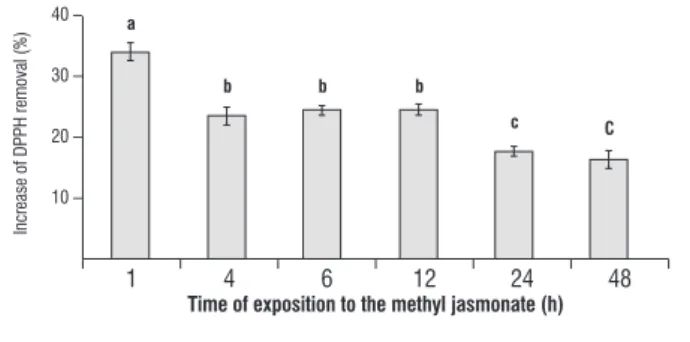
Positive control, rutin, removed 100% of the initial DPPH free radicals.
40 –
30 –
20 –
10 –
Increase of DPPH removal (%)
1 4 6 12 24 48
time of exposition to the methyl jasmonate (h)
a
b b b
c c
figure 7. Detection of reactive oxygen species in the slurry of Ricinus communis leaves after treatment with methyl jasmonate, determined as the fold increase from control at 1 to 48h after treatment. Data are means ± S.D. (n=3).
DiscUssion
The role of JA in plants has received considerable attention and various modes of JA and JAME action have been discussed (Farmer and Ryan, 1990; Farmer, 2007). This study aimed at evaluating the ability of JAME, exogenously applied
to Ricinus communis leaves, to counteract stress-induced
responses involving antioxidative enzymes and ROS.
Compounds that are able to reduce the damaging effects of certain stresses may be of great importance from both theoretical and practical points of view. ROS are dangerous cytotoxic molecules, but also act as intermediate signaling molecules to regulate the expression of genes associated with antioxidant defense mechanisms (Vranova et al., 2002; Neill et al., 2002). To counteract the toxicity of reactive oxygen species, plants have developed a highly efficient antioxidant enzymes defense system, mainly including SOD, APX, CAT and Prx, increasing tolerance to different stress factors (Jiang and Zhang 2002).
In our studies, three SOD isoforms were found (Mn-SOD, Fe-SOD and Cu/Zn-SOD), however just one presented alteration in its activity after JAME treatment. JAME resulted in a significant decrease in the activity of one Mn-SOD isoform, compared to the control, in R. communis leaves, 24 and 48 h after treatment. Similar results were observed by Lijun et al. (2005), who reported inhibition of SOD by high concentrations of cadmium. In addition, Hernández et al. (1993) demonstrated that saline stress causes a reduction of Mn-SOD and
Cu/Zn-SOD in susceptive cultivars of Pisum sativum. In Arabidopsis
thaliana, the total SOD activity decreased three days after
JAME treatment and increased two fold after seven days of treatment (Jung, 2004).
Furthermore, in the present study, catalase activity showed a transient behavior at 1 h to 12 h of JAME treatment and than remained at a level similar to that of the control. The catalase reduction at 1 h after treatment could be related to an accumulation of H2O2 in the peroxissomes, as a strategy to
preserve it from total degradation. Thus, H2O2 could be used
as a defense signalizing molecule in other parts of the plant (Thordal-Christensen et al., 1997). Therefore, an increase in CAT activity 12 h after JAME treatment and its return to control levels at 24 and 48 h after treatment could be related to a negative feedback, where the JA and JAME could be stabilized. Other reports have registered the decrease in catalase activity, as in nodules of Phaseolus vulgares roots under saline stress, whereas in other parts of the roots, CAT activity was not detected (Jebara et al., 2005). Chamnongpol et al. (1998) reported catalase as a mediator of defense response, because the reduction in its activity was related to the accumulation of H2O2 in tobacco. Jung (2004) verified
that catalase activity did not modify between the 3rd and
5th day after exposure to treatment with methyl jasmonate,
but increased by 58%, seven days after the treatment in A.
thaliana. Depending on the concentration of jasmonic acid
used in the induction of the defense response, the activity of antioxidant enzymes is modified in a distinct manner. In
Morinda elliptica, catalase activity was observed to increase
after 1 day and was then drastically reduced after 3 and 6 days of treatment with 50 µM JA.
When JA was used at a concentration of 100 µM, no significant alteration in the enzyme was observed until the third day, but the activity was reduced after 6 days of treatment (Chong et al. 2005). The catalase activity reduction was also registered in Catharanthus roseus cultures, induced by fungi (Zhao et al. 2001) and by water stress in strawberry (Wang, 1999).
in which the ascorbate peroxidase activity did not suffer any alteration 1 day after 50 µM JAME treatment, however, it increased at 3 days and reduced drastically at 6 days after treatment with JAME. These authors also observed an increase in APX activity after 3 days of 100 µM JAME treatment and a reduction of 50% in the activity after 6 days of treatment.
Guaiacol is often used as a substrate for the measurement of peroxidase activity (Hiraga et al., 2001). The enzyme, guaiacol peroxidase, is involved in protective actions in plants, participates in the lignification of the cellular wall, biosynthesis of ethylene, reestablishment after wound and defense against pests (Gazaryan et al. 1996). In our study, GPX presented a gradual decrease in activity until 6 h and a reestablishment of control values 12 h after the treatment. After this period, the activity increases significantly, reaching nearly 50% at 48 h after JAME treatment. The gel analysis of GPX revealed an activity behavior similar to that obtained by crude extract analysis. Jung (2004) reported that GPX activity increases by about 100 fold after seven days of treatment with JAME in A. thaliana and suggested that most of the protection against oxidative stresses in plants treated with JAME may be the result of enzymatic antioxidant substances, particularly GPX. Our results indicate a gradual and significant increase in GPX activity at 24 and 48 h, however the reduced activity of this enzyme at the initial moments after the treatment with JAME may be due to the generation of ROS, possibly involved with the mediation of stress defense signaling. The delayed increase in GPX activity is probably related to the return to normal metabolism. Jebara et al. (2005) showed the activity reduction of three GPX isoforms (one acid isoform and two basic ones) in roots of Phaseolus vulgaris, while in the nodules, a reduction of activity GPX occurs under saline stress, suggesting that GPX has an important role in detoxify H2O2 under saline stress.
The variation in antioxidant activity within the cell can modify the ROS content and lead to death or an acclimatization response. With the objective of verifying whether the alteration in the antioxidant status in Ricinus communis is in accordance with variations in the ROS formation, we carried out experiments to detect the H2O2 content and free radicals. A
gradual accumulation of H2O2 was observed to occur between
1 and 6h after the treatment with JAME. In this period, the staining with DAB disclosed circular injuries that spread over the surface of the leaf. At 12 and 24h after the treatment, DAB
staining was less intense and was restricted to the central vein and, at 48h, there was no apparent formation of H2O2.
Other studies have documented the hydrogen peroxide production in stress. Rao et al. (1997) observed that salicylic acid (SA) increases the production of H2O2, the
lipidic peroxidation and the oxidative damage to the proteins
in A. thaliana. Cho and Seo (2005) related an increase of
H2O2 content in A. thaliana treated with Cadmium. Our data
referring to the detection of free radical content, in leaves treated with methyl jasmonate, complement results regarding the production of H2O2 in R. communis leaves. An increase of
33% was observed to occur in the content of free radicals at 1h after treatment with JAME; this value decreased, returning to control levels by 48h. Apparently, the sharp formation of ROS at the initial moments of JAME exposure is related to the decrease in the activities of CAT and GPX.
In conclusion, the modification of antioxidants enzymes (SOD, APX, GPX, CAT) can play important protective roles avoiding the deleterious effects triggered by elevated levels of ROS observed at initial moments after JAME exposure. In general, plant defense signaling is studied using model plants. However, different plant species probably have particularities in their responses. Therefore, reinforce the relevance to study plant defense signaling in Ricinus communis, an important tropical oil-plant and a promising bioenergy source.
acknowledgements: We thank FAPERJ and CNPq for the financial support and fellowships.
references
Azevedo RA, Alas RM, Smith RJ, Lea PJ (1998) Response of antioxidant enzymes to transfer from elevated carbon dioxide to air and ozone fumigation, in the leaves and roots of wild-type and a catalase-deficient mutant of barley. Physiol. Plant. 104:280 –292.
Barka EA (2001) Protective enzymes against reactive oxygen species during ripening of tomato (Lycopersicon esculentum) fruits in response to low amounts of UV-C. Aust. J. Plant Physiol. 28:785–791.
Becana M, Paris FJ, Sandalio LM, del Rio LA (1989) Isoenzymes of superoxide dismutase in nodules of Phaseolus vulgaris L; Pisum sativum L; and Vigna unguiculata (L.) Walp. Plant Physiol. 90:1286–92.
Bradford MM (1976) A rapid and sensitive method for the quantification of microgram quanties of protein utilizing the principle of protein-dye binding. Anal. Biochem. 72:248–254.
Brand-Williams W, Cuvelier ME, Berset C (1995) Use of a free radical method to evaluate antioxidant activity. LWT Food Sci. Technol. 28:25–30.
Chamnongpol S, Willekens H, Moeder W, Langebartels C, Sandermann HJr, Van Montagu M, Inze D, Van Camp W (1998) Defense activation and enhanced pathogen tolerance induced by H2O2 in transgenic tobacco. Proc.
Natl. Acad. Sci. USA. 95:5818–5823.
Cho UH, Seo NH (2005) Oxidative stress in Arabidopsis thaliana exposed to cadmium is due to hydrogen peroxide accumulation. Plant Sci. 168:113– 120.
Chong TM, Abdullah, MA, Fadzillah NM, Lai OM, Lajis NH (2005) Jasmonic acid elicitation of anthraquinones with some associates enzymic and non enzymic antioxidant responses in Morinda elliptica. Biochem. J. 36:469– 477.
Dalton DA, Joyner SL, Becana M, Iturbe-Ormaetxe I, Chatfield JM (1998) Antioxidant defences in the peripheral cell layers of legume root nodules. Plant Physiol. 116:37–43.
Davis BJ (1964) Disc electrophoresis, part II. Ann. Ny. Acad. Sci. 121:404– 427.
Farmer EE (2007) Plant biology - Jasmonate perception machines. Nature. 448:659-660.
Farmer EE, Ryan CA (1990) Interplant communication - airborne methyl jasmonate induces synthesis of proteinase-inhibitors in plant-leaves. Proc. Natl. Acad. Sci. USA. 87:7713-7716.
Faurie B, Cluzet S, Merillon, JM (2009) Implication of signaling pathways involving calcium, phosphorylation and active oxygen species in methyl jasmonate-induced defense responses in grapevine cell cultures. J. Plant Physiol. 166:1863-1877.
Gazaryan IG, Lagrimini LM, Ashby GA, Thorneley RNF (1996) Mechanism of indole-3-acetic acid oxidation by plant peroxidases: anaerobic stopped-flow spectrophotometric studies on horseradish and tobacco peroxidases. Biochem. J. 313:841–847.
Hiraga S, Sasaki K, Ito H, Ohashi y, Matsui H (2001) A large family of class III plant peroxidases. Plant Cell Physiol. 42:462–468.
Hernández JA, Corpas FJ, Gómez M, Rio LA, Sevilla F (1993) Salt-induced oxidative stress mediated by activated oxygen species in pea leaf mitochondria. Physiol. Plant. 89:103–110.
Hossain Z, Lopez-Climent MF, Arbona V, Perez-Clemente RM, Gomez-Cadenas A (2009) Modulation of the antioxidant system in citrus under waterlogging and subsequent drainage. J. Plant Physiol. 166: 1391-1404.
Jebara S, Jebara M, Limam F, Aouani ME (2005) Changes in ascorbate peroxidase, catalase, guaiacol peroxidase and superoxide dismutase activities in common bean (Phaseolus vulagis) nodules under salt stress. J. Plant Physiol. 162:929–936.
Jiang My, Zhang JH (2002) Water stress-induced abscisic accumulation triggers the increased generation of reactive oxygen species and up-regulates the activities of antioxidant enzymes in mayze leaves. J. Exp. Bot. 379:2401– 2410.
Jubany-Mari T, Prinsen E, Munne-Bosch S, Alegre L (2010) The timing of methyl jasmonate, hydrogen peroxide and ascorbate accumulation during water deficit and subsequent recovery in the Mediterranean shrub Cistus albidus L. Environ. Exp. Bot. 69:47-55.
Jung S (2004) Effect of chlorophyll reduction in Arabidopsis thaliana by methyl jasmonate or norflurazon on antioxidant systems. Plant Physiol. Biochem. 42:225–231.
Kumari GJ, Reddy AM, Naik ST, Kumar SG, Prasanthi J, Sriranganayakulu G, Reddy PC, Sudhakar C (2006) Jasmonic acid induced changes in protein pattern, antioxidative enzyme activities and peroxidase isozymes in peanut seedlings. Biol. Plant. 50:219–226.
Lijun L, Xuemei L, yaping G, Enbo M (2005) Activity of the enzymes of the antioxidative system in cadmium-treated Oxya chinesis (Orthoptera Acridoidae). Environ. Toxicol. Pharmacol. 20:412–416.
Ma ZL, Gao KS (2010) Spiral breakage and photoinhibition of Arthrospira platensis (Cyanophyta) caused by accumulation of reactive oxygen species under solar radiation. Environ. Exp. Bot. 68:208-213.
Matamoros MA, Dalton DA, Ramos J, Clemente MR, Rubio MC, Becana M (2003) Biochemistry and molecular biology of antioxidants in the rhizobia-legume symbiosis. Plant Physiol. 133:499–509.
Miller G, Shulaev V, Mittler R (2008) Reactive oxygen signaling and abiotic stress. Physiol. Plant. 133:481–489.
Mittler R, Zilinskas BA (1993) Detection of ascorbate peroxidase activity in native gels by inhibition of the ascorbate-dependent reduction of nitroblue tetrazolium. Anal. Biochem. 212:540–546.
Mittler R (2002) Oxidative stress, antioxidants and stress tolerance. Trends Plant Sci. 7:405–410.
Mittler R, Vanderauwera S, Gollery M, Van Breusegem F (2004) Reactive oxygen gene network of plants. Trends Plant Sci. 9:490-498.
Neill SJ, Desikan R, Clarke A, Hurst RD, Hencock JT (2002) Hydrogen Peroxide and nitric oxide as signaling molecules in plants. J. Exp. Bot. 53:1237–1247. Noctor G, Foyer CH (1998) Ascorbate and glutathione keeping active oxygen under control. Annu. Rev. Plant Physiol. Plant Mol. Biol. 49:249–79. Parra-Lobato MC, Fernandez-Garcia N, Olmos E, Alvarez-Tinaut MC, Gomez-Jimenez MC (2009) Methyl jasmonate-induced antioxidant defence in root apoplast from sunflower seedlings. Environ. Exp. Bot. 66: 9-17.
Pearce G, Moura D, Stratmann J, Ryan CA (2001) Production of multiple plant hormones from a single polyprotein precursor. Nature, 411: 817-820. Rao MV, Paliyath G, Ormrod DP, Murr DP, Watkins, CB (1997) Influence of salicylic acid on H2O2 production, oxidative stress, and H2O2 -metabolizing
enzymes. Plant Physiol. 115:137–149.
Tanou G, Molassiotis A, Diamantidis, G (2009) Induction of reactive oxygen species and necrotic death-like destruction in strawberry leaves by salinity. Environ. Exp. Bot. 65:270-281.
Thordal-Christensen H, Zhang Z, Wei y, Collinge DB (1997) Subcellular localization of H2O2 in plants. H2O2 accumulation in papillae and hypersensitive
response during the barley-powdery mildew interaction. Plant J. 11:1187– 1194.
Vranova E, Inze D, Van Breusegen F (2002) Signal transduction during oxidative stress. J. Exp. Bot. 53:1227–1236.
Wang Sy (1999) Methyl jasmonate reduces water stress in strawberry. Plant Growth Regul. 18:127–134.