Session editor: Alfredo José Mansur
Associate editors: Desiderio Favarato Vera Demarchi Aiello
Mailing address: Alfredo José Mansur - Incor - Av. Dr. Enéas C. Aguiar, 44 - 05403-000 - São Paulo, SP, Brazil
Case 6/2000 – Dyspnea and pain in the left lower limb in a 52-year-old male patient (Instituto do Coração of Hospital das Clínicas - FMUSP - São Paulo)
Clinicopathologic Session 6/2000
A 52-year-old male patient was admitted to the hospi-tal complaining of dyspnea and pain in the left lower limb.
The patient reported that he sought medical care 7 years earlier due to dyspnea triggered by moderate exertion, which lasted for 3 months. He also reported at that time an episode of dizziness and an episode of nocturnal paroxys-mal dyspnea.
Two months before, he was diagnosed with hyperten-sion. He reported smoking until 10 years ago.
On physical examination, the patient was in good con-dition, his pulse was regular, his heart rate was 72bpm, and his blood pressure was 170/110mmHg. His lung examination revealed no abnormalities. His heart examination showed normal first and second heart sounds, and a systolic mur-mur (+/4) in the aortic area. His liver was palpable 2cm from the right costal margin. No edema existed in the lower limbs. The electrocardiogram showed sinus rhythm, a heart rate of 72bpm, a QRS axis of -10° backwards, left chamber hy-pertrophy, and alterations of ventricular repolarization (fig. 1). The echocardiogram (Table I) revealed diffuse left ven-tricular hypokinesia, calcification of the aortic valve, and double dysfunction of the aortic valve.
Serology for the diagnosis of Chagas’ disease was negative.
The following diagnoses were established: hyperten-sion, mild aortic stenosis, and ventricular dysfunction.
The continuous treadmill stress test showed a good physical capacity, a duration of exertion of 8 minutes, and initial and final velocities of 2 miles/hour and 5 miles/hour, respectively. The patient’s heart rate at rest was 100bpm and on maximum exertion 143bpm; his blood pressure ranged from 160/99mmHg to 220/74mmHg.
Rest-exercise equilibrium radionuclide ventriculo-graphy with red blood cells labeled with technecium-99m revealed diffuse left ventricular hypokinesia. Left ventri-cular ejection fraction at rest was 0.30 and on exertion (25 W), 0.37.
The patient remained asymptomatic until 4 years ago, when he experienced severe precordial pain, which lasted for 2 hours, and he sought medical care on an emergency
basis. The electrocardiogram revealed at that time elevation of the ST segment in II, III, aVF, V1, V2, and from V5 to V8. The patient was diagnosed with inferodorsal myocardial in-farction, and 1,500,000 units of streptokinase were admi-nistered intravenously. Elevation of the ST segment regressed, and the maximum value of the MB fraction of creatine kinase was 131 U/L 6 hours after pain onset. The patient continued his treatment for infarction at another medical facility.
The patient evolved asymptomatically, and 4 months later he underwent hemodynamic (table II) and coronary angiographic studies, which depicted no obstruction in the coronary arteries. Left ventriculography showed diffuse hypokinesia.
The patient remained asymptomatic until 1 year and 4 months ago, when he had a cerebral stroke with motor mani-festation that regressed without sequelae.
Five months ago, the patient was hospitalized in ano-ther medical facility due to non-Q-wave myocardial infarc-tion. He was then referred to InCor for assessment of the in-dication for surgical treatment for probable coronary heart disease. However, after medical evaluation, we chose to continue the clinical treatment.
Three months ago, the patient returned to InCor, searching for treatment for his dyspnea.
The laboratory tests were as follows: hemogram with 5,100,000 red blood cells/mm3; hematocrit, 45%;
hemo-globin, 15g/dL; 12,000 leukocytes/mm3 (4% band
leukocy-tes, 68% segmented leukocyleukocy-tes, 21% lymphocyleukocy-tes, and 7% monocytes); 368,000 platelets/mm3. The serum level of
gluco-se was 96mg/dL; urea, 40mg/dL; creatinine, 1.1mg/dL; cho-lesterol, 199mg/dL; triglycerides, 136mg/dL; potassium, 4.1mEq/L; and sodium, 145mEq/L. Prothrombin time was 12.5s (observed/control ratio of 1.1), activated partial throm-boplastin time was 28.2s (observed/control ratio of 0.94), and thrombin time was 10.9s (normal up to 10s).
The electrocardiogram (9/6/94) revealed sinus rhythm, a heart rate of 95bpm, QRS axis -30° backwards, left atrial and ventricular hypertrophies, and a probable inferior inactive area (fig. 2).
One month ago the patient returned due to aggrava-tion of the dyspnea and pain in the left lower limb with
resthesia of the left foot. On physical examination the pa-tient was pale and sweating profusely. His pulse was regu-lar, of low amplitude, with a frequency of 120bpm, and was not palpable distally to the popliteal fossa. The differential between systolic and diastolic pressures was reduced and audible around 100mmHg. The lung examination showed crepitant rales up to the apices of both sides of the thorax. The heart examination showed irregular rhythm and pre-sence of the third cardiac sound in the mitral area. The abdo-minal examination was normal. The laboratory tests are listed in table III.
The patient was diagnosed with acute pulmonary ede-ma, cardiogenic shock, and thromboembolism to the left lo-wer limb. Left femoral embolectomy was indicated. During the surgery, however, the exploration with the Fogarty catheter failed to retrieve thromboemboli in the common, su-perficial, and deep left femoral arteries. The patient evolved in the postoperative period with cardiogenic shock. The
fol-Fig. 1 - Electrocardiogram. Hypertrophy of left chambers.
Table I – Echocardiographic data
9/8/88 9/11/88 8/93* 6/9/94 Interventricular septum (mm) 13 11 - 8 Left ventricular posterior 12 11 -8 wall (mm)
Left ventricular diastolic 68 70 80 75 diameter (mm)
Left ventricular systolic - - - 68 diameter (mm)
Left ventricular 0.55 0.46 0.33 0.25 ejection fraction
Aorta (mm) 42 40 - 35
Left atrium (mm) 26 34 - 48
Right ventricular 20 19 - 25
diastolic diameter (mm)
Aortic insufficiency Mild Intense Mild Mild
Transvalvar aortic 22 23 19
gradient (mmHg)
Aortic valvar area (cm2) 1.5 1.2
Left ventricular thrombus - - Não Sim * transesophageal echocardiogram
Table II – Hemodynamic data
Pressures (mm Hg) 6/12/90 14/12/94 14/12/94
Vasodilator N o N o Yes
Right atrium (mean) 14 14 5
Pulmonary trunk 86/40/57 65/40/48 55/25/35 (systole/diastole/mean)
Pulmonary occlusion (mean) 42 40 20 Left ventricle (systole
/initial diastole/ 137/20/42 100/20/40 80/10/25 telediastole)
Aorta (systole/ 130/98/109 90/70/76 65/45/52 diastole/mean)
Cardiac output (L/min) - 3.1 4.2
Systemic vascular - 20 11.2
resistance (Wood)
Pulmonary vascula - 2.58 3.6
r resistance (Wood)
Fig. 2 - Electrocardiogram. Electrically inactive area in the inferior wall and hypertrophy of left chambers.
Tabela III – Laboratory findings
Tests/ Dates 3/12/94 16/12/94 29/12/94 Red blood cells (106/mm3) - 4.2 4.2 Hemoglobin (g/dL) 12.8 12.1 11.9
Hematocrit (%) 40 37 37
Leukocytes (103/mm3) 15 17.8 19.9
Band leukocytes (%) - 7 16
Segmented leukocytes (%) - 76 69
Eosinophils (%) - 0 0
Lymphocytes (%) - 15 11
Monocytes (%) - 2 3
Platelets (103/mm3) 203 284 330
Urea (mg/dL) 42 43 72
Creatinine (mg/dL) 1.2 1.3 1.3
Glucose (mg/dL) 129 141 275
Sodium (mEq/L) 140 124 149
Potassium (mEq/L) 2.9 3.7 3.8
Venous pH 7.42 7.5 7.39
Venous pCO
2 (mmHg) 41 39 50.5
Venous pO2 (mmHg) 33,5 28 33 Venous sat O
2 (%) 66 59.5 62.7 Venous HCO3 (mEq/L) 26.5 30.3 30.3 Venous “Excess of bases” (mEq/L) +2.2 +6.4 +4.7 Prothrombin time (s) 14.8 14.85 16.5
INR 1.59 1.53 1.93
TTPA (s) 24.7 29.5 38.52
graphy; hematological thrombotic disorders; increase in oxygen need; hypotension due to septicemia, bleeding, and drugs; anatomic variations of the coronary arteries, and, perhaps, myocardial bridges.
Among the causes of myocardial infarction without coronary atherosclerosis, but with obstruction, we can cite the following diseases: Arteritis, such as the syphilitic arteritis, granulomatous arteritis (giant cell arteritis, and Ta-kayasu’s arteritis), polyarteritis nodosa, Kawasaki’s disease, systemic lupus erythematosus, rheumatoid arthritis, an-kylosing spondylitis; trauma (iatrogenic lacerations, ra-diation); metabolic diseases or diseases with intimal proli-feration (Hurler’s disease, homocystinuria, Fabry’s disease, amyloidosis, intimal hyperplasia associated with oral contra-ception or the postpartum period, elastic pseudoxanthoma, coronary artery fibrosis due to radiation); narrowing of the arterial lumen due to other mechanisms (spasms, aortic or coronary dissection); infectious endocarditis, mitral valve prolapse, intracavitary thrombi, valvar prostheses, and left atrial myxoma; paradoxical embolism, papillary fibroelastoma of the aortic valve, thrombosis of intracavitary catheters).
Among the causes of myocardial infarction without coronary obstruction, we can cite: congenital anomalies (anomalous origin of the left coronary artery from the pulmo-nary artery, of the left coropulmo-nary artery from the anterior Val-salva’s sinus, arteriovenous or arterioluminal fistulae, coro-nary artery aneurysms); disproportionate myocardial need of oxygen (aortic stenosis, aortic valve dysplasia, aortic in-sufficiency, carbon monoxide poisoning, thyrotoxicosis, prolonged hypotension).
In regard to the causes of myocardial infarction rela-ted to altered states of coagulation we can cite those relarela-ted to thromboses as follows: polycythemia vera, thrombo-cytosis, disseminated intravascular coagulation, hypercoa-gulability, and thrombocytopenic purpura.
And last but not least, among other causes of myocar-dial infarction, we can cite the use of cocaine, whose most common findings include intimal fibroproliferating disease, cardiac contusion, and complications on arteriography.
Now, analyzing these causes with more detail: I- A) Arteritis – Among the forms of arteritis, we may highlight the following: luetic arteritis, granulomatous arteritis, polyarteritis nodosa (PAN), arteritis of Kawasaki’s disease, lupus arteritis, arteritis of rheumatoid arthritis (RA), and arteritis of ankylosing spondylitis.
Kawasaki’s disease occurs in children between 2 and 10 years of age and is accompanied by a febrile syndrome 3.
Syphilis may damage the heart in the following 4 wa-ys: uncomplicated aortitis; aortic aneurysm; aortic valvulitis with regurgitation; and stenosis of the coronary ostium. Usually, a history of syphilis or another tertiary manifesta-tion is present. Thirty to fifty percent of patients have a ne-gative serology. Angiography may reveal an aortic aneu-rysm and allows evaluation of the degree of aortic regurgita-tion and the anatomy of coronary ostia 4.
One mandatory criterion for diagnosing Takayasu’s arteritis is a patient under 40 years of age, which excludes lowing drugs were administered: dobutamine, furosemide,
captopril, digoxin, heparin, and warfarin.
The patient once more underwent hemodynamic and coronary angiographic studies (Table II), which revealed no obstruction in the coronary arteries. Left ventriculogra-phy showed diffuse hypokinesia and a negative image sug-gestive of left ventricular intracavitary thrombus.
During evolution, the patient was got a pulmonary infection, and the use of 2g of imipenem/cilastatin was started. Laboratory tests are shown in table III. Blood cultures (12/23/95) were negative. The patient evolved with worsening of the consciousness status due to respiratory insufficiency, was intubated for respiratory support, but he remained in shock and died.
Discussion
Clinical features – The patient is a 52-year-old male
with repetitive ischemic episodes in the cerebral, coronary, and peripheral vascular regions.
The patient’s history comprises 2 myocardial infarctions, 1 cerebral stroke, and ischemia in the left lower limb. The atherosclerotic process usually leads to plaque formation, which associated or not with thrombus, results in narro-wing or occlusion of the vessel involved. A reduction in blood flow causes ischemia that, when prolonged, may cau-se irreversible damage, such as acute myocardial infarction or necrosis of the lower limbs1. The patient had the following
risk factors for atherosclerosis: male sex, age, smoking, and systemic hypertension. Atherosclerosis alone could explain the ischemic heart episodes. However, we could not identify the presence of arterial lesions in any of the 2 coronary an-giographies performed. As we can not attribute the occur-rences to an atherosclerotic cause, we will study the nona-therosclerotic causes of arterial obstructions.
Most infarctions result from coronary atherosclerosis, usually superimposed on thrombosis 1. Nonatherogenic
forms with no coronary obstruction are less frequent. In 2 to 4% of the patients with myocardial infarction the coronary arteries have no obstructive lesions on arteriography or au-topsy. In individuals less than 40 years of age 20% of myo-cardial infarctions have coronary arteriography with no obstructive lesions. Women are more prone to myocardial infarction with no obstructive coronary lesions than men are. In a retrospective study 2, in which 48 patients with
in-farction and normal coronary angiography were compared with 80 patients with infarction and coronary obstructions, no difference was found regarding age, familial history of is-chemic heart disease or sudden death, obesity, use of illicit drugs, contraceptive drugs, migraine, and Raynaud’s phe-nomenon. Only smoking was more prevalent among pa-tients with infarction and coronary arteries without obstruc-tive lesions.
arterio-our patient, because his symptoms began after he was 40 years old 5.
Polyarteritis nodosa is a systemic necrotizing vasculitis of small- and medium-sized vessels that mainly affects mid-dle-aged men. The cardiac lesions of polyarteritis nodosa may be primary or secondary due to systemic hypertension, renal impairment, or corticoid use. The most common cardiac complication is heart failure secondary to coronary arteritis or systemic hypertension, or both. In polyarteritis nodosa, 85% of the myocardial infarctions have alterations on arteriogra-phy. Myocarditis occurs in 3% of the individuals with poly-arteritis nodosa and may be the cause of myocardial in-farction. However, the myocardial infarction in patients with polyarteritis nodosa and normal coronary arteries is attribu-ted to vasospasm 6. Neurological involvement occurs in 60%
of patients. Vasculitis of the central nervous system in polyarteritis nodosa occurs in 20 to 40% of cases and has a focal or multifocal form that may cause convulsive crises and hemorrhagic or ischemic cerebral stroke. The diagnosis of polyarteritis nodosa is based on biopsy findings 7. However,
vascular aneurysms and circulating B antigen serve as circumstantial evidence to support diagnosis. Polyarteritis nodosa is a plausible hypothesis for our patient; however, its diagnostic confirmation is difficult 8.
The basic anatomic lesion of lupus is diffuse vasculitis of the microvessels. Cardiovascular lupus is characterized by pericarditis. Reports of rare cases of myocardial infarc-tion probably due to coronary arteritis have been published. Lupus is a disease of women of childbearing age, and it har-dly fits our patient’s characteristics 9,10.
Cardiac causes accounted for 17% of the deaths of patients with rheumatoid arthritis, which may cause corona-ritis with inflammation and intimal edema that may even oc-clude the arterial lumen. However, myocardial infarction se-condary to this arteritis is rare. Cerebrovascular events in rheumatoid arteritis result from vasculitis 11,12.
In ankylosing spondylitis, the most common cardiac lesions are aortic regurgitation, blocks, and disorders of heart conduction. Myocardial lesions consist of fibrosis, perivascular lymphocytic inflammation, and increases in mucinous substances 13,14.
B) Trauma: no iatrogenic actions or radiotherapy were reported in the patient’s history.
C) Metabolic diseases: among all metabolic diseases, we will analyze Hurler’s disease, homocystinuria, Fabry’s disease, and amyloidosis.
The most common form of homocystinuria results in a deficiency of cystathionine beta-synthase. More than 80% of the homozygous individuals have dislocation of the ocu-lar lens. Mental retardation occurs in 50% of the cases. The major causes of morbidity and mortality are vascular com-plications. Occlusion of coronary, cerebral, and renal arte-ries may occur during the first decade of life. Twenty-five percent of patients die before 30 years of age. Vascular com-plications are due to accelerated atherogenesis 15.
Hurler’s disease is a deficiency of iduronidase. In this disease, hydrocephalus and cardiovascular disease with
coronary occlusion occur, and death takes place in the first decade 16,17.
Fabry’s disease is an X-linked recessive disorder due to deficiency of alpha-galactosidase. It usually manifests during adulthood. Corneal dystrophy, progressive renal disorder, vascular thromboses, and painful neuropathy are particularities of this disease. From the cardiac point of view, pseudohypertrophy and systemic hypertension oc-cur and may result in left ventricular dysfunction. Myo– cardial infarction is caused by narrowing of the arterial lumen. Medium-sized arteries are involved in this disease, and cerebral stroke may happen. Fabry’s disease could ex-plain the clinical findings of the case being discussed, but renal impairment in Fabry’s disease is marked and death usually is due to renal failure 18,19.
Cases of primary or secondary amyloidosis may affect the heart. The symptoms usually comprise heart failure, hypo-tension, arrhythmias, and disorders of conduction. The echocardiographic findings in amyloidosis are characterized by the following: thickening of the left and right ventricles, which have a shinning granular and refringent appearance, different from the echocardiographic findings in our patient 20.
D) Narrowing of the arterial lumen – Aortic and co-ronary artery dissections were not observed on the arterio-graphies and echocardioarterio-graphies of the patient.
Spasm is undoubtedly a great hypothesis for the ischemic findings of the patient. Spasms may produce endo-thelial damage and generate a local thrombotic process that may or may not undergo spontaneous lysis. In addition, smoking is known to be closely related to vascular spasm 21.
II) Coronary embolisms – Clinical and echocardio-graphic evidence gathered in this case do not support mitral valve prolapse, myxoma, or endocarditis as causes of disea-se in our patient.
Intracavitary thrombi are worthy of note. The presen-ce of a thrombus in the left ventricle was detected during clinical evolution, and it may have originated from embolic phenomena and may have been the cause of the left lower limb ischemia. However, no emboli were found on explora-tion with the Fogarty catheter.
We could assume the existence of intracavitary throm-bi that would disseminate emboli to the coronary arteries, the brain, and left lower limb. These intracavitary thrombi could undergo spontaneous lysis and not be visualized on echocardiography. Thrombi may exist in the left atrium and not be detected on transthoracic echocardiography, but the transesophageal method has an 80% sensitivity. On the other hand, left ventricular thrombi would rarely not be vi-sualized on echocardiography.
III) Congenital anomalies – No data suggesting a con-genital cause exist.
IV) Disproportionate myocardial oxygen need – Here we will limit our evaluation to the possibility of aortic ste-nosis and systemic hypertension with left ventricular hy-pertrophy.
Myocar-dial infarctions in patients with aortic valve stenosis were described in the presence of left ventricular hypertrophy 22.
The patient being discussed, despite the mild left ventri-cular dysfunction, had mild aortic stenosis. This aortic ste-nosis may be masked by left ventricular dysfunction; howe-ver, the valvar area (1.5-1.2cm2) and the gradient
(22-23mmHg) suggest mild aortic valve stenosis.
Systemic hypertension increases tension in the left ventricular wall, leads to hypertrophy, and accelerates the development of coronary atherosclerosis. Studies in ani-mals have shown the effects of left ventricular hypertrophy on coronary circulation. Autoregulation is abnormal in the subendocardium of animals with systemic hypertension and ventricular hypertrophy. A marked increase in mortality and extension of myocardial infarction occurs with syste-mic hypertension and left ventricular hypertrophy 23,24.
V) Hematologic causes – Thromboses.
Polycythemia vera is a myeloproliferative disease cha-racterized by splenomegaly and an increase in hemoglobin concentration. Symptoms depend on the increase in red blood cells and in blood volume. Angina, intermittent clau-dication, and systemic hypertension frequently occur. Thromboses may affect coronary, cerebral, and peripheral arteries. Red blood cells, platelets, and leukocytes of the pa-tient, however, remained normal, except for during the septic period 25,26.
Disseminated intravascular coagulation manifests with the consumption of clotting factors resulting from intra-vascular activation of the clotting process with secondary activation of fibrinolysis. Disseminated intravascular coa-gulation may cause thromboses or hemorrhage. In acute disseminated intravascular coagulation, the following events occur: thrombocytopenia; an increase in the prothrombin ti-me and in the activated partial thromboplastin titi-me; a reduc-tion in fibrinogen, and in factors V and VIII; and an increase in the products of fibrin degradation. In chronic disseminated intravascular coagulation, the levels of fibrinogen, factors V and VIII, and platelets are normal, while the prothrombin and the activated partial thromboplastin times are normal or even decreased. If our patient had disseminated intravascular coa-gulation, it would probably have been chronic, and the clini-cal findings involving chronic disseminated intravascular coagulation are malignant neoplasia, great arteriovenous malformations, toxemia, retention of a dead fetus, malignant hypertension, and severe hepatic cirrhosis, which do not fit the patient’s clinical history.
Among the hypercoagulable disorders, the following are worth noting: a deficiency in proteins C and S, a defi-ciency in antithrombin III, and antiphospholipid antibody syndrome.
Antithrombin III deficiency is associated with venous thrombosis in adults under 40 years of age, and the defici-encies of proteins C and S, both vitamin K-dependent, are characterized by venous thromboembolism.
Another cause of thrombosis is the antiphospholipid antibody syndrome, which comprise anticardiolipin and lupus anticoagulant antibodies.
Antiphospholipid antibody syndrome may be primary or secondary (lupus, infections, drugs). It is characterized by thrombocytopenia, levedo reticular, neurologic symp-toms, and repetitive abortions. The pathophysiology of the antiphospholipid antibody syndrome seems to comprise an endothelial lesion superimposed on thrombocytopenia and exposure of phosphodiesterase, leading to platelet ab-normality. Lupus anticoagulant is the most common cause of the increase in activated partial thromboplastin time in asymptomatic individuals 27-30.
Even though no significant alterations in the coagu-lation tests and platelets were found in our patient, defici-encies in antithrombin III, proteins C and S, and antiphos-pholipid antibody syndrome are very plausible hypotheses for the ischemic findings, which in our case would be secon-dary to thromboses with spontaneous lysis.
VI) Other causes - We have no data in the patient’s history in regard to cocaine use, myocardial contusion, and complications from catheterization.
(Dr. Glaura Souza Alvarenga)
Diagnostic hypotheses – 1) Polyarteritis nodosa; 2)
Hypercoagulable syndromes.
Autopsy
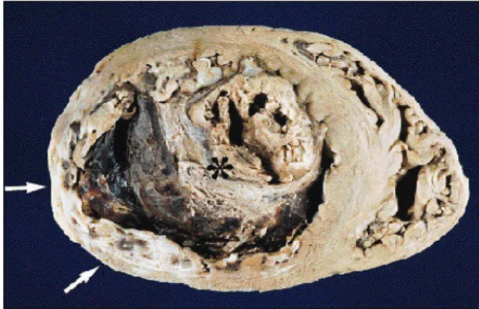
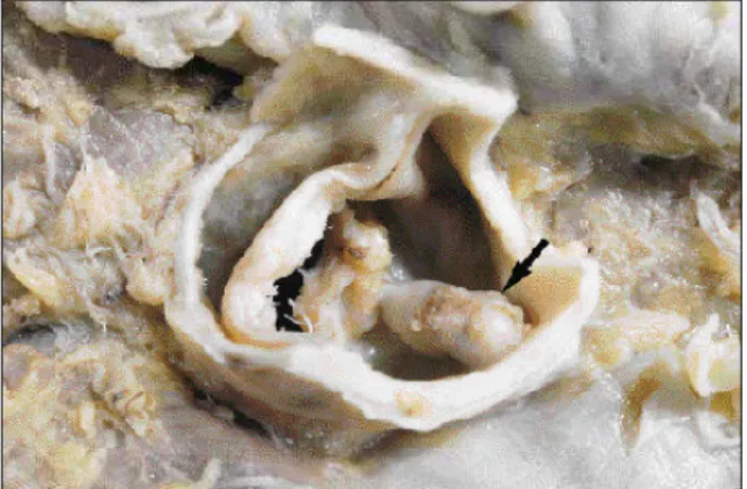
The heart weighed 660 g and showed dilation of the 4 cavities, particularly the left ventricle. We noticed a trans-mural infarction scar in the left ventricular posterolateral wall, which was thinned, and an extensive organizing thrombosis in the left ventricular cavity, which was semioc-clusive and particularly exuberant in the apex and outlet flow (fig. 3). The aortic valve was bivalvular, calcified, and pervious (approximately 2cm), with a stenosis grossly assessed as mild to moderate (fig. 4). The epicardial coronary arteries were dissected and underwent histologic examination, which proved they were normal (fig. 5). Multiple small scars of infarction in the spleen and kidneys were noticed, the latter ones depicting infarctions in an
advanced phase of organization. The liver and lungs showed chronic passive congestion. The aorta showed mild atherosclerosis. Pulmonary thromboembolism was also present, with recent hemorrhagic infarcts in the right
Fig. 4 – Aortic valve seen from its arterial face, composed of two thickened leaflets with a dense calcification at the raphe (arrow).
Fig. 5 – Histologic section of the circumflex artery (6º cm) with no pathological alterations and wide lumen (asterisk). Hematoxylin and eosin, X50.
Fig. 6 – Histologic section of the lung showing hemorrhagic infarct (HI) and arterial obstruction due to emboli (asterisk). Hematoxylin and eosin, X50.
medium and lower lobes (fig. 6), which constituted the final cause of death.
(Dr. Luis Alberto Benvenuti)
Anatomicopathological diagnoses – 1) Ischemic heart
disease with normal coronary arteries; 2) Calcified and ste-notic bivalvular aortic valve; 3) Semiocclusive thrombosis of the left ventricular cavity; 4) Pulmonary thromboem-bolism and infarcts (final cause of death).
Comments
The presence of ischemic heart disease with normal coronary arteries associated with dilation and thrombosis of the left ventricular cavity and calcified aortic stenosis of a mild to moderate degree (in a congenitally bivalvular valve) allows 3 sequential anatomicopathological possibilities.
As the first hypothesis, we could consider that the calcified aortic stenosis would be related to the initial left ventricular dysfunction. Therefore, we would have a case of valvar cardiomyopathy, according to the last classification of the World Heath Organization 31, because the ventricular
dysfunction would be disproportionate to the ventricular overload, because the aortic stenosis was mild to moderate. Ventricular dilation would favor thrombosis of the cavity and the myocardial infarction could be consequent to coro-nary embolism. In dilated cardiomyopathy, acute transmural infarcts are reported in which coronary embolism due to ca-vitary thrombi are speculated 32. The fact that no
obstructi-on in any corobstructi-onary segment was found could be explained by complete resorption of the embolus.
As a second hypothesis, we could consider that the process had begun as an ischemic heart disease with normal coronary arteries, stressing then, the probable role played by coronary spasm in the genesis of ischemia. Left ventricu-lar dilation would be secondary to ischemic heart disease with superimposition of cavitary thrombosis.
As a third hypothesis, we could consider the existence of an undiagnosed coagulation disorder that would explain the myocardial infarction due to coronary thrombosis (later rea-bsorbed) and the extension of the left ventricular cavity throm-bosis (semiocclusion), which would be very uncommon.
According to the last two hypotheses, the calcified aortic stenosis would be only an autopsy finding with no importance in the pathogenesis of heart failure, because it was of a mild to moderate degree.
The cause of death was pulmonary thromboembolism and infarction, frequent complications in the evolution of congestive heart failure.
1. Kawai C. Pathogenesis of acute myocardial infarction. Novel regulatory systems of bioactive substances in the vessel wall. Circulation 1994; 90: 1033-43. 2. Lozano C, Villegas M, Gutiérrez J, et al - Rasgos clínicos del infarto del
miocar-dio con coronariografia “normal”. Experiencia del instituto nacional de carmiocar-dio- cardio-logia. Arch Inst Cardiol Méx 1991; 61: 357-64.
3. Applegate BL. Kawasaki syndrome. An important consideration in the febrile child. Postgrad Med 1995; 97: 121-6.
4. Jackman JD Jr, Radolf JD. Cardiovascular syphilis. Am J Med 1989; 87: 425-33 5. Arend WP, Michel BA, Bloch DA, et al. The American College of Rheumatology 1990 criteria for the classification of Takayasu arteritis. Arthritis Rheum 1990; 33: 1129-34.
6. Rajani RM, Dalvi BV, D’Silva AS, et al. Acute myocardial infarction with normal coronary arteries in a case of polyarteritis nodosa: possible role of coronary arte-ry spasm. Postgard Med J 1991; 67: 78-80.
7. Guillevin L, Le Thi Huong Du, Godeau P, Jais P, Wechsler B. Clinical findings and prognosis of polyarteritis nodosa and Churg-Strauss angiitis: a study in 165 patients. Br J Rheumatol 1988; 27: 258-64.
8. Fauci AS, Haynes B, Katz P. The spectrum of vasculitis: clinical, pathologic, im-munologic and therapeutic considerations. Ann Intern Med 1978; 89: 660-76. 9. Beeson PB. Age and sex associations of 40 autoimmune diseases. Am J Med
1994; 96: 457-62
10. Moroni G, La Marchesina U, Banfi G, et al. Cardiologic abnormalities in patients with long-term lupus nephritis. Clin Nephrol 1995; 43: 20-8.
11. Suzuki A, Ohosone Y, Obana M, et al. Cause of death in 81 autopsied patients with rheumatoid arthritis. J Rheumatol 1994; 21: 33-6.
12. Ohno T, Matsuda I, Furukawa H, Kanoh T. Recovery from rheumatoid cerebral vasculitis by low-dose methotrexate. Intern Med 1994; 33: 615-20. 13. Nagyhegyi G, Nadas I, Banyai F. Cardiac and cardiopulmonary disorders in
pa-tients with ankylosing spondylitis and rheumatoid arthritis. Clin Exp Rheuma-tol 1988; 6: 17-26.
14. Alves MG, Espirito-Santo J, Queiroz MV, Madeira H, Macieira-Coelho E. Car-diac alterations in ankylosing spondylitis. Angiology 1988; 39: 567-71. 15. Fortin LJ, Genest J Jr. Measurement of homocyst(e)ine in the prediction of
arteri-osclerosis. Clin Biochem 1995; 28: 155-62.
16. Schieken RM, Kerber RE, Ionasescu VV, Zellweger H.Cardiac manifestations of the mucopolysaccharidoses.Circulation197; 52: 700-5.
17. Goldfischer S, Coltoff-Schiller B, Biempica L, Wolinsky H.Lysosomes and the sclerotic arterial lesion in Hurler’s disease. Hum Pathol 1975; 6: 633-7.
References
18. Nagao Y, Nakashima H, Fukuhara Y, et al. Hypertrophic cardiomyopathy in late-onset variant of Fabry disease with high residual activity of alpha-galactosidase A. Clin Genet 1991; 39: 233-7.
19. Donati D, Novario R, Gastaldi L. Natural history and treatment of uremia secon-dary to Fabry’s disease: an European experience. Nephron 1987; 46: 353-9. 20. Hamer JP, Janssen S, van Rijswijk MH, Lie KI. Amyloid cardiomyopathy in
systemic non-hereditary amyloidosis. Clinical, echocardiographic and electro-cardiographic findings in 30 patients with AA and 24 patients with AL amyloi-dosis. Eur HeartJ 1992; 13: 623-7.
21. Sugiishi M, Takatsu F. Cigarette smoking is a major risk factor for coronary spasm. Circulation 1993; 87: 76-9.
22. Jondeau G, Dubourg O, Partovian C, et al. Acute myocardial infarction in a patient with severe aortic stenosis and normal coronary arteries. Eur Heart J 1994; 15: 715-7.
23. Dellsperger KC, Marcus ML. Effects of left ventricular hypertrophy on the coro-nary circulation. Am J Cardiol 1990; 65: 1504-10.
24. Larsen GC, Griffith JL, Beshansky JR, D’Agostino RB, Selker HP. Electrocar-diographic left ventricular hypertrophy in patients with suspected acute cardiac ischemia- its influence on diagnosis, triage, and short-term prognosis: a multi-center study. J Gen Intern Med 1994; 9: 666-73.
25. Orlandi E, Castelli G, Brusamolino E, et al. Hemorrhagic and thrombotic compli-cations in polycythemia vera. A clinical study. Haematologica 1989; 74: 45-9. 26. Benjamin D, Yeshurun D, Charnilas J, Pinkhas J. Hyperlipidemia and myocar-dial infarction among 118 patients with polycythemia vera. Am J Med Sci 1978; 276: 23
27. Lockshirn MD. Answers to the antiphospholipid - antibody syndrome? N Engl J Med 1995; 332: 1025-7.
28. Dacosta A, Guy JM, Rousset H, et al. Painless Infarction in Antiphospholipid Syndrome. Arch Mal Coeur Vaiss 1933; 86: 1773-5.
29. Romero FB, Galve BE, Ordi RJ, et al. Coronary trombosis as the manifestation of the antiphospholipid syndrome. Rev Esp Cardiol 1994; 47: 327-9. 30. Christensen K, Herskind AM, Junker P. Antiphospholipid antibodies and
oc-clusive vascular disease. Ugeskr Laeger 1993; 155: 2896-2900.
31. Richardson P, McKenna W, Bristow M, et al. Report of the 1995 World Health Or-ganization/International Society and Federation of Cardiology Task Force on the definition and classification of cardiomyopathies. Circulation 1996; 93: 841-2. 32. Aiello VD, Mansur AJ, Favarato D. Rupure of posteromedial papillary muscle as