Case Report
Cardiovascular Complications in a Child with Chronic Renal Failure
Gesmar Volga Haddad Herdy, Vânia Gloria Silami Lopes, Maria Cecília Olivaes, Isabele Coelho Mota, Marcio Moacyr
Vasconcelos
Universidade Federal Fluminense - Niterói, RJ - Brazil
Mailing Address: Gesmar Volga Haddad Herdy •
Travessa Antonio Pedro, 10/301 24230-030 – Niterói, RJ - Brazil E-mail: [email protected]
Manuscript received February 03, 2006; revised manuscript received April 03, 2006; accepted May 9, 2006.
Introduction
Cardiac complications are the major cause of death in 25% of children with advanced chronic renal failure1. Visceral
calcifications – which include heart and vessels – may occur in adults with primary or secondary hyperparathyroidism, but are rarely seen in children2-4. The purpose of the present
report is to call attention for the occurrence of early severe cardiovascular disease in pediatric patients with chronic renal failure (CRF) and secondary hyperparathyroidism.
Case Report
A male, mulatto, 11-year-old patient was admitted to the Pediatric Unit at the University Hospital due to generalized tonic-clonic convulsive crises. The patient had no recent febrile or infectious condition. Previous pathologic history showed he had been diagnosed with CRF at the age of 4, and had his follow-up at a different hospital. Prescriptions included: calcitriol, calcium carbonate, and erythropoietin, with daily peritoneal dialysis having been started at home. At 8, the patient had a fracture of collum femoris. A year before hospital admission at this point in time the patient had an ischemic stroke, which developed into paralysis of his right limbs. Two months before current admission the patient had peritonitis with purulent secretion from dialysis catheter, and was medicated with cephalothin. The latest clinical occurrences were reported by the mother and treated at a different hospital.
Physical examination at admission showed severe nutritional deficiency – body weight, 14 kg (< 5th percentile),
height, 98 cm (< 5th percentile) – and bone malformations
This is the report of an 11-year-old boy with chronic renal disease and secondary hyperparathyroidism. The child had been on dialysis, calcitriol, calcium carbonate, and presented dyslipidemia and calcified thrombi in various vessels and organs in the course of his condition. Pathological examination showed ischemic cerebral necrosis, calcification in coronary arteries, and myocardial infarction.
Key words
Renal insufficiency, chronic / complications; cerebrovascular accident; hyperparathyroidism.
– severe dorsolumbar scoliosis – and his limbs were spastic. Blood pressure was 110/60 mm Hg at upper right limb, respiratory rate 40 breaths per minute, and heart rate 112 beats per minute. The patient had respiratory discomfort, fine rales on the right thorax, generalized convulsions. Chest X-ray showed condensation on right upper lobe.
The patient was treated with ceftriaxone, was kept on dialysis, and received phenytoin, phenobarbital, and diazepam for intercurrent convulsive seizures.
Doppler echocardiogram showed calcifications in left ventricle endocardium (papillary muscles). Vascular ultrasound showed calcified thrombi in right internal carotid artery (90% obstruction). Complete blood count showed leukocytosis and a left shift in differential count. Normal calcium plasma level, mild hyperphosphatemia – 7.6 mg/dL (reference values: 4 – 6.9mg/dL), with increased values: urea - 111 mg/dL (reference values: 15 - 40mg/dL), creatinine – 3.5mg/dL (reference values: 0.8 – 1.4mg/dL), uric acid – 11.4mg/dL (reference values: 3 - 7mg/dL), alkaline phosphatase - 949 U/L (maximum reference value: 350 - 227mg/dL), total cholesterol – 227 mg/dL (maximum reference value: 200mg/dL) and triglycerides - 245mg/dL (maximum reference value: 40 ± 170 mg/dL).
On day 6 the patient showed purulent conjunctivitis, stupor, anisocoria, and spastic limbs when flexing. Head CT scan showed an extensive ischemic infarction in left anterior cerebral artery, subfalcial and uncal herniation, due to possible ischemic encephalic vascular accident in left middle cerebral artery and obstructive hydrocephalus from herniation with compression of the Monro’s foramen. Early treatment for cerebral infarction included manitol, orotracheal intubation, and strict control of fluid intake. On day 8, the patient reported respiratory alkalosis and was in a coma (Glasgow 6). Cutaneous mucosal paleness followed, with progressive dyspnea. As a result of no success from clinical treatment, the patient was sent for decompression craniectomy. The patient presented hypovolemic shock, and was treated with intravenous fluids, packed red blood cells, and dopamine infusion. Death occurred on day 10.
Diagnosis hypothesis was: chronic renal failure, with renal osteodystrophy, secondary hyperparathyroidism, and secondary calcifications in various organs. The cerebrovascular accident occurrence could be explained both by the atherosclerotic disease and artery obstruction from calcification. Evidence was found on the vascular impairment of right carotid and left anterior cerebral arteries.
Pathological exam showed:
Gross exam: Thoracolumbar scoliosis, malnutrition, bone malformations. Extensive cerebral edema, right cerebral
Case Report
Herdy et al
CARDIOVASCULAR COMPLICATIONS IN A CHILD WITH CHRONIC RENAL FAILURE
Arq Bras Cardiol 2007; 88(2) : e31-e34
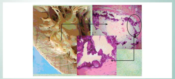
ischemic infarction. Lungs: Calcification area at upper lobe apex and pulmonary atelectasia. Heart: Weight and volume increase; calcifications at right and left atriums, left ventricle endocardium, papillary muscle and tendon chords; abscess at right ventricle myocardium; infarction in left ventricle anterior wall (Fig. 1). Kidneys with diffuse microondulations, right kidney rather hypotrophic. Volume increase in parathyroids, with diffuse distribution of nodules. Metastatic calcifications in vessels, brain, and kidneys. Renal rickets.
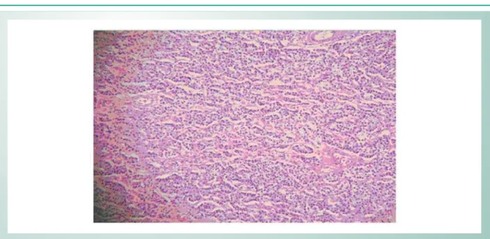
Microscopy: Brain: Recent infarction of right parietal lobe; ischemic necrosis of left parietal lobe (left carotid artery territory); previous infarction in left parietal lobe (anterior cerebral artery territory). Lung: Bronchitis, septal thickening with mixed inflammatory afflux in parenchyma, calcification at alveolar sacs and capillary walls, with thrombi formation. Heart: Endocarditis, pericarditis, infarction at the anterior wall, right ventricle myocardium abscess. The anterior descending coronary artery showed intimal thickening, an area with atheroma plaque with collagen, inflammatory infiltrate with mononuclear and polymorphonuclear leukocytes, macrophages, fibrin, necrotic center and calcium deposition. Right internal carotid artery showed calcified thrombus with intimal thickening and inflammatory infiltrate similar to that found in the coronary. Parathyroids: Hyperplasia of major cells (Fig. 2). Kidneys: Glomerular atrophy, hyalinized glomeruli and segmental glomerular fibrosis (Fig. 3); urate crystals in tubules and interstitial calcifications; chronic glomerulonephritis (end-stage kidney).
Causes of death were cerebral edema and right cerebral ischemic infarction.
Pathological conclusion: Chronic glomerulonephritis, secondary hyperparathyroidism, renal osteodystrophy, metastatic calcifications in different organs and vessels. Myocardial infarction. Cerebral ischemic necrosis.
Discussion
In the present case, the long lasting (seven years) CRF triggered the whole pathologic process. An infarction area and thrombosis with calcifications in different vessels were described in the heart. The cardiovascular complications described in patients with CRF are: Arrhythmias from metabolic disorder, uremic pericarditis, hypertension, atherosclerosis (secondary to dyslipidemia), and calcifications in vessels and in the heart 1,5,6. As for our case, dyslipidemia
was present, which could explain the previous stroke. Children prepared for transplant with triglycerides level above 150 mg/dL report lesions in the endothelium and in arteries, which include middle layer calcification7. Pediatric patients with
chronic renal disease in advanced stage are under the risk of accelerated atherosclerosis, associated with coronary lesions, as well as insulin resistance – which explains why statins are recommended6. Endothelium integrity rupture triggers a
cascade of inflammatory factors that lead to the formation of atheroma plaques; parathyroid hormone elevation contributes to artery calcification8,9. No drug was administered
to our patient for dyslipidemia control. Therefore, that pathophysiologic sequence could explain the many calcified lesions that have been found.
Renal disease is known to retain phosphates. In doing so, it decreases serum concentration of total and ionized calcium, in addition to decreasing renal production of 1D.25-dihydroxyvitamin D. It is known that 80% of Vitamin D is produced in the skin by the transformation of dihydrocholecalciferol under the influence of ultraviolet rays. Vitamin D is then hydroxylated to 25-hydroxyvitamin D in the liver and then in the kidney, resulting in 1D.25-dihydroxyvitamin D or 1D.25-(OH) 2-D10. Low concentration of ionized calcium
is the stimulus for parathyroid cells hypertrophy, as described in the necropsy for our case. When ionized calcium serum
Fig. 1 -A. Heart Macroscopy: ischemic area that impairs mural endocardium and myocardium. In the inset: microscopy to show myocardial necrosis area (B) and darker areas due to calcium deposit (C). (H.E. 40×0,25).
A
B
C
Case Report
Herdy et al CARDIOVASCULAR COMPLICATIONS IN A CHILD WITH CHRONIC RENAL FAILURE
Arq Bras Cardiol 2007; 88(2) : e31-e34
Fig. 2 -Parathyroid Microscopy: diffuse hyperplasia of major cells (H.E. 20×0,25).
Fig. 3 -Kidney Microscopy: atrophy and segmental glomerular fibrosis. Bowman capsule thickening. Interstitial fibrosis and tubular dilation (T. Gomori 40×0,25).
level is decreased to 4 mg/dL (1 mmol/L) there is stimulus for secretion of parathyroid hormone2. Increased production of
that hormone is body’s attempt to normalize calcium and phosphate serum levels through the activation of osteoblasts and osteoclasts. Therefore, bone calcium mobilization and an increased tubular resorption occur.
The patient described showed significant bone changes that had been developed years earlier through femoral fracture and severe scoliosis. Therefore, he had renal osteodystrophy11,
which includes all bone abnormalities and deranged mineral metabolism that result from renal failure. Secondary hyperparathyroidism is still a challenge for the treatment of pediatric CRF through peritoneal dialysis or hemodialysis, due to the adverse effects in growing patients12.
In our case growth had been interrupted, since body weight and height were compatible with a 3 or 4-year-old child, which is frequently seen in CRF12-14. The treatment carried
out (calcium carbonate and calcitriol) is recommended by the authors13,14. Those drugs have the purpose to increase calcium
concentration, which in turn improves hypophosphatemia. However, calcitriol was shown to inhibit the proliferation of condrocytes and to change the trophic actions of growth hormone in pre-pubertal patients’ bones12.
Oral or IV administration of calcitriol combined with calcium supplements has been implicated in the causation of episodes of hypercalcemia and/or hyperphosphatemia associated with osteodystrophy (adynamic bone disease), growth interruption,
Case Report
Herdy et al
CARDIOVASCULAR COMPLICATIONS IN A CHILD WITH CHRONIC RENAL FAILURE
Arq Bras Cardiol 2007; 88(2) : e31-e34
References
1. Parekh RS, Carroll CE, Wolfe RA, Port FK. Cardiovascular mortality in children and young adults with end-stage kidney disease. J Pediatr. 2002; 141: 191-7.
2. Stefenelli T, Abela C. Cardiac abnormalities in patients with primary hyperparathyroidism: implications for follow-up. J Clin Endocrinol Metab. 1997; 82: 106-12.
3. Moraes CR. Calcification of heart: a rare manifestation of chronic renal failure. Pediatr Radiol. 1986; 16: 422-4.
4. Marx SJ. Hyperparathyroidism and hypoparathyroidism disorders. N Engl J Med. 2000; 343: 1863-75.
5. Zaidi AN, Ceneviva GD, Phipps LM, Dettorre MD, Mait CR, Thomas NJ. Myocardial calcification caused by secondary hyperparathyroidism due to dietary deficiency of calcium and vitamin D. Pediatr Cardiol. 2005; 26: 460-3.
6. Parekh RS, Gidding SS. Cardiovascular complications in pediatric end-stage renal disease. Ped Nephrol. 2005; 20: 125-31.
7. Nayir A, Bilge I, Kilicaslan I, Ander H, Emre S, Sirin A. Arterial changes in paediatric haemodialysis patients undergoing renal transplantation. Nephrol Dial Transplant. 2001; 16: 2041-7.
8. Goodman WG, Goldin J, Kuizon BD, Yoon C, Gales B, Sider D. Coronary-artery calcification in young adults with end-stage renal disease who are undergoing dialisys. N Engl J Med. 2000; 342: 1478-83.
9. Oh J, Wunsch R, Bahner M, Turzer M, Raggi P, Querfeld U. Advanced coronary and carotid arteriopathy in young adults with childhood-onset chronic renal failure. Circulation. 2002; 106: 100-5.
10. Ringe JD, Schacht E. Prevention and therapy of osteoporosis: the roles of plain vitamin D and alfacalcidol. Rheumatol Int. 2004; 24: 189-97.
11. Corsi A, Collins MT, Riminucci M, Howell PG, Boyde A, Robey PG. Osteomalacic and hyperparathyroid changes in fibrous displasia of bone: core biopsy studies and clinical correlations. J Bone Miner Res. 2003; 18: 1235-46.
12. Kuizon BD, Salusky IB. Cell biology of renal osteodiostrophy. Pediatr Nephrol. 2002; 17: 777-89.
13. Waller S, Lederman S, Trompeter K, Van Hoff W. Catch-up growth with normal parathyroid hormon levels in chronic renal failure. Ped Nephrol. 2003; 18: 1236-42.
14. Schmitt CP, Ardissino G, Testa S, Claris-Appiani A, Mehls O. Growth in children with chronic renal failure on intermittent versus daily calcitriol. Pediatr Nephrol. 2003; 18: 440-4.
15. Sanchez CP, Kuizon BD, Adbulla PA, Salusky IB. Impaired growth, delayed ossification, and reduced osteoclastic activity in the growth plate of calcium-supplemented rats with renal failure. Endocrinology. 2000; 141: 1536-44.
16. Salusky IB. Are new vitamin D analogues in renal bone disease superior to calcitriol? Ped Nephrol. 2005; 20: 393-8.
and vascular calcifications13-16. New vitamin D analogues
have been developed recently (19-nor-paricalcitol and doxercalciferol) to control hyperparathyroidism without the biochemical changes secondary to calcitriol16. The case here
described was at high risk for cardiovascular disease, and calcitriol may have played an adjuvant role in the calcifications that were found. Myopericarditis and myocardial abscess may have been the result of previous bacterial infection (peritonitis), not treated with adequate doses of antibiotics.
Conclusion
The changes observed at necropsy were proof of clinical diagnosis. Therefore, treating CRF did not prevent complications such as hyperparathyroidism, osteodystrophy, dyslipidemia, and the serious cardiovascular changes.
Potential Conflict of Interest
No potential conflict of interest relevant to this article was reported.