DOI: 10.18410/jebmh/2015/827
ORIGINAL ARTICLE
J of Evidence Based Med & Hlthcare, pISSN- 2349-2562, eISSN- 2349-2570/ Vol. 2/Issue 38/Sept. 21, 2015 Page 5997
PORTAL SUPPLY TO CAUDATE LOBE AND QUADRATE LOBE OF
LIVER
K. Maheswari1
HOW TO CITE THIS ARTICLE:
K. Maheswari. “Portal Supply to Caudate Lobe and Quadrate Lobe of Liver”. Journal of Evidence based Medicine and Healthcare; Volume 2, Issue 38, September 21, 2015; Page: 5997-6000,
DOI: 10.18410/jebmh/2015/827
ABSTRACT: The precise knowledge of intra hepatic branching pattern of portal vein to caudate lobe and quadrate lobe is important for Gastroenterologist during hepatic segmental and subsegmental resection. The study was done in 47 adult human liver specimens. In this study methods like Manual dissection and Contrast study were used. During this study the portal branches to caudate lobe, Quadrate lobe and accessory branches to segment IV in addition to its branches were observed. The results were compared with previous studies.
KEYWORDS: Portal supply, Caudate lobe, Quadrate lobe, Accessory portal branches.
INTRODUCTION: The knowledge of portal branches to caudate lobe and Quadrate lobe are important while planning for Hepatic surgery like Segmental and Subsegmental resection. Caudate lobe has three parts are Right portion, Left portion of caudate lobe and Caudate process. Right portion of caudate lobe receives portal branches from Right branch, Left branch of portal vein and portal trunk
Left portion of caudate lobe receives portal branches only from Pars transversalis part of left branch of portal vein.
Caudate process receives portal branches from Right branch of portal vein and from portal trunk.
The different types of portal supply to caudate lobe and caudate process were observed in the present study1,2,3 (Elias & Petty 1952, Michel 1955, Gupta et al 1977).
Quadrate lobe received portal supply from right side of Left branch of portal vein3,4 (Gupta et al 1977, Osamu Matsui et al 1977)
ACCESSORY BRANCHES: In the present study, for Quadrate lobe, accessory branch was also observed from Right anterior segmental branch in addition to its portal branch from Left branch of portal vein. (Osamu Matsui et al 1977 by Helical computed tomographic hepatic arterioportography).
MATERIALS AND METHODS: This study was done in 47 adult liver specimens. The methods like Manual dissection and Contrast study were used.
OBSERVATIONS:
DOI: 10.18410/jebmh/2015/827
ORIGINAL ARTICLE
J of Evidence Based Med & Hlthcare, pISSN- 2349-2562, eISSN- 2349-2570/ Vol. 2/Issue 38/Sept. 21, 2015 Page 5998
Right Portion of Caudate Lobe: Received branches from;
Parstransversalis part of Left branch of portal vein – in 63.8%.
Right branch of portal vein – in 8.5%.
Portal trunk – in 27.7%.
In one specimen a branch arose from portal trunk divided into two branches, one to supply Right portion of caudate lobe and other to caudate process. (Fig. 2)
Caudate Process: All 47 liver specimens received portal branches from;
Right branch of portal vein – in 70%.
Portal trunk – in 30%.
Left portion of caudate Lobe: In all 47 liver specimens it received its branches only from Parstransversalis part of Left branch of portal vein. (Fig. 3)
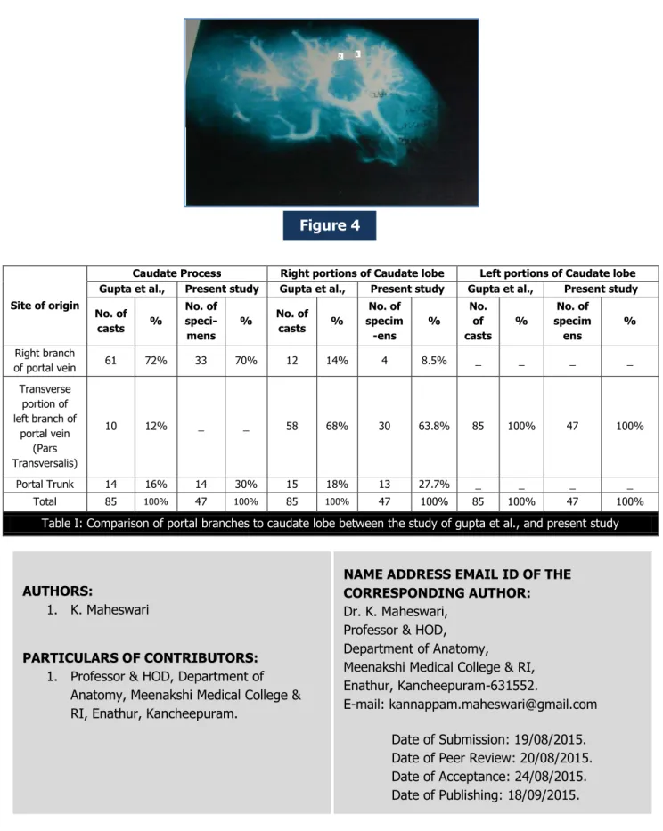
QUADRATE LOBE: In all 47 liver specimens, the quadrate lobe received its branches from right side of Left branch of portal vein. In four specimens (three by manual dissection and one by contrast study), it was observed that the quadrate lobe received accessory branch from Right anterior segmental branch in addition to its branch from Left branch of portal vein. (Fig. 4) (Osamu Matsui et al).4
DISCUSSION: In the present study the portal branches to caudate lobe and Quadrate lobe were observed. The study was discussed with previous study done by corrosion cast2 in Table I. Apart from its usual branches the accessory branch to quadrate lobe was also observed in this study. In the previous study, found accessary branches from right branch of portal vein to segment IV by helical computed tomographing arterioportography in patients4. The precise knowledge of this portal branches are clinically important for the operating Gastroenterologist before planning for hepatic segmental and sub segmental resection.
REFERENCES:
1. Elias, Hans and David Petty 1952, Gross anatomy of the blood vessels and ducts within human liver. Am. J. of Anatomy 90: 59-111.
2. Gupta S.C., CD. Gupta, AK. Arora 1977, Intra hepatic branching pattern of Portal vein. A study by corrosion cast. Gastroenterology 72; issue 4, 621 – 624.
3. Nicholas A. Michels Blood supply and anatomy of upper abdominal organs with a descriptive atlas, 1955 philadelphia: lipincott 39: 6; pp-1052.
DOI: 10.18410/jebmh/2015/827
ORIGINAL ARTICLE
J of Evidence Based Med & Hlthcare, pISSN- 2349-2562, eISSN- 2349-2570/ Vol. 2/Issue 38/Sept. 21, 2015 Page 5999
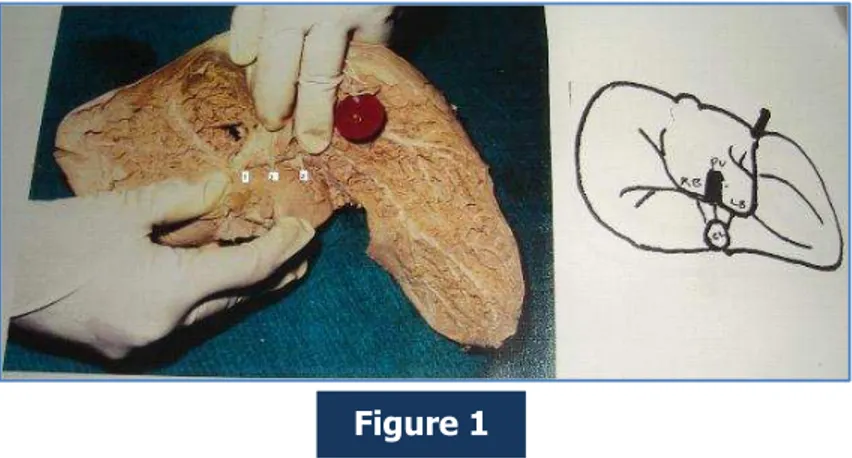
Figure 1: Portal branches to Caudate Lobe. 1. From right branch of Portal vein, 2. From Portal trunk 3. From Left branch.
Figure 2: Branches to right portion of Caudate Lobe. 1. Branch from portal trunk divided into two. 2. Left branch of Portal vein.
Figure 3: Branch to left portion of Caudate Lobe. Figure 1
Figure 2
DOI: 10.18410/jebmh/2015/827
ORIGINAL ARTICLE
J of Evidence Based Med & Hlthcare, pISSN- 2349-2562, eISSN- 2349-2570/ Vol. 2/Issue 38/Sept. 21, 2015 Page 6000
Figure 4: Accessory Branch to Quadrate Lobe. 1. From left branch of portal vein 2.From right anterior segmental branch of right branch of portal vein.
Site of origin
Caudate Process Right portions of Caudate lobe Left portions of Caudate lobe Gupta et al., Present study Gupta et al., Present study Gupta et al., Present study
No. of
casts %
No. of speci-mens
% No. of
casts %
No. of specim -ens % No. of casts % No. of specim ens % Right branch
of portal vein 61 72% 33 70% 12 14% 4 8.5% _ _ _ _
Transverse portion of left branch of
portal vein (Pars Transversalis)
10 12% _ _ 58 68% 30 63.8% 85 100% 47 100%
Portal Trunk 14 16% 14 30% 15 18% 13 27.7% _ _ _ _
Total 85 100% 47 100% 85 100% 47 100% 85 100% 47 100%
Table I: Comparison of portal branches to caudate lobe between the study of gupta et al., and present study
NAME ADDRESS EMAIL ID OF THE CORRESPONDING AUTHOR:
Dr. K. Maheswari, Professor & HOD, Department of Anatomy,
Meenakshi Medical College & RI, Enathur, Kancheepuram-631552.
E-mail: [email protected]
Date of Submission: 19/08/2015. Date of Peer Review: 20/08/2015. Date of Acceptance: 24/08/2015. Date of Publishing: 18/09/2015.
AUTHORS:
1. K. Maheswari
PARTICULARS OF CONTRIBUTORS:
1. Professor & HOD, Department of Anatomy, Meenakshi Medical College & RI, Enathur, Kancheepuram.