Severe Heart Failure at Intensive Therapy Unit - Is
there an Ideal Prognostic Index?
Cristina Bueno Terzi, Silvia G. Lage, Desanka Dragosavac, Renato G. G. Terzi
,QVWLWXWRGR&RUDomRGR+RVSLWDOGDV&OtQLFDVGD)0863H8QLYHUVLGDGH(VWDGXDOGH&DPSLQDV81,&$03 São Paulo, SP - Campinas, SP - Brazil
Mailing Address: Silvia H. G. Lage Ć 5XD &DSRWH 9DOHQWH ă ă 6mR 3DXOR 63 %UD]LO
(PDLOVJODJH#LQFRUXVSEU 5HFHLYHGRQĆ$FFHSWHGRQ
O
BJECTIVETo assess the applicability of three prognostic indexes - APACHE II, SAPS II and UNICAMP II – in a subgroup of critical heart failure (HF) patients.
M
ETHODSNinety patients were studied, being 12 females and 78 males. Mean age was 56 (18-83). Patients were ranked in functional class IV (NYHA) or cardiogenic shock secondary to cardiomyopathies: dilated (44%), chagasic (25.5%), ischemic (18%), hypertensive (1.1%), hypertrophic (1.1%), alcoholic (1.1%), and secondary to valvopathies after surgical correction (7.7%). Tables with frequency of categorical variables and descriptive statistics of continuous variables were created in order to describe sample profile for the different variables under study. In order to analyze the relationship between prognostic indexes levels and course towards death, an analysis of the ROC curve, as well as Hosmer and Lemeshow Test of Goodness of Fit calculated, and Standardized Mortality Ratio (SMR) were carried out.
R
ESULTSThe statistical analysis showed low sensitivity, specificity, and accuracy of the three prognostic indexes for HF patients. Mortality was underestimated in this group. Pulmonary thromboembolism (PTE) was a major factor of mortality rate in severe HF.
C
ONCLUSIONSThe three prognostic indexes under study did not prove to be appropriate for the assessment of cardiopathy patients at Intensive Care Unit (ICU). For HF patients, PTE played a major role in mortality of heart failure. Specific prognostic indexes for cardiopathy patients with severe HF should be proposed, and the discussion on anticoagulation on those patients should be expanded.
K
EY WORDSPrognostic disease severity indexes; cardiogenic shock, intensive care unit; heart failure, pulmonary embolism.
Different prognostic indexes have been developed for the assessment of patient’s severity level as well as to estimate mortality rate in Intensive Care Units. The application of such indexes aims at identifying patients with higher recovery potential; which is to say, those to benefit from ICU assistance, leading to proper screening for hospital admission and discharge. Additionally, those indexes should allow the comparison of different intensive care units.
Among the systems proposed, the one most frequently used in many countries is the APACHE (Acute Physiology and Chronic Health Evaluation), originally developed by Knaus, in 19811, and modified in 1985, into APACHE
II2. Another system available is the SAPS (Simplified
Acute Physiology Score)3, which differs from APACHE II
in that it contains other clinical variables, such as diuresis, serum bicarbonate and bilirrubine, and also for not including admission diagnosis. In addition to those, the UNICAMP II4model - developed at the ICU, Hospital das
Clínicas-UNICAMP - uses the same computer techniques of step-by-step linear discriminating function and logistic regression analysis previously described by Lemenshow et al5, and tries to identify the most significant variables
reported for intensive care mortality rate.
Other systems are also available, with application for specific subgroups, such as trauma6, hemodynamic
instability7, myocardial ischemia8, heart surgery
post-surgery9, multiple organic dysfunction (SOFA)10 and
sepsis11 (Sepsis-related Organ Failure Assessment). At this
point in time, however, few studies did focus the subgroup of heart failure (HF) cardiopathy patients.
Keeping in mind the large number of hospital admissions for severe refractory heart failure in our unit, our purpose was to assess the applicability of three prognostic indexes (APACHE II, SAPS II, UNICAMP II) in this subgroup, with reports specific clinical characteristics not included in the variables of those indexes.
M
ETHODSNinety patients diagnosed with heart failure were investigated after their first emergency care.
Exclusion criteria were: patients under 18, those who stayed less than 24 hours in the Intensive Care Unit, those after heart surgery and those reporting heart structural lesions that could be corrected surgically, acute heart failure and other acute cardiac pathologies.
Data collection was carried out by one of the physicians in the ICU staff and included clinical history information on worst clinical and laboratory data in the first 24 hours of hospitalization in the ICU, in addition to results of echodopplercardiogram and electrocardiogram. Pulmonar y thromboembolism was confirmed by ventilation-perfusion cintigraphy and/or necropsy. Records included course towards death or discharge, and date. Based on those data, the score systems APACHE II, SAPS II and UNICAMP II were applied following the protocols as previously defined in literature2,3,4. Individual mortality
risks pointed out by the three prognostic indexes were obtained based on the equation Pr(y=1/logit)= elogit/1
+ elogit where e = 2.7182818, which converts scoring
into death risk.
Considering that APACHE II and SAPS II are the most widely known indexes, the present paper will present further details of UNICAMP II index.
The UNICAMP model was developed at the ICU, Hospital das Clínicas-UNICAMP, uses the same computer techniques of step-by-step linear discriminant function and logistic regression analysis previously described by Lemenshow et al, and tries to identify the most expressive factors reported for intensive care mortality rate.
The UNICAMP II4equation was based on the analysis
of 819 patients admitted at the ICU Hospital das Clínicas - UNICAMP in the period between March, 1988 and September, 1989. It is essentially based on APACHE II scoring, added by other variables, such as the use of a respirator (for over 24 hours), with or without renal failure (serum creatinine higher or equal to 1.6 mg/dl), and whether hospital admission was elective or due to emergency. The system does not use any coefficient related to admission diagnosis, which makes bedside application easier, even by paramedics. UNICAMP II equation was validated by recent publication4.
UNICAMP II Equation
Death calculated risk = 1/1[1+ EXP(-Y)] Where y = - 3.7594
+ 0.1162 x APACHE II scoring + 0.7178 if mechanical ventilation + 0.7318 if renal failure
+ 0.8367 if emergency / urgency
The present study was carried out at the Heart Institute Cardiology Intensive Care Unit in São Paulo, Brazil (InCor). Data were collected at the ICU in a 2-year period after the first emergency admission. Ninety patients diagnosed with heart failure were selected: 12 females and 78 males, mean age 56 (18-83).
From the 90 patients, 33 were ranked functional class IV (NYHA) and 57 with cardiogenic shock secondary to: 1) idiopathic dilated cardiomyopathy: 40 (44%); 2) Chagas disease: 23 (25.5%); 3) alcoholic cardiomyopathy: 1 (1.1%); 4) ischemic cardiomyopathy: 17 (18.9%); 5) hypertensive cardiomyopathy: 1 (1.1%); 6) hypertrophic cardiomyopathy: 1 ( 1.1%); 7) valvopathy: 7 (7.7%).
In order to describe sample profile in compliance with the different variables under study, categorical variables frequency tables (gender, diagnoses, heart rate) and continuous variables descriptive statistics were created (age, left ventricle ejection fraction, days of hospitalization, serum sodium and disease time course).
For the sake of comparison of categorical variables among groups the chi-square test was used, or else,
whenever necessary (values under 5), Fisher exact test. For the sake of comparison of continuous variables among groups, the Mann-whitney test was used as a result of data asymmetry or lack of normal distribution.
Hosmer and Lemeshow5’s Goodness of Fit adjustment
statistics were also calculated for each prognostic index in order to describe the accuracy of the different models. Death and discharge rates were compared, having been estimated and observed in ten bands, with 10% death intervals calculation (ƨg statistics). P > 0.05 values accept the assumption that estimated mortality = observed mortality, which is to say, stands for good calibration. In order to avoid a small number of patients in some of the risk bands, Ƙg statistics was also carried out by dividing the total number of patients into 10 risk bands, with a similar number of patients in each band.
Discriminatory power of each index was assessed by AUC (area under the curve) of ROC (Receiver Operating Characteristic Curve)12.
SMR (Standardized Mortality Ratio) was calculated by dividing observed mortality by predicted mortality for each of the models. SMR=1 means observed mortality = predicted mortality. SMR > 1 means death rate is higher than expected.
Significance level adopted for statistical tests was 5% (p< 0.05). Illustration 1 shows the ROC curves of the three models used.
R
ESULTSThe descriptive analyses of variables used and the comparison among groups of survivals and deaths are presented in Tables 1 to VII. High mortality rate was reported in this group of severe patients. Close to 2/3 of patients with class IV HF or cardiogenic shock died in ICU (Table 1).
Other factors such as gender (p = 0.75), type of cardiopathy (p = 0.32), left ventricle ejection fraction (p = 0.95) and days in hospital (p=0.097) did not differ between survival and death groups. Therefore, those were not considered additional risk factors (Tables 3, 4, 5 and 6).
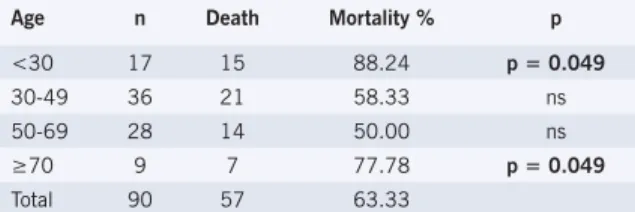
Higher mortality rate was observed among patients under 30 or over 70 (Table 2). Age group <30 and >70 were considered death risk factor (p = 0.004).
Factors such as disease time course (p < 0.001) and natremia (p = 0.097) were analyzed, and statistic difference was reported. Mortality rate was higher in those with longer than 2-year time course of disease and serum sodium lower than 129 mEq/l. Another factor – cardiac rhythm at the time of hospitalization (p=0.595) – did not report difference between the group for deaths or for survivors, and was not, therefore, considered as additional risk (Tables 7, 8 and 9).
Table 1 – Mortality ratio - overall
Course n Rate %
Survival 33 36.7
Death 57 63.3
Total 90 100.0
Table 2 – Descriptive and comparative analysis of variables between age
related survival and death rates
Age n Death Mortality % p
<30 17 15 88.24 p = 0.049
30-49 36 21 58.33 ns
50-69 28 14 50.00 ns
9 7 77.78 p = 0.049
Total 90 57 63.33
ns- non-significant difference.
Table 3 – Gender related variables between survival and death
Gender n Death Mortality %
Females 12 7 58.33
Males 78 50 64.10
Total 90 57 63.33
p=0.75.
Table 4 – Course of patients following diagnosis
Cardiomyopathy n Deaths Mortality %
Idiopathic 40 26 65.00
Chagasic 23 13 56.50
Hypertrophic 1 1 100.00
Ischemic 17 11 64.70
Alcoholic 1 0 0.00
Hypertensive 1 1 100.00
Valvar 7 4 57.10
p = 0.32
Table 5 – Descriptive and comparative analysis of 51 patients, when it was possible to analyse
left ventricle ejection fraction (LVEF) by echodopplercardiography. No patient reported
LVEF above 50%
LVEF n Deaths Mortality %
<30% 20 15 75.00
30-50% 31 23 74.19
TOTAL 51 38 74.51
p=0.95 (LVEF- left ventricle ejection fraction).
Table 6 – Descriptive and comparative
analysis of course as compared to days at the Intensive Care Unit
Days in hospital n Deaths Mortality %
1 to 5 19 16 84.21
6 to 10 23 14 60.87
>10 48 27 56.25
TOTAL 90 57 63.33
High rate of pulmonary embolism stands out. Twelve out of 90 patients (13.3%) had their diagnosis confirmed by imaging or necropsy, which stood for 21% of all patients who died (Table 10).
Indices used showed low sensitivity and specificity to assess death risks.
Hosmer and Lemeshow’s Goodness of Fit statistics is presented in Table 11. The difference between predicted risk and observed risk was statistically significant for all groups, and for all indices (p < 0.0001). Predicted risk for deaths was 19.03 with APACHE II; 15.50 with SAPS II; and 39.16 with UNICAMP II. Global mortality rate observed was 63.3% (57/90). Ratio between observed mortality and predicted risk (SMR - Standard Mortality Ratio) for each model shows that observed mortality was higher than predicted mortality for all models. However, the lowest SMR was reported for the UNICAMP II model (Table 11).
ROC curves data can be found in Table 12. It should be pointed out that the only model that showed significant AUC was APACHE II.
Illustration 1 shows results for ROC curves for APACHE II, SAPS II and UNICAMP II prognostic indices.
D
ISCUSSIONAs of 1981, different severity indexes have been proposed for patients in Intensive Care1. At a first moment,
given the limited number of ICU hospital beds, the purpose was to screen those patients with higher recovery potential. At a later stage, prognostic indexes were used to assess the performance of different units based on the equivalence of resources available as well as the profile of patients at those units. Finally, prognostic indexes have been used to stratify patients in order to assess efficacy of new treatments.
In 1967, levels of severity began to be described related to the severity of acute myocardial infarction (AMI) patients, as the KILLIP13 classification, and later on, the
Table 7 - Descriptive and comparative analysis of variables between survivals and deaths as compared
to disease time course (HF) (p < 0.001)
Disease time course n Deaths Mortality %
\HDU 21 4 42.9
2 to 5 years 6 4 58.8
\HDUV 17 15 81.8
Total 44 23
Table 8 - Descriptive and comparative analysis of variables between survivals and deaths as compared
to heart rhythm at the time of admission at the Intensive Care Unit. (TAVB = total atrioventricular blocking, HP = permanent heart pacer) (p = 0.595)
Heart rhythm n Deaths Mortality %
Sinus 18 17 94.4
TAVB (HP 3 3 100.0
Atrial fibrillation 7 6 85.7
Total 28 26
Table 9 – Analysis of natremia as compared to mortality rate. It has been shown that plasma sodium under 129 mEq/l is associated to twice the mortality rate as
compared to normal sodium levels (p = 0.011)
Natremia (mEq/l) n Deaths Mortality %
135-145 21 9 42.9
130-134 36 21 58.8
33 27 81.8
Total 90 57
Table 10 – Rate of thrombopulmonary embolism
Total number of patients 90
Total deaths 57
Deaths resulting from thrombopulmonary embolism (TPE) 12
General mortality rate from TPE (%) 13.33
TPE incidence among all deaths (%) 21.05
Table 11 - Hosmer-Lemeshow’s Goodness of Fit Tests. ƨg = observed and estimated mortality of patients according to 10% risk bands. Ƙg = observed and estimated mortality of patients according to predicted
death risk bands against the same number of patients
Model ƨg p Ƙg P SMR
APACHE II 143.6 p<0.0001 143.9 p<0.0001 3,327496
SAPS II 287.5 p<0.0001 424.8 p<0.0001 4,080172
Unicamp II 39.89 p<0.0001 41.76 p<0.0001 1,616563
Table 12 – Statistic data on ROC curves
Model AUC p S S. CI 95% Cut-off
APACHE II 0.632±0.061 0.03 63.2%; 66.7% 0.511-0.752
SAPS II 0.571±0.061 0.265 35.1% 84.8% 0.451-0.690
Unicamp II 0.595±0.061 0.136 63.2% 57.6% 0.475-0.714
AUC- Area Under the ROC Curve; p- significant statistic level; S- Sensitivity; S- Specificity; (CI) 95% = Confidence Interval 95%; Cut point- model cutting point.
6-minute walk test, or Oxygen (VO2) consumption test, or the left ventricle ejection fraction (LVEF). As HFSS was lower as compared to the other tests in the ability to predict survival, the authors suggest that a simplified model be applied, with LVEF and the 6-minute walk test as variables18. For outpatients, or for non-acute patients
in the wards, the natriuretic peptide should also be used for measurement19.
The different prognostic indexes have tried to assess long-term survival of HF patients.20-22. In those patients,
LVEF has been an important prognostic parameter. However, no known indexes consider the acute decompensation of patients with severe HF resistant to outpatient treatment. It should also be pointed out that our patients invariably report reduced LVEF in regard to HF Functional Class IV or cardiogenic shock. In the present research, all patients submitted to echodopplercardiography (n = 51) reported LVEF under 50%; from those, 39% reported LVEF under 30%. In this group of severe, unstable patients, in contrast with stable patients - at the outpatient unit or in the hospital wards - LVEF does not succeed in prognosing death, since patients reporting LVEF either lower than 30% or in the 30% to 50% range exhibited the same mortality rate - 75%. No statistical difference was shown regarding ejection fraction (p = 0.949).
From our total number of 90 patients, 57 reported cardiogenic shock, and 33 were Functional Class IV, which high mortality rate (61.4% and 66.6%, respectively), which characterized an extremely severe group. Such high mortality rate has still been reported in recent publications, despite the advances in the medical area 23.
Younger and older patients reported higher mortality AMI hemodynamic classification14. For other pathologies,
such as traumatic brain injury, the Glasgow15 trauma scale
is widely used. The first prognostic index proposed for patients in Intensive Care was APACHE (Acute Physiology and Chronic Health Evaluation)1, developed by Knaus et
al in the US, and later improved into APACHE II2. Today, it
is the most widely used among all severity indexes. More recently, APACHE III16was introduced, using seventeen
variables – a higher number as compared to variables used in the previous version. Another index - SAPS17 (Simplified
Acute Physiology Score) – was developed in France by Le Gall and later evolved into the SAPS II versions.
The three models under study used different weights for each variable of interest, such as age, acute physiologic dysfunctions, clinical or surgical hospitalization, and previous disease conditions.
APACHE II has been somewhat criticized in regard to diagnostic coefficients designed for subgroups of patients, since diagnostic classification at the moment of hospital admission not always corresponds to final diagnosis. A second relevant factor is the fact that those coefficients are quite general, because the number of diagnoses available is very limited. The coefficient is to be chosen in the first 24 hours; but differential diagnosis between HF - Functional Class IV and cardiogenic shock may be questioned. This was one of the reasons to lead Le Gall et al to develop the SAPS3 model, since this model does
not require the choice for the admission diagnosis. The UNICAMP II system does not require admission diagnosis either4.
For outpatient unit patients, the Heart Failure Survival Score (HFSS)18 has tried to identify the prognosis for
heart failure. However, that prognostic index has not managed to prove more effective when compared to the
ROC CURVES
Fig. 1 – HF Patients: Analysis of sensitivity and specificity for UNICAMP II, SAPS II and APACHE II systems, represented by ROC (Receiver Operator
Characteristic) curves.
0,0 0,1 0,2 0,3 0,4 0,5 0,6 0,7 0,8 0,9 1,0 0,0
0,1 0,2 0,3 0,4 0,5 0,6 0,7 0,8 0,9 1,0
APACHE II UNICAMP II
SAPS II
S
e
n
si
tiv
ity
1-Specificity
rate (p = 0.049) as compared to patients in the 30 to 70 years old range (Table 2). Advanced age is an aggravating prognostic factor, although not restricted to severe cardiopathy patients, but it has been patients admitted to the Intensive Care Unit. The high incidence of HF in the elderly may be due to age related changes, such as ventricular function, as well as risk factors associated to HF, such as hypertensive, diabetes and dyslipidemia24. This factor is so relevant that has been
incorporated as a significant variable for the calculation of prognostic indexes.
As for high mortality rate in patients under 30 years old, such finding has been described in literature25-27.
The explanation would be higher severity of cardiopathies in children, such as rheumatic conditions or inborn cardiopathies, as well as lower tolerance to HF.
Out of the 16 patients under 30, 13 had death as their outcome. From those, 4 had reported rheumatic cardiopathy from childhood, and 2, chagasic cardiopathy. A progression from the acute phase into chronic cardiopathy is known to take place. Severe evolution of the condition may result in death28. One patient had
had lymphoma for 15 years as a base disease. Two other patients reported complex ventricular arrhythmia; and lastly, two deaths resulted from TPE: both confirmed at necropsy.
No statistical difference was reported regarding gender related mortality rate (p = 0.753), as in the EPICA Niterói29 study, among the different cardiomyopathies
(p=0.324). As for the etiology, only 18.8% showed ischemic cardiopathy. That low incidence – if compared to other statistic data – can be explained by the fact that the hospital has a cardiac unit which is separate from the Intensive Care Unit. Additionally, Chagas disease incidence is practically absent in international statistics. A trend (p < 0.10) was reported towards higher mortality rate (84.21%) among patients with shorter hospitalization time (5 days or less) as compared to longer hospitalization periods (56.25% to 60.87%) (Table 6). It should be assumed that the severity level of early hemodynamic and systemic impairments may have contributed for higher mortality among patients in their first days after hospitalization.
As both Functional Class IV HF and cardiogenic shock have important systemic repercussion on organs and tissues, prognostic indexes to allow global assessment of patients seem to be more powerful tools as compared to the assessment limited to cardiovascular performance.
The present paper used three models of prognostic indexes (APACHE II, SAPS II, UNICAMP II). The models were proposed to assess critical patients, and their applicability was studied for the specific subgroup of cardiopathy patients admitted at the Intensive Care Unit.
Results show that the three prognostic indexes under study have not properly evaluated patients’ course, thus underestimating mortality rate of severe cardiopathy patients. Therefore, the 3.33 SMR for APACHE II, the 4.08 for SAPS II, and the 1.62 for the UNICAMP II
model show higher mortality rate than that estimated by the models (Table 7).
A number of factors may have interfered in the assessment of APACHE II, such as the type of assistance to patient before hospital admission30 and delayed transfer
to Intensive Care31. Particularly in developing countries,
those factors are not to be underestimated. That explains the inclusion of a model based on a data base collected from a University public hospital in Brazil. Here, it is important to point out that those factors are not dependent on the performance of Intensive care assistance, but rather, of the inherent health system deficiencies in this country. It could be argued whether it would be pertinent if the system developed were applied in other countries, with significant populational, nutritional and health differences, and with pre-hospital care different from the one in our country. That is why the UNICAMP4 model
was included: it uses an equation generated by logistic regression and based on data collected from National Health System (SUS) patients, developed at the University of Campinas and later validated32. Similarly to results in
literature, and obtained from other prognostic indexes applied to patients at general Intensive Care Units (case mix), the UNICAMP model reported SMR close to 1. In contrast, while analyzing cardiopathy patients the present paper reported higher SMR, although not as high as in the APACHE II and SAPS II models.
In a multicenter study using APACHE III, Bastos et al33
reported quite variable SMRs at 13 Brazilian hospitals. Mean SMR = 1.67. The differences pointed out between the hospitals ranged from 1.01 to 1.30. Technology resources were accounted for those differences at the different Units. SMR variation in the APACHE III project in Brazil was said to have been the result of technology availability. However, it is arguable whether this would be the only reason, or even the most relevant.
Any way, even the highest SMR levels were quite below the SMRs found in this group of severe cardiopathy patients, according to the study by Bastos et al34, using
the APACHE II and SAPS II models.
APACHE II was also used in a Brazilian population of 208 patients at General Intensive Care Unit4. APACHE
SMR resulted 1.31, and UNICAMP II SMR resulted in 0.85.
Such discrepant data regarding patients at the General Intensive Care Unit - even in our setting, with a developed system in the country - do suggests that some specific factor not predicted by prognostic indexes may influence mortality rate in those patients.
Therefore, deep sedation, neuromuscular blocking, and mechanical ventilation may mask abnormalities35. Acute
Physiological Score (APS) data may express distinctive physiological variables depending not only on how early treatment starts with volume and vasoactive drugs, but also at the point in time mechanical ventilation is used. Additionally, Glasgow`s coma scale assessment is jeopardized if the patient is sedated and/or under orotracheal intubation when admitted to the Intensive Care.
Although arrhythmia 35,36, hyponatremia 37,38, the
size of ventricular cavities39,40 , and serum catecholamin
levels41 do play key roles in the long term course of
cardiopathy patients, those variables may also influence decompensation in the acute phase. In our population we have observed that hyponatremia, and serum sodium lower than 129 mEq/l double mortality rate as compared to patients reporting normal sodium levels (Table 5). Heart rhythm analyzed at hospital admission was divided into three groups: sinus rhythm, atrial fibrillation, and those with previous heart pacer implantation due to atrial-ventricular blocking. Mortality rate was 94.4%, 85.7%, and 100%, respectively (p = 0.595). Heart rhythm usually alternates in a short time span under advanced cardiopathy condition, which makes accurate analysis harder.
The duration of HF proved to be a prognostic factor (p = <0.001) for all patients under study.
However, most important, in authors’ view, is the fact that prognostic indexes are based on the first 24 hours after hospital admission at the Intensive Care Unit. Out of the 90 patients, 71 (79%) stayed in the hospital over 5 days. Forty-eight patients (over 50%) stayed in the hospital for over 10 days, and from those, over 50% died. It is understandable that although prognostic indexes are kept unchanged as calculated at the time of admission, real death risk increases as time spent in hospital increases. Complications arise in the course of hospitalization in such a susceptible group of critical and unstable patients. Therefore, those patients require a higher degree of invasive procedures, which may result in systemic infections. Intensive care doctors are aware that Systemic Inflammatory Response Syndrome (SIRS) is common and lethal in severe patients, especially the elderly42. As a rule, conditions such as SIRS, Septic
Shock, and Multiple Organ Failure are not present at the moment of admission at the Intensive Care. As a result, they are not included in prognostic indexes scoring. This had been pointed out by Cerra et al43for surgical
patients admitted at the Intensive Care post-surgery. More recently, it was also pointed out by Lim et al, for patients under cardiogenic shock24. Those authors
describe that despite normalized hemodynamics, those patients die from distributive shock. Considering that 50% of patients who die report no clinical evidence of clear infection, the authors assume that cytokines and other inflammatory mediators may be released during a cardiogenic shock episode. The authors also suggest that mesenteric ischemia and pulmonary embolism may not be recognized as causes for death in patients whose
hemodynamics has been normalized24.
Curiously, the population under study reported high incidence of thrombopulmonary embolism of high clinical repercussion in the group of patients whose outcome was death. The clinical diagnosis of TPE is usually difficult to reach, and many times it is underestimated in HF patients44, thus leading to acute decompensation – even
when facing sub segmental pulmonary thromboembolic episodes36.
No variables to quantify the risk of thrombopulmonary embolism have been applied in the prognostic indexes. Despite patients’ prophylactic anticoagulation, twelve of them had their TPE confirmed through ventilation-perfusion cintigraphy or necropsy. All of these patients died. Those patients accounted for 21% of global mortality rate.
These data points out the relevance of complications that occur after hospital admission. Those complications have not been anticipated by conventional prognostic system for this unique group of patients. Out of all complications, the thromboembolic phenomenon stands out for its high incidence. And the thromboembolic phenomenon is usually diagnosed at the autopsy, usually carried out only at university hospitals. Although classic TPE may not be clinically relevant, it may – even when not diagnosed – worsen symptoms and lead to treatment resistance. It may eventually lead to death of these severe cardiopathy patients at the Intensive Care.
In conclusion: APACHE II, SAPS II and UNICAMP II have not been appropriate models to express the prognosis of cardiopathy patients at the Intensive Care Unit. Mortality rate was seen to be higher than anticipated by HF prognostic indexes. Factors not included in the prognostic indexes presented in this paper may have influenced the discrepancy of results obtained. However, the high incidence of PTE in our population – despite prophylactic anticoagulation - points out to the need for a review of full anticoagulation criteria. New systems should be investigated to encompass specific variables for HF patients at Intensive Care Units.
Acknowledgments
Ms Mônica Moreira Julião, for her technical support. Mr Helymar Machado for the statistical analyses.
Potential Conflict of Interest
No potential conflict of Interest relevant to this article
R
EFERENCES1. Knaus WA, Zimmerman JE, Wagner DP, et al. APACHE – Acute Physiology and Chronic Health Evaluation: A physiologically based classification system. Crit Care Med 1981; 9: 591-597.
2. Knaus WA, Draper EA, Wagner DP, Zimmerman J. APACHE II: A severity of disease classification system. Crit Care Med 1985; 13: 818-29.
3. Le Gall JR, Lemeshow S, Saulnier F. A new simplified acute physiology score. J Am Coll Cardiol 1993; 270: 2957-63.
4. Terzi RGG, Gomes MI, Araújo S, Dragosavac D, Falcão ALE, Machado HC. Índices prognósticos em Medicina Intensiva III Modelo UNICAMP. Rev Bras Terap Intens 2002; 14: 6-21.
5. Lemeshow S, Teres D, Pastides H, Avrunin JS, Steingrub J. A method for predicting survival and mortality of ICU patients using objectively derived weights. Crit Care Med 1985; 13: 519-25.
6. Stewart TC, Lane PL, Stefanits T. An evaluation of patients outcomes before and after Trauma Center designation using Trauma and injury Severity Score analysis. J Trauma 1995; 39: 1036-401.
7. Yeung C, Ming-Weilu, Martinez EG, Puri VK. Critical Care Scoring System-New concept based on hemodynamic data. Crit Care Med 1990; 18: 1347-52.
8. Norris RM, Brant PWT, Caughey DE, Lee AJ, Scott PJ. A new coronary prognostic index. Lancet 1969: 1(7589): 274-8.
9. Tuman KJ, McCarthy RJ, March RJ, Najafi H, Ivankovich AD. Morbidity and duration of ICU stay after cardiac surgery. Chest 1992; 102: 36-44.
10. Vincent JL, Moreno R,Takala J, et al. The SOFA (sepsis-related organ failure assessment) score to describe organ dysfunction / failure. On behalf of the Working Group on sepsis-related problems of the European Society of intensive care Medicine. Intensive Care Med 1996; 22: 707-710.
11. Barriere SL, Lowry S. An overview of mortality risk prediction in sepsis. Crit Care Med 1995; 23: 376-93.
12. Fleiss, JL. Statistical Methods for Rates and Proportions. 2nd ed. New York: John Wiley & Sons, 1981.
13. Killip T, Kimbal JT. Treatment of miocardial infarction in a coronary care unit. A two year experience with 2150 patients. Am J Cardiol 1967; 2: 457-64.
14. Forrester JS, Diamond GA, Swan HJC. Correlative classification of clinical and hemodynamic function after acute myocardial infarction. Am J Cardiol 1977; 39: 137.
15. Teasdale G, Jennet B. Assessment of coma and impaired consciousness. A practical scale. Lancet 1974; 13: 81-6.
16. Knaus AW, Wagner DP, Draper EA, et al. APACHE III prognostic System. Risk Prediction of Hospital Mortality for Critically ill Hospitalized Adults. Chest 1991; 100: 1619-36.
17. Le Gall JR, Loirat P, AlperovitchL A, et al. A simplified acute physiology score for ICU patients. Crit Care Med 1984; 12: 975-7.
18. Zugck C, Krüger C, Kell R, et al. Risk stratification in middle-aged patients with congestive heart failure: prospective comparison of the Heart Failure Survival score(HFSS) and a simplified two-variable model. Eur J Heart Fail 2001; 3: 577-85.
19. Maisel AS, Krishnaswamy P, Nowak RM, et al. Rapid measurement of B-type natriuretic peptide in the emergency diagnosis of heart failure. N Eng J Med 2002; 347: 161-7.
20. The CONSENSUS Trial Study Group.Effects of Enalapril on mortality in severe congestive heart failure: results of the cooperative North Scandinavian enalapril survival study. N Engl J Med 1987; 316: 1429-35.
21. SOLVD - Effects of enalapril on survival in patients with reduced left ventricular ejection and congestive heart failure. N Engl J Med 1991; 325: 293-302.
22. Cintron G, Johnson G, Francis G, Colb JN. Prognostic significance of serial changes in left ventricular ejection in patients with congestive heart failure. Circulation 1993; 87(Suppl): VI 17-VI 23.
23. Lim N, Dubois M-J, De Backer D, Vincent J-L. Do All nonsurvivors of cardiogenic shock die with a low cardiac index? Chest 2003; 124: 1885-91.
24. Hunt SA, Baker DW, Chin MH, et al. American College of Cardiology/ American Heart Association Task Force on Practice Guidelines (Committee to Revise the 1995. Guidelines for the Evaluation and Management of Heart Failure). Circulation 2001; 104(24): 2996-3007.
25. Schocken DD, Arrieta MI, Leaverton PE, et al. Prevalence and mortality rate of congestive heart failure in United States. J Am Cardiol Coll 1992; 20: 301-06.
26. Ho KKL, Pinsky JL, Kannel WB, et al. The epidemiology of heart failure: the framigham study. J Am Cardiol Coll 1993; 22(Supll A): 6A-13A.
27. Barretto ACP, Nobre MRC, Wajngarten M, Canesin MF, Ballas D, Serro-Azul JB. Insuficiência em grande Hospital Terciário de São Paulo. Arq Bras Cradiol 1998; 71: 1-11.
28. Dias JCP. História natural da doença de Chagas. Arq Bras Cardiol 1995; 65: 359-66.
29. Tavares LR, Victer H, Linhares JM, et al. Epidemiologia da insuficiência cardíaca descompensada em Niterói: Projeto EPICA - Niterói. Arq Bras Cardiol 2004; 82: 121-4.
30. Rapaport J, Teres D, Lemeshow S, Harris D. Timing of intensive care unit admission in relation to ICU outcome. Crit Care Med 1990; 18: 1231-5.
31. Dragsted L, Jorgensen J, Jensen NH, et al. Interhospital comparisons of patients outcome from intensive care:importance of lead-time bia. Crit Care Med 1989; 17: 418-22.
32. Alves CJ, Terzi RGG, Franco GPP, Malheiros WMP. Comparação entre o modelo UNICAMP II e APACHE II em uma UTI Geral. Rev Bras Terap Intens 2003; 15: 144-52.
33. Bastos PG, Sun X, Wagner DP, Knaus WA, Zimmerman JE. Application of the APACHE III prognostic system in Brazilian intensive care units: a prospective multicenter study. Intensive Care Med 1996; 22: 564-70.
34. Bastos PG, Knaus WA, Zimmerman JE, Magalhães A Jr, Sun X, Wagner. The importance of technology for achieving superior outcomes from intensive care. Brazil APACHE III Study Group. Intensive Care Med 1996; 22: 664.
35. Lengyel M, Kokeny M. Follow-up study in congestive cardiomyopathy. Cardiologica 1981; 36: 35-48.
36. Reese DB, Silverman ME, Gold MR, Gottlieb SS. Prognostic importance of the length of ventricular tachycardia in patients with non ischemic congestive heart failure. Am Heart J 1995; 130: 489-93.
37. Lee WH, Packer M. Prognostic importance of serum sodium concentration and its modification by converting-enzyme inhibition in patients with severe chronic heart failure. Circulation 1986; 73: 257-67.
38. Oster J, Materson BJ. Renal and eletrolyte complication of congestive heart failure and effects of therapy with angiotensin converting enzyme inhibitors. Arch Intern Med 1992; 152: 704-10.
39. Lee TH, Hamilton MA, Stevenson LW, et al. Impact of left ventricular cavity size on survival on in advanced heart failure. Am J Cardiol 1993; 72: 672-76.
40. Fuster V, Gersh BJ, Giuliani ER, Tajik AJ, Brandenburg O, Fryl RL. The natural course of idiopathic dilated cardiomyopathy. Am J Cardiol 1981; 47: 525-31.
41. Cohn JN, Levine TB, Garberg V, Francis GS, Simon AB, Rector T. Plasma norepinephrine as a guide to prognosis in patients with chronic heart failure. N Engl J Med 1984; 311: 819-23.
42. Bone RC. A continuing evolution in our understanding of the Systemic Inflammatory Response Syndrome (SIRS) and the Multiple Organ Dysfunction Syndrome (MODS). Ann Intern Med 1996; 125: 680-7.
43. Cerra NF, Abrams J. APACHE II score does not predict multiple organ failure or mortality in postoperative surgical patients Arch Surg 1990; 125: 519.
44. Lage SG, Ramires JAF, Bellotti G, et al. Análise da repercussão clínico-hemodinâmico do tromboembolismo pulmonar agudo lobar e segmentar nos cardiopatas. Arq Bras Cardiol 1983; 41(supl1): 86.