The diagnosis is difficult because the initial signs and symptoms, especially those of a respi-ratory nature, can present a broad spectrum of severity—ranging from mild stridor to severe respiratory failure. The symptoms typically occur in the first months of life, after a bacterial
Introduction
Congenital tracheal stenosis is a rare and potentially lethal malformation including a variety of entities that, although pathogeni-cally distinct, are all associated with a reduction in airway diameter.(1) As of 1999, fewer than 200 cases had been described in the literature.(2)
Surgical treatment of congenital tracheal stenoses*
Tratamento cirúrgico das estenoses traqueais congênitas
Ricardo Mingarini Terra, Helio Minamoto, Lívia Caroline Barbosa Mariano, Angelo Fernandez, José Pinhata Otoch, Fabio Biscegli Jatene
Abstract
Objective: To analyze the outcomes of patients undergoing repair of congenital tracheal stenosis. Methods: This
was a retrospective review of congenital tracheal stenosis patients treated between 2001 and 2007 at the University of São Paulo School of Medicine Hospital das Clínicas in São Paulo, Brazil. Results: Six boys and one girl (age at diagnosis ranging from 28 days to 3 years) were included. Five of the patients also had cardiac or major vessel malformations. The stenosis length was short in three patients, medium in one and long in three. The techniques used were pericardial patch tracheoplasty in three patients, resection and anastomosis in two, slide tracheoplasty in one and vascular ring correction in one. One patient died during surgery due to hypoxia and hemodynamic instability, and one died from septic shock on postoperative day 11. Other complications included pneumonia, arrhythmia, stenosis at the anastomosis level, residual stenosis, granuloma formation and malacia. The mean follow-up period was 31 months; four patients were cured, and one required the use of a T-tube to maintain airway patency. Conclusions: Congenital tracheal stenosis is a curable disease. However, its repair is complex and is associated with high rates of morbidity and mortality.
Keywords: Tracheal stenosis/congenital; Tracheal diseases; Surgical procedures, operative.
Resumo
Objetivo: Analisar os desfechos dos pacientes submetidos ao reparo de estenose congênita de traqueia.
Métodos: Análise retrospectiva dos pacientes com estenose traqueal congênita tratados no Hospital das Clínicas da
Faculdade de Medicina da Universidade de São Paulo entre 2001 e 2007. Resultados: Seis meninos e uma menina (idade ao diagnóstico entre 28 dias e 3 anos) foram incluídos. Cinco pacientes apresentavam malformações intra-cardíacas e/ou de grandes vasos associadas. A extensão das estenoses foi curta em três pacientes, média em um e longa em três. As técnicas utilizadas foram traqueoplastia com enxerto de pericárdio em três pacientes, ressecção e anastomose em dois, traqueoplastia em bisel em um e correção de anel vascular em um. Um paciente morreu no intraoperatório por hipóxia e instabilidade hemodinâmica e outro no 11º dia pós-operatório por choque séptico. Outras complicações observadas foram pneumonia, arritmia, estenose na anastomose e estenose residual, malácia e formação de granulomas. O tempo médio de seguimento pós-operatório foi de 31 meses; quatro pacientes ficaram livres da doença e um necessitou de tubo T para manter a via aérea pérvia. Conclusões: A estenose congênita de traqueia é uma doença curável. Entretanto, seu reparo é complexo e está associado a taxas de morbidade e mortalidade significativas.
Descritores: Estenose traqueal/congênita; Doenças da traqueia; Procedimentos cirúrgicos operatórios.
* Study carried out in the Department of Thoracic Surgery, University of São Paulo School of Medicine Hospital das Clínicas, São Paulo, Brazil.
Correspondence to: Ricardo Mingarini Terra. Alameda Fernão Cardim, 161, apto. 61, Jardim Paulista, CEP 01403-020, São Paulo, SP, Brasil.
Tel 55 11 3214-6661. E-mail: rmterra@uol.com.br Financial support: None.
tracheal length); and long (two thirds or more of the total tracheal length). This classification, suggested by Elliot et al.(9) in 2003, is similar to that devised by Cantrell & Guild, in which congenital stenoses are divided into three types: segmental stenosis; funnel-shaped stenosis; and generalized hypoplasia. The classification by Elliot et al. was chosen because it was consid-ered simpler to use in clinical practice.
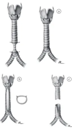
At our facility, the surgical strategy for the treatment of congenital tracheal stenosis is based on the stenosis length. In the cases in which the stenosis length was short less than one third of the total tracheal length), the patients were treated with resection and tracheal reconstruc-tion with end-to-end anastomosis (Figure 1a). Patients with long-segment stenosis underwent tracheoplasty with autologous pericardium, a procedure that consists in making a longitudinal incision on the anterior wall of the whole sten-osed segment in order to enlarge the tracheal lumen and covering the resulting defect with a pericardial flap; this flap is fixed to the medias-tinal tissue by sutures in order to prevent airway collapse (Figure 1b). Medium length-segment stenoses were treated by slide tracheoplasty, in which, initially, the stenosed area of the trachea is divided axially. After this section, the posterior wall of the cranial segment and the anterior wall of the distal portion are divided by means of median longitudinal incisions. Subsequently, the proximal and distal portions are slid and sutured, promoting tracheal shortening and enlargement of tracheal lumen (Figure 1c). All of these tech-niques have been previously described.(10,11) Extracorporeal circulation was used on a case-by-case basis, and correction of cardiac or vascular anomalies was performed when neces-sary, preferably during the same operation.
Results
Six boys and one girl were included. The median age at diagnosis was 2 months (range, 28 days to 3 years). The patients presented a wide variety of respiratory signs and symptoms, the most common being dyspnea, cyanosis and wheezing. Two patients had respiratory infec-tion at diagnosis.
Based on the bronchoscopy and CT findings, the stenosis length was classified as short in three patients, as medium in one and as long in three. The following intracardiac anomalies were infection or after intubation, when the fragile
respiratory system of such patients decompen-sates due to the reduction in airflow.(2,3)
The diagnosis is frequently delayed due to the rarity and diversity of manifestations of the disease. In addition, other malformations often divert the attention of the team of attending physicians. Experience and clinical suspicion are essential for accurate and early diagnosis. Unfortunately, prenatal diagnosis is still not possible, since the accuracy of intrauterine examination remains limited.
Congenital stenosis and its treatment are classically associated with high rates of morbidity and mortality. Endoscopic and surgical techniques developed in recent decades have contributed to better outcomes in such patients.(1) However, a highly specialized team and individual management are essential for good results. No technique—resection and anas-tomosis; pericardial patch tracheoplasty; or slide tracheoplasty—is accepted as definitive, and no surgical technique corrects all of the anatomic variants of this disease.(4-7) The treatment choice is a controversial issue, and the approach adopted at each facility varies, mainly depending on the clinical status of patients and the char-acteristics of the stenosis.(8) The objective of the present study was to analyze the evolution and outcomes of patients undergoing repair of congenital tracheal stenosis at our facility.
Methods
This was a retrospective study of patients treated between January of 2002 and December of 2005. Patient charts were reviewed for the following data: gender; age; comorbidities; symptoms; surgical procedures; complications; and outcomes. The present study was approved by the Ethics Committee of the University of São Paulo School of Medicine Hospital das Clínicas.
tomatic. The three patients with long-segment stenosis underwent pericardial patch tracheo-plasty. Extracorporeal circulation was used in two patients who presented cardiac malformations, which were corrected during the same operation. One of the patients died of severe intracardiac anomalies occurring during the procedure. In the remaining patients, the immediate post-operative complications were pneumonia (in one) and arrhythmia (in one). During the medium- and long-term follow-up period, one of those two patients remained asymptomatic, without the need for additional procedures. The other developed pronounced granulation in the trachea, accompanied by malacia at the patch site and residual stenosis at the carina. Various endoscopic procedures were needed in order to maintain airway patency. Granulomas were resected, dilatations were performed, and, finally, a T-tube was implanted. At the time of the final data collection, the T-tube remained in place, the patient was asymptomatic, and peri-odic endoscopic follow-up examinations were being performed in an outpatient setting.
Slide tracheoplasty was performed in one patient with tracheal and subglottic stenosis. In that case, extracorporeal circulation was also used. The patient developed extensive tracheal necrosis, together with tracheal fistula and pneumopericardium. In addition, the patient developed pulmonary atelectasis and pneu-monia, dying on postoperative day 11 from sepsis secondary to infection at the surgical site.
Resection and end-to-end anastomosis were used in two of the patients with short-segment stenosis. One of those two had a cerebrovas-cular accident and developed renal failure in the postoperative period. However, despite a long stay in the intensive care unit, that patient was discharged in good clinical condition after 120 days. That same patient had stenosis at the anastomosis site, and two endoscopic dilatations were needed in the first 6 months. Subsequently, no other procedures were necessary. For the other patient, the postoperative course was excellent, without complications.
One of the patients presented congen-ital tracheal stenosis due to extrinsic vascular compression. In that patient, the anomalous aortic arch was ligated and sectioned. After the arch had been sectioned, the extrinsic compression ceased to exist—as confirmed by found: tetralogy of Fallot in one patient; and
interventricular communication accompanied by interatrial communication in another. Other congenital anomalies were as follows: dextro-cardia and left superior vena cava, both in the same patient; double aortic arch (type 1A/Mayo classification) in one; and imperforate anus in one. Two patients presented pulmonary artery sling. The demographic characteristics of the patients are described in Table 1.
Surgical treatment was necessary in all cases, since all seven of the patients were highly
symp-a
b c
Figure 1 - In a), resection and end-to-end
(57%)—one patient presented malformation of the pulmonary artery and cardiac malformation.
There is no standard procedure for the treat-ment of congenital tracheal stenosis, and there have been no controlled studies comparing the different methods described (which would be quite difficult to compare due to the rarity of the disease, as well as to the severity of its distinct manifestations and associated diseases). The conservative approach is an option recom-mended for cases in which the stenosis is short and the diameter of the stenotic segment is more than 60% of the normal tracheal diam-eter.(12) Such cases have been described in the literature. However, in the present study, none of the patients met those criteria and therefore all underwent surgical treatment.
The morbidity and mortality rates associated with the surgical treatment of congenital tracheal stenosis are high, and despite the growing world-wide experience, the latest publications show that the mortality rate is 18% in patients under-going pericardial patch tracheoplasty and 24% in those undergoing slide tracheoplasty.(13,14) In our study, the overall surgical mortality was 28.5%. This higher mortality rate might be due to the fact that our sample comprised children in whom the condition was more severe, chil-dren who were underweight and chilchil-dren whose diagnosis was delayed. The results of a recent meta-analysis tend to confirm this suspicion, intraoperative bronchoscopy—and, therefore, no
other additional tracheal procedures were neces-sary. The postoperative course was favorable, without complications.
The mean length of hospital stay was 45 days, and, excluding the inpatient deaths, the mean postoperative follow-up period was 31.6 months (range, 13-84 months). Our 30-day survival rate was 71%. By the end of the data collection period, all five of the surviving patients were asymptomatic and completely free of disease, although, in one case, the T-tube remained in place (Table 2).
Discussion
In congenital tracheal stenosis, the onset of symptoms can be quite dramatic. In our study, all patients presented at least one severe respi-ratory event prior to treatment. The presence of congenital malformations (especially cardiac malformations) makes diagnosis difficult. In this context, clinical suspicion is fundamental and should be followed by sophisticated assess-ment. All patients underwent echocardiography, bronchoscopy and CT of the chest.(9) Those tests allowed us to confirm the diagnosis, as well as to characterize the stenosis and the malforma-tions. In our study, echocardiography revealed cardiac malformations in two patients (28.5%), and chest CT detected vascular anomalies in four
Table 1 - Characteristics of the patients studied (patients included in the table by date of surgical procedure).
Patient Gender Age Other diagnoses Type of tracheal stenosis
Surgical procedure
Other procedures ECC
01 F 3 years PAS Short-segment stenosis (PAS)
Resection and anastomosis
Pulmonary artery reimplantation
Yes
02 M 28 days PAS, IAC, IVC, imperforate anus
Long-segment stenosis
Pericardial patch tracheoplasty
Pulmonary artery reimplantation
Yes
03 M 2 mos. GER Long-segment stenosis
Pericardial patch tracheoplasty
None No
04 M 1.3 mos. TF, GER Long-segment stenosis
Pericardial patch tracheoplasty
Correction of TF Yes
05 M 29 days Dextrocardia, LSVC
Medium length-segment stenosis
Slide tracheoplasty None Yes
06 M 3.7 mos. DAA Short-segment stenosis (DAA)
Vascular ring correction
None No
07 M 2 mos. None Short-segment stenosis
Resection and anastomosis
None No
factors previously described as increasing the risk for an unfavorable outcome: being less than 1 month of age; and presenting severe intracar-diac anomalies (single ventricle and pulmonary stenosis). Two patients had postoperative tracheal stenosis, and, in both, the stenosis was treated endoscopically without difficulties. One of the patients presented reduction in the diam-eter of the anastomotic line, which evolved well after two dilatation sessions. Another patient presented residual stenosis at the carina and developed pronounced granulation tissue along the cranial edge of the suture. The residual stenosis was treated with hydrostatic balloon dilatation, the granulation tissue was resected by rigid bronchoscopy, and a T-tube was used in order to maintain the airway. It is of note that, during the follow-up period, the distal stenosis stabilized, and it was possible to progressively decrease the caudal extent of the T-tube.
The present study represents the experience of a referral facility for pediatric laryngotracheal and cardiac surgery in the treatment of congen-ital tracheal stenosis, a complex entity that requires specialized treatment and an adequate infrastructure. Our results show that, although surgical treatment of congenital tracheal sten-osis is possible, it is associated with significant morbidity and mortality in high-risk patients.
References
1. Antón-Pacheco JL, Cano I, García A, Martínez A, Cuadros J, Berchi FJ. Patterns of management of congenital tracheal stenosis. J Pediatr Surg. 2003;38(10):1452-8. 2. Lang FJ, Hurni M, Monnier P. Long-segment congenital
tracheal stenosis: treatment by slide-tracheoplasty. J Pediatr Surg. 1999;34(8):1216-22.
3. Hoffer ME, Tom LW, Wetmore RF, Handler SD, Potsic WP. Congenital tracheal stenosis. The otolaryngologist’s
since two significant risk factors for morbidity and mortality were identified in such children: being less than 1 month of age and presenting associated intracardiac malformations.(13) In the present study, 57% of the patients were at high risk according to these factors (two patients were less than 1 month of age, and two presented intracardiac anomalies), confirming the severity of the cases comprising our sample.
It is of note that few studies have addressed short-segment stenosis,(9) which suggests that this is an uncommon characteristic. However, three of our patients had short-segment stenosis. The therapeutic option in these cases was for resection of the stenosis and end-to-end anas-tomosis (except for one patient with a vascular ring). Although some authors discourage the use of this procedure, in our study, we found that late evolution was favorable, suggesting that resection and anastomosis are a good option for this specific population.(15,16)
Based on the experiences mentioned in the literature, we realized that complications are common after surgical treatment of congenital tracheal stenosis. The consequences of this fact can be measured by the long mean length of hospital stay found in the present study. Such complications are typically related to associated cardiac malformations, infectious profiles (espe-cially pneumonia or mediastinitis) and tracheal reconstruction (restenosis, malacia or granula-tion tissue formagranula-tion).(4,5,14) In our study, two patients had severe complications leading to death: one died during surgery (this patient had developed hemodynamic instability and hypox-emia, and weaning from mechanical ventilation had been impossible); and one died on postoper-ative day 11 from septic shock. The patient who died during the procedure presented the two
Table 2 - Surgical treatment results.
Patient Hospital stay, days
Postoperative complications
Reintubation Outcome Follow-up
01 120 Dialytic renal failure Yes Outpatient decannulation 7 years
02 NA NA NA Intraoperative death
03 55 Pneumonia, multiple intubations Yes Outpatient decannulation 26 months 04 25 Arrhythmia, granuloma Yes Outpatient T-tube use 18 months 05 NA Infection at the incision site,
hypoxia, atelectasis.
Yes Death on postoperative day 11
extensive tracheal stenosis in infants and children. J Thorac Cardiovasc Surg. 1984;88(4):527-36.
11. Tsang V, Murday A, Gillbe C, Goldstraw P. Slide tracheoplasty for congenital funnel-shaped tracheal stenosis. Ann Thorac Surg. 1989;48(5):632-5.
12. Cheng W, Manson DE, Forte V, Ein SH, MacLusky I, Papsin BC, et al. The role of conservative management in congenital tracheal stenosis: an evidence-based long-term follow-up study. J Pediatr Surg. 2006;41(7):1203-7.
13. Chiu PP, Kim PC. Prognostic factors in the surgical treatment of congenital tracheal stenosis: a multicenter analysis of the literature. J Pediatr Surg. 2006;41(1):221-5; discussion 221-5.
14. Airway Reconstruction Team. Recent challenges in the management of congenital tracheal stenosis: an individualized approach. J Pediatr Surg. 2005;40(5):774-80.
15. Beierlein W, Elliott MJ. Variations in the technique of slide tracheoplasty to repair complex forms of long-segment congenital tracheal stenoses. Ann Thorac Surg. 2006;82(4):1540-2.
16. Hasaniya N, elZein CF, Mara S, Barth MJ, Ilbawi M. Alternative approach to the surgical management of congenital tracheal stenosis. Ann Thorac Surg. 2006;82(6):2305-7.
perspective. Arch Otolaryngol Head Neck Surg. 1994;120(4):449-53.
4. Tsugawa C, Nishijima E, Muraji T, Satoh S, Takamizawa S, Yamaguchi M, et al. Tracheoplasty for long segment congenital tracheal stenosis: analysis of 29 patients over two decades. J Pediatr Surg. 2003;38(12):1703-6. 5. Backer CL, Mavroudis C, Dunham ME, Holinger L.
Intermediate-term results of the free tracheal autograft for long segment congenital tracheal stenosis. J Pediatr Surg. 2000;35(6):813-8; discussion 818-9.
6. Backer CL, Mavroudis C, Dunham ME, Holinger LD. Reoperation after pericardial patch tracheoplasty. J Pediatr Surg. 1997;32(7):1108-11; discussion 1111-2. 7. Kim HK, Kim YT, Sung SW, Park JD, Kang CH, Kim JH,
et al. Management of congenital tracheal stenosis. Eur J Cardiothorac Surg. 2004;25(6):1065-71.
8. Antón-Pacheco JL, Cano I, Comas J, Galletti L, Polo L, García A, et al. Management of congenital tracheal stenosis in infancy. Eur J Cardiothorac Surg. 2006;29(6):991-6.
9. Elliott M, Roebuck D, Noctor C, McLaren C, Hartley B, Mok Q, et al. The management of congenital tracheal stenosis. Int J Pediatr Otorhinolaryngol. 2003;67 Suppl 1:S183-92.
10. Idriss FS, DeLeon SY, Ilbawi MN, Gerson CR, Tucker GF, Holinger L. Tracheoplasty with pericardial patch for
About the authors
Ricardo Mingarini Terra
Attending Physician. Department of Thoracic Surgery, University of São Paulo School of Medicine Hospital das Clínicas, São Paulo, Brazil.
Helio Minamoto
Attending Physician. Department of Thoracic Surgery, University of São Paulo School of Medicine Hospital das Clínicas, São Paulo, Brazil.
Lívia Caroline Barbosa Mariano
Medical Student. University of São Paulo School of Medicine, São Paulo, Brazil.
Angelo Fernandez
Attending Physician. Department of Thoracic Surgery, University of São Paulo School of Medicine Hospital das Clínicas, São Paulo, Brazil.
José Pinhata Otoch
Associate Professor. University of São Paulo School of Medicine Hospital das Clínicas, São Paulo, Brazil.
Fabio Biscegli Jatene