Prevalence and Prognostic Impact of Diastolic Dysfunction in
Patients with Chronic Kidney Disease on Hemodialysis
Silvio H. Barberato
1,2, Sérgio G. E. Bucharles
1, Admar M. Sousa
2, Costantino O. Costantini
2, Costantino R. F.
Costantini
2, Roberto Pecoits-Filho
1Pontifícia Universidade Católica do Paraná1; Hospital Cardiológico Costantini2, Curitiba, PR - Brazil
Mailing address: Silvio Henrique Barberato •
Rua Saint Hilaire, 122/203 - Água Verde - 80240-140 - Curitiba, PR - Brazil E-mail: silviohb@cardiol.br, msbarberato@terra.com.br
Manuscript received February 17, 2009; revised manuscript received August 18, 2009; accepted August 27, 2009.
Abstract
Background: Diastolic dysfunction (DD) is frequent in patients on hemodialysis (HD), but its impact on the clinical evolution is yet to be established.
Objective: To evaluate the prevalence and prognostic impact of left ventricular (LV) advanced diastolic dysfunction (ADD) in patients on hemodialysis.
Methods: The echocardiograms were performed during the first year of HD therapy, in patients with sinus rhythm, with no evidence of cardiovascular disease, excluding those with significant valvopathy or pericardial effusion. The combined assessment of the Doppler echocardiographic data classified the diastolic dysfunction as: 1) normal diastolic function; 2) mild DD (relaxation alteration) and 3) ADD (pseudonormalization and restrictive flow pattern). The assessed outcomes were general mortality and cardiovascular events.
Results: A total of 129 patients (78 males), aged 52 ± 16 years, with a DD prevalence of 73% (50% with mild DD and 23% with ADD) were included in the study. The group with ADD was older (p < 0.01) and presented higher systolic (p < 0.01) and diastolic BP (p = 0.043), LV mass (p < 0.01), left atrial volume index (p < 0.01) and number of diabetic patients (p = 0.019), as well as lower ejection fraction (EF) (p < 0.01). After 17 ± 7 months, the general mortality was significantly higher in individuals with ADD, when compared to those with normal function and mild DD (p = 0.012, log rank test). At Cox multivariate analysis, ADD was predictive of cardiovascular events (hazard ratio 2.2; confidence interval: 1.1-4.3; p = 0.021) after adjusted for age, gender, diabetes, LV mass and EF.
Conclusion: The subclinical ADD was identified in approximately 25% of the patients undergoing hemodialysis and had a prognostic impact, regardless of other clinical and echocardiographic data. (Arq Bras Cardiol 2010; 94(4):431-436)
Key words: Kidney diseases; renal dialysis; ventricular dysfunction, left/physiopathology/therapeutic.
for the onset of heart failure, whatever the underlying cause is5. Therefore, it becomes important to detect the presence of
advanced DD (ADD), characterized by the increase in the filling pressures, especially at the subclinical phase.
In this context, the Doppler echocardiographic pre-load independent techniques, such as tissue Doppler6 and left
atrial volume7, represent an important advancement in the
assessment of diastolic function.
The objectives of the present study are to estimate the prevalence, identify predisposing factors and evaluate the prognostic impact of left ventricular ADD in individuals on HD with no clinical evidence of cardiovascular disease.
Methods
Population
Patients eligible for the study were those with chronic kidney disease (CKD) undergoing maintenance hemodialysis (4-hour sessions, three times a week), at two Services of Nephrology in University hospitals.
Introduction
Cardiovascular complications are the main cause of death in patients with chronic kidney disease (CKD) undergoing renal replacement therapy through hemodialysis (HD)1.
In these individuals, the finding of left ventricular (LV) echocardiographic alterations, such as LV hypertrophy, dilation and systolic dysfunction, results in a 3-fold higher risk of heart failure, regardless of age, diabetes and coronary failure2. On
the other hand, although the LV filling alterations are often detected in this group, the actual prevalence of diastolic dysfunction (DD) and its impact on the clinical evolution are yet to be established3,4.
The inclusion criteria were: to have been undergoing HD for a period between 1 and 12 months, no previous cardiovascular diseases (congestive heart failure, acute myocardial infarction, cerebrovascular accident or peripheral arterial failure) and sign the free and informed consent form. The exclusion criteria were: malignant diseases, active infection, non-sinus rhythm, significant valvopathy (valve prosthesis, any degree of mitral or aortic stenosis and moderate to significant mitral, aortic or tricuspid regurgitation), pericardial effusion, and technically unsatisfactory echocardiogram. All patients were submitted to HD in an Altra Touch equipment (Althin, Miami, Florida, Fl, USA) , containing a cellulose acetate dialyzer with a minimum blood flow regulation of 200 ml/minute and dialysate flow of 300 to 500 ml/minute. The “dry-weight” estimation (volume to be removed by ultrafiltration) was carried out by clinical signs of hydration, blood pressure behavior during the HD session and electrical bioimpedance8. The body surface was
calculated using the equation proposed by Du Bois and Du Bois (0.20247 x weight0.425 x height0.725). The body mass index
(BMI) was calculated by dividing the weight in kilograms by the height in meters squared; obesity was considered when BMI > 30. The systolic and diastolic arterial pressures, heart rate, weight and height were measured at the time of the assessment. The Ethical Committee in Research of our institution approved the study protocol and informed consent was obtained from all patients.
Doppler echocardiogram
The assessments were carried out on interdialytic days, between 12 PM and 6 PM. The patients were placed on left lateral decubitus, using an Envisor C echocardiographic equipment (Phillips Inc., Andover, Massachusetts, EUA) equipped with 2 to 4 MHz transducers to undergo the complete echocardiographic assessment through the M-mode, two-dimensional and Doppler techniques (pulsed, continuous, color and tissue).
The following parameters were obtained in the M-mode: anteroposterior diameter of the left atrium, thickness of the interventricular septum and LV posterior wall in diastole, LV-end diastolic diameter (dilatation when > 54 mm for women and > 56 mm for men) and LV-end systolic diameter. LV mass was estimated using Devereux equation9 and index
by height to the power of 2.710. LV hypertrophy was defined
as the presence of mass/height2.7 ≥ 45 g/m2.7 for women
and ≥ 49 g/m2.7 for men11. Systolic function was assessed by
calculating ejection fraction (EF) using Simpson’s method11.
Mitral transvalvular flow was recorded in 4-chamber apical view with the pulsed Doppler sampling positioned between the extremities of the mitral cusps, and we measured the early filling velocity (E wave), atrial contraction velocity (A wave) and E/A ratio. The tissue Doppler velocities were recorded in the 4-chamber apical view with a volume sampling positioned consecutively on the junction between the LV lateral and septal walls with the mitral annulus12.
The early (E’) and late (A’) diastolic mitral annular velocities were analyzed and the E’/A’ and E/E’ (average of septal and lateral annulus sides) ratios were calculated. The left atrial volume was determined at the two-dimensional Doppler using Simpson’s biplane technique11 and indexed for body
surface. A total of three cardiac cycles were considered for all measurements.
Diastolic function assessment
LV diastolic function was classified by considering the interpretation of all conventional and tissue-Doppler derived indices of the mitral annulus in four patterns: normal, relaxation alteration (E/A ratio < 1 and E´ < 10 cm/s), pseudonormal and restrictive flow pattern ( E/A ratio > 2 and E´ < 8 cm/s)13.
The diagnosis of the pseudonormal pattern (differentiating it from the actual normal one) used the following criteria: E/E’ ratio > 1513; or b) E/E’ ratio ≥ 1112,associated to a left atrial
volume index > 35 ml/m2,14. The sample was subsequently
classified in three groups, according to the hemodynamic pattern of LV filling: 1) normal diastolic function, 2) mild DD (MDD) (relaxation alteration) and 3) ADD (comprehending restrictive flow and pseudonormalization).
Analysis of survival
Demographic data, comorbidities (diabetes mellitus, arterial hypertension, dyslipidemia, smoking), current medications and routine laboratory findings were obtained after a careful analysis of the patients’ files, in addition to an interview with the physician in charge of the patient’s treatment, when necessary. The primary and secondary outcomes were, respectively, general mortality and a combination of fatal and nonfatal cardiovascular events. The analyzed events were cardiovascular death (including sudden death, acute myocardial infarction, cerebrovascular accident), nonfatal myocardial infarction and decompensated congestive heart failure (requiring hospitalization). The follow-up was based on the analysis of the outcomes, periodically performed at the dialysis clinic. Patients submitted to kidney transplant or that changed the type of dialysis were censored.
Statistical analysis
An adequate descriptive analysis was applied to continuous (means and standard deviations) and categorical (percentages) variables. Comparisons between the groups were carried out by analysis of variance (ANOVA), with Dunnett’s post hoc correction for continuous variables and Chi-square test for categorical variables. The logistic regression analysis determined the independent predictors of DD. Kaplan-Meier survival curves for the diastolic function status were constructed and compared by the log-rank test. The independent prognostic impact of DD was evaluated by the multivariate analysis of survival using Cox’s model, considering established risk predictors in this population. The level of statistical significance was set at p < 0.05. The SPSS 13 for Windows and JMP IN 5.1 statistical packages were used for all statistical analyses.
Results
Basal characteristics
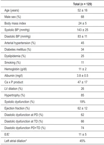
formed the study population are shown in Table 1. The group consisted of 78 men and 51 women, with a mean age of 52 ± 16 years and time of dialysis of 7 ± 4 months. The etiology of the CKD was attributed to hypertensive nephrosclerosis (39%), diabetic nephropathy (21%), an association of the former two (15%), chronic glomerulonephritis (14%), polycystic kidney (5%), lithiasis (3%), chronic pyelonephritis (2%) and other diseases (1%). Most patients (77%) used anti-hypertensive medications, especially angiotensin-converting enzyme inhibitors (46%), beta-blockers (16%), alpha-blockers (13%), calcium-channel antagonists (12%) and angiotensin-receptor blockers (11%), either alone or in combination.
Clinical and doppler echocardiographic alterations
At the study enrollment, 45% of the patients had arterial hypertension, 34% had diabetes mellitus, 25% had dyslipidemia and 11% were smokers. The prevalence of obesity (BMI > 30) was 9%. LV dilatation was present in 34 patients (26%), LV hypertrophy in 110 (85%) and systolic dysfunction in 24 patients (19%), either alone or in combination. Only 19 patients (15%) presented normal LV dimensions. Significant left atrial dilatation (volume > 32 ml/m2) was observed in 58
individuals (45%).
Table 1 - Main basal characteristics of the study population
Total (n = 129)
Age (years) 52 ± 16
Male sex (%) 68
Body mass index 24 ± 5
Systolic BP (mmHg) 143 ± 25
Diastolic BP (mmHg) 83 ± 11 Arterial hypertension (%) 45
Diabetes mellitus (%) 34
Dyslipidemia (%) 25
Smoking (%) 11
Hemoglobin (g/dl) 11 ± 2
Albumin (mg/l) 3.8 ± 0.5
Ca x P product 47 ± 17
LV dilation (%) 26
Hypertrophy (%) 85
Systolic dysfunction (%) 19%
Ejection fraction (%) 62 ± 12
Diastolic dysfunction at PD (%) 62
Diastolic dysfunction at TD (%) 66
Diastolic dysfunction PD+TD (%) 74
E/E’ 11 ± 5
Left atrial dilation* 45%
Data presented as means ± SD, percentages or medians with range; BP - blood pressure; Ca x P - calcium x phosphorus; LV - left ventricle; E/E’ - ratio of the
early transmitral low velocity to early diastolic mitral annular velocity; PD -
pulsed doppler; TD - tissue doppler; * - Left atrial volume index > 32 ml/m²).
Table 2 - Comparison of demographic, clinical, biochemical and doppler echocardiographic characteristics of patients with and without diastolic dysfunction
Variable Normal Mild DD Advanced DD valuep
Number of patients 35 64 30
Age (years) 42 ± 16 56 ± 14 52 ± 14 < 0.01
Male sex (%) 60 59 63 0.93
BMI 23 ± 5 25 ± 6 24 ± 4 0.41
Time of HD (months) 7 ± 4 8 ± 4 8 ± 4 0.38
Hemoglobin (g/dl) 11 ± 2 11 ± 2 9.6 ± 1 0.20
Albumin (mg/l) 3.9 ± 0.4 3.7 ± 0.6 3.7 ± 0.6 0.74
Ca x P product 44 ± 12 49 ± 17 45 ± 21 0.55
Systolic BP (mmHg) 125 ± 21 146 ± 22 160 ± 23 < 0.01
Diastolic BP (mmHg) 76 ± 12 84 ± 9 88 ± 9 0.043 Arterial hypertension
(%) 31 50 50 0.17
Diabetes mellitus
(%) 17 36 50 0.019
LVDD (mm) 50 ± 7 51 ± 6 54 ± 7 0.044
LVMI (g/altura2,7) 59 ± 23 88 ± 36 100 ± 32 < 0.01
EF (%) 63 ± 5 60 ± 6 52 ± 9 < 0.01
E’ (cm/s) 12± 3 7.3 ± 2 6.9 ± 2 < 0.01 E/E’ ratio 8 ± 2 10 ± 4 17 ± 5 < 0.01
LAVI (ml/m2) 27 ± 10 31 ± 11 44 ± 17 < 0.01
Data presented as means ± SD, percentages or medians and range. DD - diastolic dysfunction; BMI - body mass index; HD - hemodialysis; Ca x P - calcium phosphorus product; BP - blood pressure; LVDD - left ventricular diastolic dimension ; LVMI - left ventricular mass index; EF - ejection fraction;
E’ - early diastolic mitral annular velocity; E - early transmitral low velocity; LAVI
- left atrial volume index.
Prevalence of diastolic dysfunction
DD was diagnosed in 94 of the 129 patients (73%), with 64 presenting mild DD - MDD (50%) and 30 advanced DD - ADD (23%). In the ADD group, 20 patients presented flow pseudonormalization and 10 presented restrictive flow. In parallel, only 22 patients (17%) presented a mean E/E’ ratio > 15. When considering solely the conventional Doppler data, only 80 (62%) of the 129 studied patients would have been diagnosed with DD.
Predictors of advanced DD
Table 3 - Comparison of outcomes in the groups with normal diastolic function, mild diastolic dysfunction and advanced diastolic dysfunction
Variable Normal Mild DD Advanced DD
General mortality 9 19 37*
CV death 3 14 27*
Non-CV death 6 5 10*
Nonfatal CV events 14 12 27*
CV death + CV Events 17 26 54*
Values expressed as percentages (%). *p < 0.05 in comparison to the other groups. DD - diastolic dysfunction; CV - cardiovascular.
1.6-5.9, p = 0.011), systolic arterial pressure (OR 1.05; CI: 1.01-1.09, p = 0.023) and LV mass (OR 1.06; CI: 1.01-1.1, p < 0.01), in addition to a tendency for the ejection fraction (p = 0.052).
Outcomes
During the mean follow-up of 17 ± 7 months, there were 26 deaths (20%) and 21 nonfatal cardiovascular events (16%). The deaths were attributed to cardiovascular (18 deaths, with 8 due to myocardial infarction, 7 due to sudden death, 2 due to documented complex ventricular arrhythmia and 1 due to hemorrhagic cerebrovascular accident) and infectious causes (8 deaths). The nonfatal cardiovascular events were myocardial infarction (5) and hospitalizations due to decompensated heart failure (16), totaling 39 combined cardiovascular events. As shown in Figure 1 and Table 3, the general mortality was higher in the group with advanced DD (37%) in comparison to the groups with normal and mild DD (9% and 19%, respectively, log rank test, p = 0.012). Several analyses were carried out using Cox multivariate models. After the evaluation of general mortality and cardiovascular events separately (models including age, male sex, diabetes, LV mass and EF),
Figure 1 -Kaplan-Meier survival curves (general mortality) based on the
diastolic function classiication: NL - normal function; MDD - mild diastolic
dysfunction; ADD - advanced diastolic dysfunction.
Su
rv
iv
a
l MDD
Follow-up (months)
ADD NL
LV mass and EF showed to be independent predictors for both outcomes, in addition to age for cardiovascular events, only. The inclusion of advanced DD in these models reached a statistically significant value for the prediction of cardiovascular events (hazard ratio [HR] 2.2; CI: 1.1-4.3, p = 0.021) and a borderline value for general mortality (HR 2.8; CI: 0.97-8.2, p = 0.056), as seen in Table 4. In contrast, there was no difference regarding the outcomes between patients with and without E/E’ ratio > 15 or E’< 8 cm/s. At an alternative analysis, advanced DD and left atrial volume index (recently described as a prognostic predictor in patients undergoing HD15) were included together at the last step of the model.
Both advanced DD (HR: 3.4; CI: 1.3-9.2, p = 0.03) and left atrial volume index (HR: 6.6; CI: 1.8-24, p = 0.004) were predictive of cardiovascular events.
Discussion
The present study suggests that almost 75% of the individuals undergoing the first year of HD present DD and that approximately 25% of them present ADD. Studies with smaller samples reported DD incidence in 50-65% of uremic patients, including pre-dialysis populations, those undergoing dialysis and post-transplant populations4 (a percentage
similar to that found in our group, if the tissue Doppler and left atrial volume data were not taken into account). Such analyses were limited by the use of transmitral flow-derived parameters and those derived from the pulmonary venous flow to categorize diastolic function16. It is known that such
methods are particularly vulnerable to preload variations6;
therefore, such approach might have induced false-negative
Table 4 - Independent prognostic predictors at Cox multivariate analysis
General mortality Cardiovascular events
HR 95%CI p value HR 95%CI p value
Age (years) ns 1.04 1.02-1.07 < 0.01
LVMI (g/m2.7) 1. 018 1.01-1.03 < 0.01 1.01 1.01-1.02 < 0.01
EF (per 1% decrease) ns ns
Advanced DD 2.8 0.97-8.2 0.056 2.2 1.1-4.3 0.021 HR - hazard ratio; CI - conidence interval; LVMI - left ventricular mass index; EF - ejection fraction; TD - time of deceleration; E/E’ - ratio of early transmitral low velocity
results, diagnosing individuals with pseudonormalization (high filling pressures “masking” the LV relaxation alteration) as normal. In this context, to combine the conventional Doppler data with techniques that are relatively independent from the preload, such as the tissue Doppler and left atrial volume adds accuracy to the diagnosis of DD and especially of the ADD, characterized by high diastolic intraventricular pressure.
To detect the presence of DD and estimate the LV filling pressures is currently of general interest to predict the risk of developing the so-called “heart failure with preserved ejection fraction”. The clinically evident heart failure represents an independent predictor of mortality in patients starting hemodialysis therapy17, but the identification of the underlying
cause can be important to direct the therapeutic management. Necropsy and experimental uremia studies have pointed out the presence of specific diffuse intermyocardiocytic fibrosis in the heart of uremic individuals, which could predispose to electrical instability (associated with sudden death) and an increase in filling pressures18,19.
The effect on the physiopathological mechanisms related to marked myocardial fibrosis, such as the activation of humoral factors (plasma angiotensin II, parathyroid hormone, endothelin, aldosterone and catecholamines)3, can represent an important
therapeutic target, especially at the subclinical phase. In spite of such considerations, whether the diagnosis of ADD collaborates to refine and stratify the cardiovascular risk in uremic patients has been little investigated. Recently, two studies tried to estimate the prognostic impact of ADD on CKD through the E/E’ ratio, an acknowledged noninvasive index of LV filling pressures in the general population12,20. In a
study with 125 candidates to kidney transplant, Sharma et al21
found an association between E/E’ > 15 and higher general mortality. However, the study population was heterogeneous (only one-third of the patients was undergoing HD) and only the univariate analysis was performed, which did not allow inferring whether E/E’ ratio adds a prognostic value to the traditional assessment of cardiovascular risk in this group.
Wang et al22 reported, in a study with 220 patients
(exclusively undergoing peritoneal dialysis), that E/E’ > 15 was able to predict general and cardiac mortality better than the classic clinical and echocardiographic data. In our population, which consisted exclusively of patients undergoing HD, the E/E’ ratio > 15 did not present a discriminating
power regarding the studied outcomes. The physiopathology of the cardiovascular disease in individuals undergoing HD is influenced by the abrupt and extensive volemic variations to which these patients are submitted23.
The accurate prediction of the filling pressures (and, consequently, of the prognosis) for a certain individual requires the inclusion of all available data, which makes the approach more consistent than a single parameter considered alone.
Among the study limitations, it is important to mention the relatively small sample size, which was compensated, however, by the attained statistical power (86% for the primary outcome and 94% for the secondary one). Other limitations were the absence of invasive measurements of LV filling pressure (practical and ethical questions would not justify a cardiac catheterism in the studied population) and the stringent exclusion criteria (which might have made our findings not applicable to all of the dialyzed population).
Conclusions
The present study indicates that ADD, diagnosed through the combined interpretation of Doppler echocardiographic information, has a prognostic impact on dialyzed patients with no previous history of cardiovascular events.
The general mortality was significantly higher in the group with ADD, when compared to those with normal diastolic function and mild DD. In parallel, the ADD was predictive of cardiovascular events, regardless of age, gender, diabetes, LV mass and ejection fraction.
Potential Conflict of Interest
No potential conflict of interest relevant to this article was reported.
Sources of Funding
The present study was partially supported by a cardiology research grant from Brazilian Society of Cardiology.
Study Association
This study is not associated with any post-graduation program.
References
1. Sarnak MJ, Levey AS, Schoolwerth AC, Coresh J, Culleton B, Hamm LL, et al. Kidney disease as a risk factor for development of cardiovascular disease: a statement from the American Heart Association Councils on Kidney in Cardiovascular Disease, High Blood Pressure Research, Clinical Cardiology, and Epidemiology and Prevention. Circulation. 2003; 108: 2154-69.
2. Parfrey PS, Foley RN, Harnett JD, Kent GM, Murray D, Barre PE. Outcome and risk factors of ischemic heart disease in chronic uremia. Kidney Int. 1996; 49: 1428-34.
3. London GM. Left ventricular alterations and end-stage renal disease. Nephrol Dial Transplant. 2002; 17 (Suppl 1): 29-36.
4. Barberato SH, Pecoits Filho R. Alterações ecocardiográficas em pacientes de hemodiálise. Arq Bras Cardiol. 2009. [In press].
5. Oh JK. Echocardiography as a noninvasive Swan-Ganz catheter. Circulation. 2005; 111: 3192-4.
6. Barberato SH, Pecoits Filho R. Influence of preload reduction on Tei index and other Doppler echocardiographic parameters of left ventricular function. Arq Bras Cardiol. 2006; 86: 425-31.
7. Barberato SH, Mantilla DE, Misocami MA, Goncalves SM, Bignelli AT, Riella MC, et al. Effect of preload reduction by hemodialysis on left atrial volume and echocardiographic Doppler parameters in patients with end-stage renal disease. Am J Cardiol. 2004; 94: 1208-10.
9. Devereux RB, Alonso DR, Lutas EM, Gottlieb GJ, Campo E, Sachs I, et al. Echocardiographic assessment of left ventricular hypertrophy: comparison to necropsy findings. Am J Cardiol. 1986; 57: 450-8.
10. de Simone G, Daniels SR, Devereux RB, Meyer RA, Roman MJ, de Divitiis O, et al. Left ventricular mass and body size in normotensive children and adults: assessment of allometric relations and impact of overweight. J Am Coll Cardiol. 1992; 20: 1251-60.
11. Lang RM, Bierig M, Devereux RB, Flachskampf FA, Foster E, Pellikka PA, et al. Recommendations for chamber quantification: a report from the American Society of Echocardiography’s Guidelines and Standards Committee and the Chamber Quantification Writing Group, developed in conjunction with the European Association of Echocardiography, a branch of the European Society of Cardiology. J Am Soc Echocardiogr. 2005; 18: 1440-63.
12. Dokainish H, Zoghbi WA, Lakkis NM, Al-Bakshy F, Dhir M, Quinones MA, et al. Optimal noninvasive assessment of left ventricular filling pressures: a comparison of tissue Doppler echocardiography and B-type natriuretic peptide in patients with pulmonary artery catheters. Circulation. 2004; 109: 2432-9.
13. Lester SJ, Tajik AJ, Nishimura RA, Oh JK, Khandheria BK, Seward JB. Unlocking the mysteries of diastolic function: deciphering the Rosetta Stone 10 years later. J Am Coll Cardiol. 2008; 51: 679-89.
14. Barberato SH, Pecoits-Filho R. Usefulness of left atrial volume for the differentiation of normal from pseudonormal diastolic function pattern in patients on hemodialysis. J Am Soc Echocardiogr. 2007; 20: 359-65.
15. Barberato SH, Pecoits Filho R. Prognostic value of left atrial volume index in hemodialysis patients. Arq Bras Cardiol. 2007; 88: 643-50.
16. Gupta S, Dev V, Kumar MV, Dash SC. Left ventricular diastolic function in end-stage renal disease and the impact of hemodialysis. Am J Cardiol. 1993; 71: 1427-30.
17. Foley RN, Parfrey PS, Harnett JD, Kent GM, Martin CJ, Murray DC, et al. Clinical and echocardiographic disease in patients starting end-stage renal disease therapy. Kidney Int. 1995; 47: 186-92.
18. Mall G, Huther W, Schneider J, Lundin P, Ritz E. Diffuse intermyocardiocytic fibrosis in uraemic patients. Nephrol Dial Transplant. 1990; 5: 39-44.
19. Ritz E, Rambausek M, Mall G, Ruffmann K, Mandelbaum A. Cardiac changes in uraemia and their possible relationship to cardiovascular instability on dialysis. Nephrol Dial Transplant. 1990; 5 (Suppl 1): 93-7.
20. Ommen SR, Nishimura RA, Appleton CP, Miller FA, Oh JK, Redfield MM, et al. Clinical utility of Doppler echocardiography and tissue Doppler imaging in the estimation of left ventricular filling pressures: a comparative simultaneous Doppler-catheterization study. Circulation. 2000; 102: 1788-94.
21. Sharma R, Pellerin D, Gaze DC, Mehta RL, Gregson H, Streather CP, et al. Mitral peak Doppler E-wave to peak mitral annulus velocity ratio is an accurate estimate of left ventricular filling pressure and predicts mortality in end-stage renal disease. J Am Soc Echocardiogr. 2006; 19: 266-73.
22. Wang AY, Wang M, Lam CW, Chan IH, Zhang Y, Sanderson JE. Left ventricular filling pressure by Doppler echocardiography in patients with end-stage renal disease. Hypertension. 2008; 52: 107-14.