Case Report
Iliopsoas Muscle Hematoma During Treatment with Warfarin
Gabriel Zago
1, Marcelo Campos Appel-da-Silva
1, Luiz Claudio Danzmann
2Hospital Nossa Senhora da Conceição, Porto Alegre, RS1; Hospital São Lucas da PUCRS2, Porto Alegre, RS - Brazil
Mailing address: Marcelo Campos Appel da Silva •
Rua Com. Caminha, 250 /902 - M Vento - 90430-030 - Porto Alegre, RS - Brazil
E-mail: [email protected]
Manuscript received February 01, 2009; revised manuscript received May 15, 2009; accepted May 15, 2009.
Key Words
blood coagulation; warfarin; hematoma; retroperitoneal space; psoas muscles.
Warfarin is a widely used drug for the prevention of thromboembolic events. Knowledge of its adverse effects is necessary for patient follow-up. Although the development of blood dyscrasias is a potential complication in these patients, retroperitoneal bleeding is rare. This article reports the case of a patient who developed iliopsoas muscle hematoma during treatment with warfarin after implantation of a metallic prosthetic aortic valve. The clinical manifestations involved important differential diagnoses.
Introduction
Continuous oral anticoagulation is indicated in several clinical conditions, and warfarin is the most widely used drug for the prevention and treatment of thromboembolic events and for the reduction of death risk resulting from acute myocardial infarction (AMI) and its complications1. In
hospitalized patients, drug administration and dose titration by means of laboratory tests allow the control and safety of the therapeutic target to be reached. From the standpoint of outpatient follow-up, the management of these patients is not always regular. When patients are lost to follow-up and the prothrombin time (PT) and international normalized ratio (INR) levels are unknown, there is an increase in the risk of adverse effects, among which intracranial or retroperitoneal hemorrhage are the most important and potentially fatal2.
The occurrence of iliopsoas muscle hematoma during anticoagulation therapy is rare. It usually manifests unilaterally, with symptoms related to compression of the nerve plexus of the lower limb, and its differential diagnosis encompasses several medical areas3.
The objective of the present article is to report the case of a patient with mechanical prosthetic aortic valve who developed a quite uncommon - and difficult to diagnose - complication associated with the use of oral anticoagulation for the prevention of thromboembolic events.
Case report
A 68-year-old Caucasian male patient who worked as a farmer and lived in the city of Canoas, State of Rio Grande do Sul, Brazil, sought medical care in the emergency department of the University Hospital of Ulbra (Canoas, RS) with complaint of pain, pretibial paresthesia and paresis of left leg for three days. On admission, the patient was in good general state of health, lucid, with mucous membranes moist and pink, and did not have a fever. Blood pressure was 130/80 mmHg, heart rate of 110 bpm. On inspection, hematomas were observed on his thighs and abdomen. Cardiac auscultation revealed normal rate and rhythm and a late systolic murmur heard all over the precordial area. Abdominal examination was unremarkable. Neurological examination revealed right crural monoparesis with strength grade 3, symmetric reflexes and normal plantar response in both legs.
His past medical history revealed a diagnosis of hypertension for 15 years, and the patient was using captopril 25 mg every eight hours; he also reported a surgical procedure for aortic valve replacement for a St. Jude prosthesis two years earlier. Since hospital discharge, the patient had been receiving warfarin sodium 5mg/day. Fourteen days earlier, his cardiologist had recommended a dose increase to 7.5 mg/day. In the past week, he suffered a fall which resulted in mild lower back trauma that neither interfered in his activities nor caused pain.
After an initial evaluation in the emergency department of the University Hospital of Ulbra, the following tests were performed for investigation: plain computed tomography (CT) of the head, resting electrocardiogram, complete blood count, coagulation tests, renal functions tests, electrolytes, LDH and bilirubins
Head CT showed no signs of acute hemorrhagic or ischemic lesions. ECG showed changes consistent with left ventricular hypertrophy. Laboratory tests also showed normal renal function and electrolyte levels, white blood cell count not suggestive of infection, platelet count of 231,000, prothrombin time of less than 10%, INR higher than 5, activated partial thromboplastin time (APTT) of 82.4 seconds, lactate dehydrogenase (LDH) of 500 mg/dl, direct bilirubin of 0.68 mg/dl, indirect bilirubin of 0.97 mg/dl, hematocrit (Ht) of 21.4% and hemoglobin (Hb) of 7.3 g/dl.
Echocardiography performed one year earlier showed left atrium with 4.3 cm, left ventricular diastolic and systolic diameters of 5.3 cm and 3.2 cm, respectively; ejection fraction of 69% and a functioning metallic aortic valve with mean transaortic gradient of 14 mmHg.
In light of these findings, warfarin was discontinued and intravenous vitamin K 10 mg once a day, as well as one unit
Case Report
Arq Bras Cardiol 2010; 94(1) : e1-e3
Zago et al Iliopsoas muscle hematoma and Warfarin
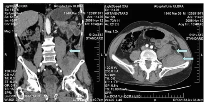
Figure 1 -Contrast-enhanced computed tomography of the abdomen showing diffuse enlargement of the left iliac and psoas muscles in sagittal and cross-sectional views.
of fresh plasma (240 ml) 8/8 hours were administered for correction of anticoagulation.
On the second day of hospitalization, there was persistence of the grade-3 crural monoparesis, in spite of which the patient’s general state of health had improved. Tests performed that day revealed Hb of 5.4g/dl, Ht of 16% and APTT and INR of 82.4 and 3.45 seconds, respectively. In light of these findings, intravenous vitamin K and fresh plasma were continued, and 2 units of red blood cell concentrate were also administered, which increased Hb values to 8.4 g/dl and Ht to 25%.
In order to investigate a possible intra-abdominal hemorrhagic complication, contrast-enhanced CT scan of the abdomen was performed, which revealed diffuse enlargement of the left iliac and psoas muscles, extending to their insertion in the left lesser femoral trochanter (Figure 1). A collection of hyperattenuating material consistent with clotted blood could be observed within the left iliac muscle, suggesting the presence of a hematoma.
In view of the clinical manifestations of sensory and motor abnormalities of the lower limb and presence of retroperitoneal hematoma in the ipsilateral iliac and psoas muscles area, the diagnosis of femoral plexus compression was postulated.
There was complete relief of the paresthesia in pretibial region, partial improvement of pain; on physical examination, the patient presented strength grade 4 in the left lower limb. In light of the INR of 1.83, vitamin K and intravenous plasma were discontinued.
On the fifth day of hospitalization, the patient presented with Hb of 10.3 g/dl, Ht of 30.5%, INR of 1.95, and strength grade 5 in the left lower limb. Warfarin was then restarted at the dose of 2.5 mg/day.
After eight days of hospitalization, the patient was asymptomatic, with INR of 2.57 and ready to be discharged. He was advised to keep on using warfarin 2.5 mg, and captopril at the dose of 25 mg 8/8 hours; a follow-up visit in the Internal Medicine outpatient service was scheduled for reassessment of prothrombin time and, if necessary, adjustment of the warfarin dose.
In the follow-up visit seven days later, the patient had no complaints and his INR was 2.55. He was given instructions on how to handle anticoagulation and to seek the emergency department of the University Hospital of Ulbra should any event occur.
Discussion
Bleeding and hemorrhage are potential and severe complications in patients receiving anticoagulation treatment, with an incidence of up to 10%, and this justifies the need for a strict and regular control of PT and INR levels. “Major hemorrhages” are those occurring especially in intracranial or retroperitoneal sites and/or those requiring transfusion, hospitalization and surgical procedures for compensation, and also those leading to death3. It has already been well established
that patients receiving warfarin and with increased levels of PT and INR, elderly individuals and those with cerebrovascular and/ or metallic prosthetic heart valves are at a higher risk of developing bleeding4. In this latter group of patients, the prevalence of
extracranial hemorrhage is of approximately 2.1%/year, with a higher tendency in the first month of treatment, and a gradual decrease in the months that follow2.
Retroperitoneal hemorrhage is a rare complication, with an incidence of 1.3% to 6.6% per year, and can occur in cases of hemophilia, trauma or anticoagulation5-7. To understand
Case Report
Arq Bras Cardiol 2010; 94(1) : e1-e3
Zago et al
Iliopsoas muscle hematoma and Warfarin
References
1. Wysowski DK, Nourjah P, Swartz L. Bleeding complications with warfarin use - a prevalent adverse effect resulting in regulatory action. Arch Intern Med. 2007; 167 (13): 1414-9.
2. Levine MN, Raskob G, Landefeld CS, Kearon C. Hemorrhagic complications of anticoagulant treatment. Chest. 2001; 119 (Suppl. 1): 108S-121S.
3. Wada Y, Yanagihara C, Nishimura Y. Bilateral Iliopsoas hematomas complicating anticoagulant therapy. Internal Medicine (Tokyo). 2005; 44: 641-3.
4. Casais P, Luceros AS, Meschengieser S, Fondevila C, Santarelli MT, Lazzari MA. Bleeding risk factors in chronic oral anticoagulation with acenocoumarol. Am J Hematol. 2000; 63: 192-6.
5. Uncini A, Tonali PL, Falappa P, Danza FM. Femoral neuropathy from iliac muscle hematoma induced by oral anticoagulation therapy. J Neurol. 1981; 226: 137-41.
6. Holscher RS, Leyten FSS, Oudenhoven LFIJ, Puylaert JBCM. Percutaneous decompression of an iliopsoas hematoma. Abdom Imaging. 1997; 22: 114-6.
7. Parmer SS, Carpenter JP, Fairman RM, Velazquez OC, Mitchell ME. Femoral neuropathy following retroperitoneal hemorrhage: case series and review of the literature. Ann Vasc Surg. 2006; 20: 536-40.
8. Seijo-Martinez M, Castro del Rio M, Fontoira E, Fontoira M. Acute femoral neuropathy secondary to an iliacus muscle hematoma. J Neurol Sci. 2003; 209: 119-22.
9. Lenchiki L, Dovgan DJ, Kier R. CT of the iliopsoas compartment: value in differentiating tumor, abscess and hematoma. Am J Roentgenol. 1994; 162: 83-6.
10. Oake N, Jennings A, Forster AJ, Fergusson D, Doucette S, Walraven CV. Anticoagulation intensity and outcomes among patients prescribed oral anticoagulant therapy: a systematic review and meta-analysis. CMAJ. 2008; 179 (3): 235-44.
the clinical manifestations of these patients, knowledge of the anatomy of this region is fundamental. The femoral nerve is formed by the union of the L2-L4 roots, which run along the tendon of the psoas and iliac muscles through the femoral canal, thus providing motor and sensitive innervation to the lower limb. Due to compression by the hematoma, the patients may develop variable symptoms, ranging from sudden low back or inguinal pain in its initial phase (with differential diagnosis including acute abdomen and musculoskeletal disorders) to paresthesia or paresis of the thigh and leg, or massive bleeding and shock3,6,8. The diagnosis is
based on the clinical manifestations and on imaging studies (computed tomography), which show enlargement of the muscle affected9.
Studies show that the risk of bleeding increases in direct proportion to INR levels, and is significantly higher when INR is greater than 3.010. In the case here reported, the patient
had INR higher than 5.0, which represents a quite high risk of hemorrhagic complications.
The treatment of cases of femoral neuropathy due to
retroperitoneal hematoma secondary to anticoagulation therapy is controversial. Conservative management with drug discontinuation and reversion of the coagulation disorder by means of the administration of vitamin K and fresh plasma may lead to full recovery. When the hematoma is extensive or the symptoms are very intense, fasciotomy with evacuation of the hematoma is indicated7.
Potential Conflict of Interest
No potential conflict of interest relevant to this article was reported.
Sources of Funding
There were no external funding sources for this study.
Study Association
This study is not associated with any post-graduation program.