UNIVERSIDADE FEDERAL DO CEARÁ – UFC
FACULDADE DE FARMÁCIA, ODONTOLOGIA E ENFERMAGEM - FFOE PROGRAMA DE PÓS-GRADUAÇÃO EM ODONTOLOGIA
RENATO QUEIROZ NOGUEIRA LIRA
RESISTÊNCIA DE UNIÃO DE UM SISTEMA ADESIVO DE
CONDICIONAMENTO TOTAL COM DIFERENTES VISCOSIDADES À DENTINA TRATADA COM LASER DE ER:YAG
RENATO QUEIROZ NOGUEIRA LIRA
RESISTÊNCIA DE UNIÃO DE UM SISTEMA ADESIVO DE
CONDICIONAMENTO TOTAL COM DIFERENTES VISCOSIDADES À DENTINA TRATADA COM LASER DE ER:YAG
Dissertação submetida à coordenação do Curso de Pós-Graduação em Odontologia, da Universidade Federal do Ceará, como requisito parcial para obtenção do grau de Mestre em Odontologia.
Área de concentração: Clínica Odontológica Orientadora: Profa. Drª. Lidiany Karla Azevedo Rodrigues
Dados Internacionais de Catalogação na Publicação Universidade Federal do Ceará
Biblioteca de Ciências da Saúde
L745r Lira, Renato Queiroz Nogueira.
Resistência de união de um sistema adesivo de condicionamento total com diferentes viscosidades à dentina tratada com laser de Er:YAG/ Renato Queiroz Nogueira Lira. – 2013. 45 f. : il.
Dissertação (Mestrado) - Universidade Federal do Ceará. Faculdade de Farmácia, Odontologia e Enfermagem. Programa de Pós-Graduação em Odontologia, Fortaleza, 2013. Área de concentração: Clinica Odontológica
Orientação: Profa. Drª. Lidiany Karla Azevedo Rodrigues.
1. Lasers 2. Dentina 3. Adesivos Dentinários I.Título.
RENATO QUEIROZ NOGUEIRA LIRA
RESISTÊNCIA DE UNIÃO DE UM SISTEMA ADESIVO DE
CONDICIONAMENTO TOTAL COM DIFERENTES VISCOSIDADES À DENTINA TRATADA COM LASER DE ER:YAG
Dissertação submetida à coordenação do Curso de Pós-Graduação em Odontologia, da Universidade Federal do Ceará, como requisito parcial para obtenção do grau de Mestre em Odontologia área de concentração Clínica Odontológica.
Aprovada em ____/_____/______
BANCA EXAMINADORA
______________________________________________________ Profa. Drª. Lidiany Karla Azevedo Rodrigues (Orientadora)
Universidade Federal do Ceará – UFC
_____________________________________________________ Prof. Dr. Haroldo César Pinheiro Beltrão
Universidade Federal do Ceará – UFC
_____________________________________________________ Profa. Drª. Vanara Florêncio Passos
Dedico esta dissertação aos meus pais (José e Helena) pelo apoio e dedicação incondicionais
AGRADECIMENTOS
A Deus, a fortaleza que torna possível o impossível e que sempre iluminou os meus passos durante esta caminhada.
Aos meus pais, José e Helena que, durante toda minha vida, me apoiaram, me aconselharam e me guiaram em todas minhas escolhas.
Às minhas irmãs, Ariane e Isabela, que sempre estiveram ao meu lado. Que apesar das distâncias, sejamos unidos.
À minha tia, Tereza, uma segunda mãe e ao meu avô José Alves, que com sua sabedoria transcendente sempre buscou o conhecimento e me fez nele espelhar-me na busca pelo mesmo.
Aos meus padrinhos, Augusto Guilherme e Liduína, pela paciência e compreensão que me ofereceu nas mais diversas situações.
À Profª. Drª. Lidiany Karla Azevedo Rodrigues, que me acolheu como orientado, tendo
bastante compreensão e paciência durante todo o mestrado. Agradeço imensamente por acreditar em mim.
Aos sempre presentes Profª. Drª. Mônica Yamauti, Prof. Dr. Francisco Fábio Oliveira Sousa e Prof. Dr. Juliano Sartori Mendonça pelas suas contribuições na realização desta
Ás colegas de mestrado e companheiras de laboratório Jacqueline Santiago Nojosa e Sarah Florindo de Figueiredo Guedes, por sua contribuição no trabalho e amizade. Somente com a
ajuda delas esse trabalho foi possível.
Ao Prof. Dr. Sérgio Lima Santiago pelos anos que compartilhamos a experiência de PET (Programa de Educação Tutorial). Fato marcante para despertar o desejo de realizar esse mestrado.
À querida amiga Jorgiana Silva de Assis pelos diversos momentos de amizade, cumplicidade e apoio durante esse mestrado, PET e vida.
Aos demais colegas de mestrado (Turma 2011.1), em especial Camila de Ataíde e Ferraz, Francisco Artur Forte Oliveira, Clarissa Pessoa Fernandes, Adriana Kelly de Sousa
Santiago, Bruna Marjorie Dias Frota, amigos que sempre me incentivaram e
acompanharam durante esta caminhada.
Aos demais colegas da pós-graduação, principalmente Jiovanne Rabelo Neri e Carolina Alexandrino de Arraes Alencar.
Aos funcionários da Universidade Federal do Ceará, principalmente ao técnico David Queiroz de Freitas.
RESUMO
Preparos cavitários dentários confeccionados com laser de Er:YAG podem resultar em uma maior conservação de tecidos sadios, maior abertura dos túbulos dentinários sem presença de lama dentinária, menor número de microrganismos na cavidade, além de menor sensibilidade dolorosa durante sua confecção. Considerando que a irradiação a laser pode diminuir a resistência de união de materiais restauradores à dentina, esse estudo analisou o efeito da viscosidade de um sistema adesivo de condicionamento total de dois passos, em dentina irradiada com laser de Er:YAG. A dentina preparada foi testada em 2 tipos de preparo de superfície (ponta diamantada e laser de Er:YAG) e a viscosidade do adesivo em 3 diferentes diluições (Adper™ Single Bond 2 [SB], SB+10% e SB+20% de etanol). Sessenta terceiros molares humanos foram cortados e polidos, expondo superfícies de dentina oclusal. Foi utilizado o sistema adesivo SB para preparar as formulações com diferentes viscosidades. As superfícies de dentina foram condicionadas com ácido fosfórico e os adesivos foram aplicados de acordo com as instruções do fabricante. Platôs de resina composta (Filtek™ Z250 XT) de 5 mm foram confeccionados. Os dentes preparados foram estocados em água destilada (37ºC) por 24 h. Os espécimes foram seccionados em palitos de cerca de 1 mm² de área transversal, os quais foram submetidos ao teste de microtração. Propriedades físico-mecânicas como ângulo de contato, taxa de evaporação, grau de conversão, sorção/solubilidade de água e viscosidade dos adesivos foram definidas. Todos os resultados foram analisados com Two-way ANOVA e teste de comparações múltiplas de Tukey (α=0.05). Não houve diferença estatística entre os valores de microtração para todos os grupos. Os níveis de viscosidade não afetaram a resistência de união da dentina irradiada com laser de Er:YAG ou tratada com ponta diamantada.
ABSTRACT
Dental cavities prepared with Er:YAG laser may result in greater conservation of sound tissue, further opening of dentinal tubules without the presence of smear layer, few microorganisms in the cavity, and lower pain sensitivity during operative procedure. Whereas laser irradiation may decrease the bond strength of resin materials to dentin, this study examined the effect of viscosity of a two-step total-etch adhesive system on the bonding to dentin irradiated with Er:YAG laser. The prepared dentin was tested in 2 types of surface preparation (diamond tip and Er:YAG laser) and the viscosity of the adhesive at 3 different dilutions (Adper™ Single Bond 2 [SB], SB+10% e SB+20% de ethanol). Sixty human third molars were sectioned and polished, exposing dentin occlusal surfaces. SB adhesive system was used to prepare formulations with different viscosities. The dentin surfaces were acid etched and the adhesives were applied according to the manufacturer's instructions. Composite resin buildups (Filtek™ Z250 XT) of 5 mm were made. Prepared teeth were stored in distilled water (37oC) for 24 h. Specimens were sectioned into sticks (1 mm² of cross sectional area), which were submitted to microtensile test. Physical-mechanical properties, such as contact angle, evaporation rate, degree of conversion, sorption/solubility of water and viscosity of the adhesives were defined. All results were compared with Two-way ANOVA and Tukey multiple comparison test (α=0.05). There was no statistical difference between the values of microtensile for all groups. The viscosity levels did not affect the bond strength of dentin treated either with Er:YAG laser or diamond bur.
SUMÁRIO
1 INTRODUÇÃO GERAL ... 10
2 PROPOSIÇÃO ... 12
2.1 Objetivo Geral ... 12
3 CAPÍTULO ... 13
4 CONCLUSÃO GERAL ... 34
REFERÊNCIAS ... 35
1 INTRODUÇÃO GERAL
Os avanços na odontologia restauradora vêm buscando procedimentos menos invasivos. O conceito moderno de “odontologia minimamente invasiva” (STAEHLE, 1999) demanda eliminação apenas da dentina cariada irreversivelmente infectada e conservação da dentina desmineralizada. Desta forma, métodos de remoção de dentina cariada idealmente deveriam ser seletivos e também promovedores da paralização da lesão, enquanto permitissem manter a resistência e a estabilidade da união à estrutura dentinária remanescente, prolongando a durabilidade da restauração (TYAS et al., 2000).
Desde o desenvolvimento do laser de rubi por Maiman (1960), diferentes tipos de lasers têm sido estudados para uso em Odontologia com aplicações distintas. Stern e Sognnaes (1964) realizaram o primeiro estudo, onde demonstraram que o esmalte exposto à irradiação com laser de rubi tinha sua resistência à desmineralização aumentada. Subsequentemente, outros tipos de lasers foram introduzidos no mercado odontológico e o número potencial de aplicações também se multiplicou. Até o momento, a Federação Dentária Americana aprovou apenas o uso do laser de ErμYAG ( = 2,λ4 m) e de ErμYSGG ( = 2,7λ
m) para remoção de tecidos dentários (BIRARDI; BOSSI; DINOI, 2004).
11
No entanto, apesar das vantagens do uso do laser de Er:YAG em preparos cavitários, a literatura científica aponta para uma menor qualidade de união entre compostos resinosos e substratos irradiados quando comparada àquela obtida com o preparo convencional com brocas ou pontas diamantadas (CEBALLOS et al., 2002; CELIK et al., 2006; TARÇIN et al., 2009; LEONETTI et al., 2012). A dentina irradiada com laser de Er:YAG apresenta um aumento da resistência à desmineralização devido às mudanças químicas e morfológicas da superfície. Essa resistência à desmineralização pode ser uma das causas para a diminuição da força de união dente-resina pelo provável decréscimo da efetividade do condicionamento ácido (MARTÍNEZ-INSUA et al., 2000). Além disso, vários fatores estão relacionados com a obtenção de uma maior ou menor resistência de união, como: distância focal, potência do feixe, frequência, tempo de aplicação do ácido, entre outros (FERREIRA et al., 2010; SCATENA et al., 2011).
Um estudo conduzido por Kameyama et al. (2009), utilizando sistemas adesivos autocondicionantes de frasco único, apontou para uma possível relação entre a viscosidade do sistema empregado e a resistência de união com a dentina preparada com laser de Er:YAG. À medida que a viscosidade diminuía os valores de resistência aumentavam. Contudo, nesse estudo, as diferentes viscosidades não foram um parâmetro de seleção ou avaliação dos materiais, sendo somente uma característica identificada e relacionada à resistência de união entre os sistemas adesivos testados, que tinham composições distintas, e a dentina irradiada.
2PROPOSIÇÃO
O presente trabalho teve como objetivos:
2.1 Objetivo Geral
13
3 CAPÍTULO
Esta dissertação está baseada no Artigo 46 do Regimento Interno do Programa de Pós-Graduação em Odontologia da Universidade Federal do Ceará que regulamenta o formato alternativo para dissertações de Mestrado e teses de Doutorado e permite a inserção de artigos científicos de autoria ou coautoria do candidato (Anexo A). Por se tratar de pesquisa envolvendo dentes humanos, o projeto de pesquisa deste trabalho foi submetido à apreciação pelo Comitê e Ética em Pesquisas da Universidade Federal do Ceará, tendo sido aprovado sob o protocolo COMEPE nº 2981/2013 de 14 de março de 2013 (Anexo B). Assim sendo, esta dissertação é composta de um capítulo contendo um artigo científico visando a publicação no periódico "Dental Materials" (Anexo C), conforme descrito abaixo:
Bond Strength of an Etch-and-rinse Adhesive System With Different Viscosities to
Er:YAG Laser-treated Dentin
Title: Bond Strength of an Etch-and-rinse Adhesive System With Different Viscosities to Er:YAG Laser-treated Dentin
Authors: Renato Q. N. Lira1, Sarah F. F. Guedes1, Jacqueline S. Nojosa1, Monica Yamauti1, Francisco Fabio O. de Sousa2, Juliano S. Mendonça3, Lidiany K. A. Rodrigues1.
1Post-Graduate Program in Dentistry, Federal University of Ceará, Fortaleza, Ceará, Brazil 2Course of Pharmacy Science, Federal University of Amapá, Macapá, Amapá, Brazil 3Department of Restorative Dentistry, Federal University of Ceará, Fortaleza, Ceará, Brazil
Short title: Bonding to laser treated dentin
Corresponding author details: Lidiany Karla Azevedo Rodrigues
Federal University of Ceara, Faculty of Pharmacy Dentistry and Nursing Rua Cap. Francisco Pedro S/N – Bairro: Rodolfo Teófilo – CEP: 60430-355 Fortaleza - CE - Brazil
15
ABSTRACT
Objectives: Considering that laser irradiation may decrease bond strength of composites to dental substrates, this study analyzed the effect of viscosity of a two-step etch-and-rinse adhesive system on the bond strength of Er:YAG laser irradiated dentin.
Methods: The dentin preparation was tested in 2 types of surface preparation (diamond bur and ErμYAG laser) and viscosity of the adhesive at 3 different dilution levels (Adper™ Single
Bond 2 Adhesive - SB, SB+10% and SB+20% of ethanol percentage in total amount). Sixty human third molars were cut and sanded until the exposure of flat occlusal dentin surfaces and randomly divided in 6 groups (n=10). Each surface was bonded with the adhesive system and buildups of composite resin (Filtek™ Z250) were made. The specimens of each group were sectioned in sticks with around 1 mm² of cross-sectional area, which were submitted to microtensile bond strength (µTBS) test. Adhesive’s physicochemical properties (contact angle, evaporation, conversion degree, water sorption/solubility and viscosity) were determined. Results were compared using two-way ANOVA and Tukey multiple comparison test (α=5%).
Results: There was no statistical difference between the µTBS values of all groups.
Significance: Viscosity levels did not affect the bond strength of adhesive to Er:YAG laser irradiated or diamond bur treated dentin.
INTRODUCTION
The modern concept of "minimally invasive dentistry" [1] has claimed for removal only of the irreversibly infected and demineralized carious dentin. Methods of removing carious dentin ideally should inhibit the development of new lesions, while maintaining strength and stability of the union to the remaining tooth structure as well as ensuring the durability of restorations [2].
Since the development of the ruby laser [3], different types of lasers have been studied for different applications in dentistry [4]. The Food and Drug Administration has approved the use of Er:YAG, Er:YSGG and Er,Cr:YSGG lasers for removal of dental tissues [5]. Due to the 2.94-µm wavelength, which exactly matches the absorption peak of water and is also absorbed by hydroxyapatite, the radiation produced by Er:YAG lasers is very efficient in removing enamel and dentin by ablation processes, and their effects are limited to the outer layers of these tissues [6].
Despite the potential advantages of Er:YAG lasers for cavity preparation, previous studies pointed out to a lower quality of bonding between composite resin and irradiated substrates when compared to that obtained with conventional bur-treated surfaces [7]. The
17
vitro study [16] that used different one-bottle adhesive systems, pointed out to a possible correlation between the viscosity of the adhesive system and the bond strength to Er:YAG laser-prepared dentin. In this case, low bond strength was related to high viscosity of the adhesive. Since the viscosity of an adhesive is dependent on the proportion of monomers [17] or solvent [18, 19], with changes in viscosity, other properties of the adhesive formulations might be affected, such as contact angle, evaporation rate, water sorption/solubility and degree of conversion [20], which can affect bond strength.
MATERIALS AND METHODS
The study was approved by the Research Ethics Committee of the Institution (002λ81/2013). The teeth were collected after the patient’s informed consent obtained under a
research protocol in accordance with the Guidelines and Standards of the National Health Council (Resolution No. 196/96). This in vitro research used a 2x3 factorial design. The tested factors were substrate treatment (diamond bur and Er:YAG laser), and viscosity of adhesives (original formulation, +10% and +20% of ethanol), comprising six experimental groups (n=10). The study was blind and randomized and the same operator performed all the experimental procedures.
Preparation of Adhesive formulations
A commercial two-step etch-and-rinse adhesive, Adper™ Single Bond 2 (3M ESPE, St. Paul, MN, USA), was used. As the adhesive final composition is confidential, the original content of solvent (ethanol) was estimated by previous studies that reported 28-30% (w/w) [21-23]. In this study, concentration 30% (w/w) of ethanol was assumed. The experimental formulations were obtained by adding pure ethanol (ethyl alcohol P.A. ACS, Vetec Química Fina, Duque de Caxias, RJ, Brazil) to the original adhesive until a total amount of 40% (SB+10%) and 50% (SB+20%) (w/w) of solvent was achieved.
Viscosity analysis
Viscosity variations of the control and diluted adhesives were measured by using a cone-plate viscometer (viscometer AR 550 - TA Instruments, New Castle, DE, USA) at 25°C where the shear rate was evaluated at 200 rpm for 15 sec [24] using one sample for each adhesive formulation [16].
Contact angle measurements
19
drop on dentin surface. For all surfaces, five pictures of each drop were obtained in time intervals of 0, 10, 20, 30 and 60 sec using a digital camera (Nikon D3000, Nikon Inc, Chiyoda-ku, Tokyo, Japan) fixed with objective lens (Medical Nikkor 120mm). Images were analyzed to measure the contact angle (θ) with computer software (ImageJ 1.46r, National Institutes of Health, Bethesda, MD, USA) [25].
Evaporation of adhesives
A glass slide was weighted before and immediately after depositing a 40-l drop of adhesive (n=5). The glass slide with the unpolymerized adhesive was stored in the dark for 60 min at room temperature (25oC) for spontaneously evaporation of components. An air syringe was positioned 15 cm from the adhesive and air flow (20 L/min) for the first 30 min in the dark was used to promote air-stream evaporation. For pre-determined time, the weight of glass slide with the adhesive drop was successively measured. The weight of residual adhesives at each time was determined by subtracting the weight of the glass slide measured initially [26].
Water sorption and solubility
Disks (7.5±0.1 mm diameter and 1±0.1 mm thick, n=10) of each adhesive formulation were prepared, using a metal matrix, placed in a desiccator and transferred to an oven at 37ºC, immediately after polymerization and weighed after 24 h intervals until a constant mass (m1) was obtained. The adhesive disks were then individually placed in vials containing distilled water at 37ºC for 10 days of storage and weighted for the second mass value (m2). Finally, out of water, reaching constant mass again, a third value (m3) was obtained [26].
Degree of conversion (DC)
Infrared Spectroscopy (FTIR, Perkin Elmer Spectrum 100, Perkin Elmer, Shelton, CT, USA) spectrum of unpolymerized adhesive solution was obtained from each sample. Then, adhesive solutions were light activated for 20 s using a conventional quartz-tungsten halogen light source (QTH, VIP Junior; Bisco Inc., Schaumburg, IL, USA, 500 mW/cm²). Additional FTIR spectra were obtained after curing. For analysis of the DC, the aliphatic carbon-to-carbon double bond absorbance peak intensity and that for the aromatic component was compared in each spectrum before and after the polymerization reaction, and monomer conversion was determined [27].
Microtensile bond strength (µTBS)
Sixty extracted sound human third molars were cleaned and stored in a solution of 0.1% thymol at 4°C for no more than six months. Coronal dentin surfaces were obtained by removing the occlusal third of crowns using a slow speed diamond saw in a cutting machine (Isomet 1000, Buehler, Lake Bluff, IL, USA).
Thirty specimens were prepared using cylindrical diamond burs with flat top (no. 2294, KG Sorensen, Barueri, SP, Brazil) in high-rotation handpiece (Kavo do Brasil, Joinville, SC, Brazil) under irrigation by 30 sec. Each bur was used for 5 samples. The other dentin surfaces were irradiated with a pulsed Er:YAG laser (Key Laser 2, Kavo, Biberach, Germany) at a wavelength of 2.94 µm (pulse duration of 250-500 µsec). The output power and repetition rate of this equipment was 250 mJ and 4 Hz, respectively. The beam diameter at the focal area for the handpiece #2065 was 0.63 mm. Samples were irradiated manually for 30 sec, perpendicular to dentin at a distance of 10 mm (focused mode), scanning the surface once in both directions. The laser irradiation was performed using 64.2 J/cm² [28] under water-cooling (5.0 ml/min).
21
USA), the adhesives were applied according to the manufacturers ‘instructions. A 5-mm resin composite (shade A3, Filtek™ Z250 XT, 3M ESPE, Dental Products; St. Paul, MN, USA)
build-up was made. Each increment was cured for 40 sec using QTH light source. After restorative procedures, teeth were stored in distilled deionized water at 37ºC for 24 h. The teeth were sectioned in two orientations to obtain sticks of about 1.0 mm² of cross-sectional area. The sticks were fixed with cyanoacrylate adhesive (Super Bonder Gel; Loctite, São Paulo, SP, Brazil) to Geraldelli’s Jig and subjected to microtensile test using a universal testing machine (Instron model 3345, Instron Corp., Canton, MA, USA, speed of 1.0 mm/min). Values were expressed in MPa. Fractured mode was determined (80x) and were classified as adhesive, cohesive in dentin, cohesive in resin or mixed failure.
Statistical Analysis
RESULTS
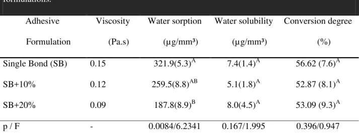
Viscosity, water sorption, water solubility and conversion degree results of each adhesive data are summarized in Table 1. The original SB formulation obtained the highest value for viscosity, followed, gradually, by formulations SB+10% and SB+20%. The higher is the level of ethanol in the adhesive the lower the water sorption. With regard to water sorption, statistical difference was found between the SB and SB+20% groups. However, no statistical difference was found for solubility between all groups. Considering the conversion degree of monomer, there was no statistical difference between adhesive formulations (p= 0.557).
Regarding the contact angle, all the formulations had similar behavior on all surfaces tested (Fig. 1A, Fig. 1B and Fig. 1C). The θ values complied with the amount of solvent formulations. The greater is the amount of solvent; the lower is the contact angle.
Figure 1D shows similar evaporation of volatile adhesive components induced by an air-stream, and Figure 1E depicts the degree of spontaneous evaporation of the volatile solvents of the adhesive formulations measured at room temperature for 60 min. In both situations, weights of all adhesives formulations gradually decreased and weight loss of SB+10% and SB+20% reached a plateau from 10-15 min. There was weight loss over the 60 min of a directed air-stream for all groups in both test situations.
23
DISCUSSION
The addition of ethanol changed the viscosity of the adhesive (Table 1), which is in agreement with a previous study [19], where this solvent successfully decreased this property. The reduction in viscosity was also evidenced by the reduction of the contact angle [29], which demonstrated an enhanced wetting ability of the diluted adhesive [30] regardless the tested surfaces of glass, teflon or dentin, as shown at Figure 1. Thus, the first hypothesis was accepted.
The extra ethanol concentration decreased the water sorption, which may be beneficial to the bond strength, since bonding can be less susceptible to deleterious action of water [31]. In addition, the results of solubility seem to confirm this trend, because enhancing ethanol concentrations; solubility was not increased, suggesting some stability of the formulations. Our results did not agree with an early study [32], which found that increasing the amount of ethanol, the sorption and solubility of water raised. However, in the latter study, solutions of dimethacrylate resins were used, not corresponding to adhesive systems as currently. The decrease in viscosity of SB did not compromise adhesive polymerization since similar degree of conversion was found among groups (Table 1), which is in accordance with Ye et al.[19]. Considering the performed tests, the second hypothesis was partially accepted, since the changes in viscosity did not cause any deleterious effect on the properties of the adhesives. This way, it can be suggested that the bond strength results may be attributed solely to different viscosities but not to the other tested properties.
however it could have influenced the bonding results. It is important to remember that the addition of solvent should be set at a threshold level of 50%, which was respected in the present investigation, inasmuch as the original content of ethanol of SB is 30% [21-23]. The evaporation rate test (Figure 1) confirmed these reports.
Neither dentin treatments nor adhesive viscosities affected bond strength results. In fact, it has been reported that there was no difference on bond strength of SB to Er:YAG laser-treated, using the same parameters or bur-treated dentin [28]. Using other laser parameters and adhesives, these results are also in accordance with other studies [33-37]. However, the results of this study are not in accordance with previous data [12, 38-39] that showed lower values for laser irradiated groups. It is possible that dental substrates were more modified by the different laser parameters used [40]. In the present study, all groups showed more mixed failures (98.2%) than the other types of failure. These results are in disagreement with others [12, 28, 34] that reported bur-prepared dentin had more mixed failures, but the laser-irradiated had more adhesive failures. This fact may be explained because those studies used shear bond test to obtain the strength bond values while, in this present study, the microtensile test was used. Cardoso et al. [41] used microtensile test and found that mixed failures were frequently observed in laser-irradiated dentin and it was ascribed to the prominent irregularities of laser-irradiated dentin.
25
CONCLUSION
REFERENCES
1. Staehle HJ. Minimally invasive restorative treatment. The journal of adhesive dentistry. 1999 Autumn;1(3):267-84.
2. Tyas MJ, Anusavice KJ, Frencken JE, Mount GJ. Minimal intervention dentistry--a review. FDI Commission Project 1-97. International dental journal. 2000 Feb;50(1):1-12. 3. Maiman TH. Stimulated optical radiation in ruby. Nature. 1960;187:493-4.
4. Stern RH, Sognnaes, RF. Laser beam effects on dental hard tissues. Journal of dental research. 1964;43:873.
5. Birardi V, Bossi L, Dinoi C. Use of the Nd:YAG laser in the treatment of Early Childhood Caries. European journal of paediatric dentistry : official journal of European Academy of Paediatric Dentistry. 2004 Jun;5(2):98-101.
6. Li ZZ, Code JE, Van De Merwe WP. Er:YAG laser ablation of enamel and dentin of human teeth: determination of ablation rates at various fluences and pulse repetition rates. Lasers in surgery and medicine. 1992;12(6):625-30.
7. Keller U, Hibst R. Effects of Er:YAG laser in caries treatment: a clinical pilot study. Lasers in surgery and medicine. 1997;20(1):32-8.
8. Hibst R, Keller U. Experimental studies of the application of the Er:YAG laser on dental hard substances: I. Measurement of the ablation rate. Lasers in surgery and medicine. 1989;9(4):338-44.
9. Keller, U.; Hibst, R. Ultrastructural changes of enamel and dentin following Er:YAG laser radiation on teeth. Proceedings of SPIE, v.1200, p.408-415, 1990.
27
11. Martinez-Insua A, Da Silva Dominguez L, Rivera FG, Santana-Penin UA. Differences in bonding to acid-etched or Er:YAG-laser-treated enamel and dentin surfaces. The Journal of prosthetic dentistry. 2000 Sep;84(3):280-8.
12. Ceballos L, Toledano M, Osorio R, Tay FR, Marshall GW. Bonding to Er-YAG-laser-treated dentin. Journal of dental research. 2002 Feb;81(2):119-22.
13. de Souza AE, Corona SA, Dibb RG, Borsatto MC, Pecora JD. Influence of Er:YAG laser on tensile bond strength of a self-etching system and a flowable resin in different dentin depths. Journal of dentistry. 2004 May;32(4):269-75.
14. Donadio-Moura J, Gouw-Soares S, de Freitas PM, Navarro RS, Powell LG, Eduardo Cde P. Tensile bond strength of a flowable composite resin to ER:YAG-laser-treated dentin. Lasers in surgery and medicine. 2005 Jun;36(5):351-5.
15. Goncalves M, Corona SA, Borsatto MC, Silva PC, Pecora JD. Tensile bond strength of dentin-resinous system interfaces conditioned with Er:YAG laser irradiation. Journal of clinical laser medicine & surgery. 2002 Apr;20(2):89-93.
16. Kameyama A, Aizawa K, Kato J, Hirai Y. Tensile bond strength of single-step self-etch adhesives to Er:YAG laser-irradiated dentin. Photomedicine and laser surgery. 2009 Feb;27(1):3-10.
17. Silikas N, Watts DC. Rheology of urethane dimethacrylate and diluent formulations. Dental materials : official publication of the Academy of Dental Materials. 1999 Jul;15(4):257-61.
18. Craig RG, Powers JM. Restorative Dental Materials. 11.ed. St. Louis: Mosby ,2002. 261-262p.
20. Bae JH, Cho BH, Kim JS, Kim MS, Lee IB, Son HH, et al. Adhesive layer properties as a determinant of dentin bond strength. Journal of biomedical materials research Part B, Applied biomaterials. 2005 Aug;74(2):822-8.
21. Reis AF, Oliveira MT, Giannini M, De Goes MF, Rueggeberg FA. The effect of organic solvents on one-bottle adhesives' bond strength to enamel and dentin. Operative dentistry. 2003 Nov-Dec;28(6):700-6
22. Hashimoto M, Tay FR, Ito S, Sano H, Kaga M, Pashley DH. Permeability of adhesive resin films. Journal of biomedical materials research Part B, Applied biomaterials. 2005 Aug;74(2):699-705.
23. Garcia G, Fernandes KB, Garcia FC, D'Alpino PH, da Rocha Svizero N, Wang L. Solvent retention of contemporary commercial dentin bonding agents in a demineralized dentin matrix. European journal of dentistry. 2010 Jul;4(3):293-7.
24. Faria ESAL, Piva E, Moraes RR. Time-dependent effect of refrigeration on viscosity and conversion kinetics of dental adhesive resins. European journal of dentistry. 2010 Apr;4(2):150-5.
25. Benetti P, Della Bona A, Kelly JR. Evaluation of thermal compatibility between core and veneer dental ceramics using shear bond strength test and contact angle measurement. Dental materials : official publication of the Academy of Dental Materials. 2010 Aug;26(8):743-50.
26. Ito S, Hoshino T, Iijima M, Tsukamoto N, Pashley DH, Saito T. Water sorption/solubility of self-etching dentin bonding agents. Dental materials : official publication of the Academy of Dental Materials. 2010 Jul;26(7):617-26.
29
comonomers in model dental adhesives. Dental materials : official publication of the Academy of Dental Materials. 2009 Oct;25(10):1275-84.
32. Celik EU, Ergucu Z, Turkun LS, Turkun M. Shear bond strength of different adhesives to Er:YAG laser-prepared dentin. The journal of adhesive dentistry. 2006 Oct;8(5):319-25.
29. Eick JD, Johnson LN, Fromer JR, Good RJ, Neumann AW. Surface topography: its influence on wetting and adhesion in a dental adhesive system. Journal of dental research. 1972 May-Jun;51(3):780-8.
30. Mittal KL. Polymer Surface Modification: Relevance to Adhesion. vol. 2. VSP, The Netherlands; 2000. P 640
31. Malacarne J, Carvalho RM, de Goes MF, Svizero N, Pashley DH, Tay FR, et al. Water sorption/solubility of dental adhesive resins. Dental materials : official publication of the Academy of Dental Materials. 2006 Oct;22(10):973-80.
32. Sideridou ID, Karabela MM. Sorption of water, ethanol or ethanol/water solutions by light-cured dental dimethacrylate resins. Dental materials : official publication of the Academy of Dental Materials. 2011 Oct;27(10):1003-10.
33. Koliniotou-Koumpia E, Kouros P, Zafiriadis L, Koumpia E, Dionysopoulos P, Karagiannis V. Bonding of adhesives to Er:YAG laser-treated dentin. European journal of dentistry. 2012 Jan;6(1):16-23.
34. Koliniotou-Koumpia E, Kouros P, Dionysopoulos D, Zafiriadis L. Bonding strength of silorane-based composite to Er-YAG laser prepared dentin. Lasers in medical science. 2013 May 26.
36. Ramos TM, Ramos-Oliveira TM, Moretto SG, de Freitas PM, Esteves-Oliveira M, de Paula Eduardo C. Microtensile bond strength analysis of adhesive systems to Er:YAG and Er,Cr:YSGG laser-treated dentin. Lasers in medical science. 2013 Jan 26.
37. Oliveira DC, Manhaes LA, Marques MM, Matos AB. Microtensile bond strength analysis of different adhesive systems and dentin prepared with high-speed and Er:YAG laser: a comparative study. Photomedicine and laser surgery. 2005 Apr;23(2):219-24.
38. De Munck J, Van Meerbeek B, Yudhira R, Lambrechts P, Vanherle G. Micro-tensile bond strength of two adhesives to Erbium:YAG-lased vs. bur-cut enamel and dentin. European journal of oral sciences. 2002 Aug;110(4):322-9.
39. Dunn WJ, Davis JT, Bush AC. Shear bond strength and SEM evaluation of composite bonded to Er:YAG laser-prepared dentin and enamel. Dental materials : official publication of the Academy of Dental Materials. 2005 Jul;21(7):616-24.
40. de Oliveira MT, Arrais CA, Aranha AC, de Paula Eduardo C, Miyake K, Rueggeberg FA, et al. Micromorphology of resin-dentin interfaces using one-bottle etch&rinse and self-etching adhesive systems on laser-treated dentin surfaces: a confocal laser scanning microscope analysis. Lasers in surgery and medicine. 2010 Sep;42(7):662-70.
41. Cardoso MV, Coutinho E, Ermis RB, Poitevin A, Van Landuyt K, De Munck J, et al. Influence of dentin cavity surface finishing on micro-tensile bond strength of adhesives. Dental materials: official publication of the Academy of Dental Materials. 2008 Apr;24(4):492-501.
31
Table 1 – Viscosity, water sorption, water solubility and conversion degree of the adhesives
formulations.
Adhesive Formulation
Viscosity (Pa.s)
Water sorption (µg/mm³)
Water solubility (µg/mm³)
Conversion degree (%) Single Bond (SB) 0.15 321.9(5.3)A 7.4(1.4)A 56.62 (7.6)A
SB+10% 0.12 259.5(8.8)AB 5.1(1.8)A 52.87 (8.1)A
SB+20% 0.09 187.8(8.9)B 8.0(4.5)A 53.09 (9.3)A
p / F - 0.0084/6.2341 0.167/1.995 0.396/0.947
Table 2 – Micro-tensile bond strength to dentin (MPa).
Adhesive Formulation Diamond Bur Laser Er:YAG
Single Bond (SB) 24.8 (7.6) A 27.9 (6.5) A
7/42 4/37
SB+10% 25.6 (8.0) A 26.9 (6.5) A
4/35 5/33
SB+20% 25.5 (8.7) A 25.1 (7.7) A
3/33 4/47
33
4 CONCLUSÃO GERAL
35
REFERÊNCIAS
BIRARDI, V.; BOSSI, L.; DINOI, C. Use of the Nd:YAG laser in the treatment of Early Childhood Caries. Eur. J. Paediatr. Dent., v. 5, n. 2, p. 98-101, June 2004.
BAE, J. H.; CHO B. H.; KIM J. S.; KIM M. S.; LEE I. B.; SON H. H.; UM C. M.; KIM C. K.; KIM O. Y. Adhesive layer properties as a determinant of dentin bond strength. J. Biomed. Mater. Res. B Appl. Biomater., v. 74, n. 2, p. 822-828, Aug. 2005.
CEBALLOS, L.; TOLEDANO M.; OSORIO R.; TAY F. R.; MARSHALL G. W. Bonding to Er-YAG-laser-treated dentin. J. Dent. Res., v. 81, n. 2, p. 119-122, Feb. 2002.
CELIK, E. U.; ERGÜCÜ Z.; TÜRKÜN L. S.; TÜRKÜN M. Shear bond strength of different adhesives to Er:YAG laser-prepared dentin. J. Adhes. Dent., v. 8, n. 5, p. 319-325, Oct. 2006. CRAIG, R. G.; POWERS, J. M. Restorative Dental Materials. 11st ed. St. louis: Mosby, 2002.
DE MOOR, R. J.; DELME, K. I. Laser-assisted cavity preparation and adhesion to erbium-lased tooth structure: part 2. present-day adhesion to erbium-erbium-lased tooth structure in
permanent teeth. J. Adhes. Dent., v. 12, n. 2, p. 91-102, Apr. 2010.
FERREIRA, L. S.; APEL C.; FRANCCI C.; SIMOES A.; EDUARDO C. P.; GUTKNECHT N. Influence of etching time on bond strength in dentin irradiated with erbium lasers. Lasers Med. Sci., v. 25, n. 6, p. 849-854, Nov. 2010.
HIBST, R.; KELLER, U. Experimental studies of the application of the Er:YAG laser on dental hard substances: I. Measurement of the ablation rate. Lasers Surg. Med., v. 9, n. 4, p. 338-344, 1989.
KAMEYAMA, A.; AIZAWA K.; KATO J.; HIRAI Y. Tensile bond strength of single-step self-etch adhesives to Er:YAG laser-irradiated dentin. Photomed Laser Surg., v. 27, n. 1, p. 3-10, Feb. 2009.
KELLER, U.; HIBST, R. Effects of Er:YAG laser in caries treatment: a clinical pilot study. Lasers Surg. Med., v. 20, n. 1, p. 32-38, 1997.
KELLER, U.; HIBST, R. Ultrastructural changes of enamel and dentin following Er:YAG laser radiation on teeth. Proc. SPIE, v.1200, p.408-415, 1990.
LEONETTI EDOS, S.; RODRIGUES J. A.; REIS A. F.; NAVARRO R. S.; ARANHA A. C.; CASSONI A. Microtensile bond strength of resin composite to dentin treated with Er:YAG laser of bleached teeth. Lasers Med. Sci., v. 27, n. 1, p. 31-38, Jan. 2012.
LI, Z. Z.; CODE, J. E.; VAN DE MERWE, W. P. Er:YAG laser ablation of enamel and dentin of human teeth: determination of ablation rates at various fluences and pulse repetition rates. Lasers Surg. Med., v. 12, n. 6, p. 625-630, 1992.
MARTINEZ-INSUA, A.; DA SILVA DOMINGUEZ L.; RIVERA F. G.; SANTANA-PENÍN U. A. Differences in bonding to acid-etched or Er:YAG-laser-treated enamel and dentin surfaces. J. Prosthet. Dent., v. 84, n. 3, p. 280-288, Sept. 2000.
NALBANTGIL, D.; OZTOPRAK M. O.; TOZLU M.; ARUN T. Effects of different
application durations of ER:YAG laser on intrapulpal temperature change during debonding. Lasers Med. Sci., v. 26, n. 6, p. 735-740, Nov. 2011.
PERITO, M. A.; JORGE A. C.; DE FREITAS P. M.; CASSONI A.; RODRIGUES J. A. Cavity preparation and influence of restorative materials on the prevention of secondary caries. Photomed. Laser Surg., v. 27, n. 5, p. 729-734, Oct. 2009.
SCATENA, C.; TORRES C. P.; GOMES-SILVA J. M.; CONTENTE M. M.; PÉCORA J. D.; PALMA-DIBB R. G.; BORSATTO M. C. Shear strength of the bond to primary dentin: influence of Er:YAG laser irradiation distance. Lasers Med. Sci., v. 26, n. 3, p. 293-297, May 2011.
SILIKAS, N.; WATTS, D. C. Rheology of urethane dimethacrylate and diluent formulations. Dent. Mater., v. 15, n. 4, p. 257-261, July 1999.
STAEHLE, H.J. Minimally invasive restorative treatment. J. Adhes. Dent., v. 1, n. 3, p. 267-84, 1999.
STERN, R. H.; SOGNNAES, R. F. Laser beam effects on dental hard tissues. J. Dent. Res., v. 43, p. 873, 1964.
TARCIN, B.; GÜNDAY M.; OVEÇOĞLU H. S.; TÜRKMEN C.;OVEÇOĞLU M. L.; OKSÜZ M.; AY M. Tensile bond strength of dentin adhesives on acid- and laser-etched dentin surfaces. Quintessence Int., v. 40, n. 10, p. 865-874, Nov./Dec. 2009.
TYAS, M. J.; ANUSAVICE K. J.; FRENCKEN J. E.; MOUNT G. J. Minimal intervention dentistry--a review. FDI Commission Project 1-97. Int. Dent. J., v. 50, n. 1, p. 1-12, Feb. 2000.
VISURI, S. R.; GILBERT J. L.; WRIGHT D. D.; WIGDOR H. A.; WALSH J. T. Shear strength of composite bonded to Er:YAG laser-prepared dentin. J. Dent. Res., v. 75, n. 1, p. 599-605, Jan. 1996.
YE, Q.; SPENCER P.; WANG Y.; MISRA A. Relationship of solvent to the
37
41
ANEXO C – NORMAS DO PERÍODICO
Guide for Authors
Authors are requested to submit their original manuscript and figures via the online submission and editorial system for Dental Materials. Using this online system, authors may submit manuscripts and track their progress through the system to publication. Reviewers can download manuscripts and submit their opinions to the editor. Editors can manage the whole submission/review/revise/publish process. Please register at: http://ees.elsevier.com/dema. Dental Materials now only accepts online submissions.
The Artwork Quality Control Tool is now available to users of the online submission system. To help authors submit high-quality artwork early in the process, this tool checks the submitted artwork and other file types against the artwork requirements outlined in the Artwork Instructions to Authors on www.elsevier.com/artworkinstructions. The Artwork Quality Control Tool automatically checks all artwork files when they are first uploaded. Each figure/file is checked only once, so further along in the process only new uploaded files will be checked.
Manuscripts
The journal is principally for publication of Original Research Reports, which should preferably investigate a defined hypothesis. Maximum length 6 journal pages (approximately 20 double-spaced typescript pages) including illustrations and tables.
Systematic Reviews will however be considered. Intending authors should communicate with the Editor beforehand, by email, outlining the proposed scope of the review. Maximum length 10 journal pages (approximately 33 double-spaced typescript pages) including figures and tables.
Three copies of the manuscript should be submitted: each accompanied by a set of illustrations. The requirements for submission are in accordance with the "Uniform Requirements for Manuscripts Submitted to Biomedical Journals", Annals of Internal Medicine, 1997,126, 36-47. All manuscripts must be written in American English. Authors are urged to write as concisely as possible.
The Editor and Publisher reserve the right to make minimal literary corrections for the sake of clarity. Authors for whom English is not the first language should have their manuscripts read by colleagues fluent in English. If extensive English corrections are needed, authors may be charged for the cost of editing. For additional reference, consult issues of Dental Materials published after January 1999 or the Council of Biology Editors Style Manual (1995 ed.). All manuscripts should be accompanied by a letter of transmittal, signed by each author, and stating that the manuscript is not concurrently under consideration for publication in another journal, that all of the named authors were involved in the work leading to the publication of the paper, and that all the named authors have read the paper before it is submitted for publication.
Always keep a backup copy of the electronic file for reference and safety.
General
• number all pages consecutively.
• type double-spaced on A4 or 8.5 x 11-inch bond paper, with margins of 30 mm. • double-space references.
• indent or space paragraphs.
• arrange article in the following order: Title, Abstract, Introduction, Materials and Methods, Results, Discussion, Conclusion, Acknowledgements, References, Tables, Figures, Captions. • start each section on a separate page.
Title page
• Title (capitalize the first letter of the first word) e.g. Comparison of the color stability of ten new composites.
• Authors (first name, middle initial, surname) e.g. Kenneth J. Anusavice 1, Victoria Marker 2 • Authors' addresses (abbreviated) e.g.
1 Department of Biomaterials, University of Florida, Gainesville, Florida, USA
2 Department of Biomaterials Science, Baylor College of Dentistry, Dallas, Texas, USA • Short Title (45 characters) e.g Color stability of composites
• Corresponding Author details (essential)μ Name, complete address, phone, fax, and E-mail numbers
Abstract (structured format) • 250 words or less.
• subheadings should appear in the text of the abstract as followsμ Objectives, Methods, Results, Significance. (For Systematic Reviews: Objectives, Data, Sources, Study selection, Conclusions). The Results section may incorporate small tabulations of data, normally 3 rows maximum.
Keywords
Up to 10 keywords should be supplied e.g. dental material, composite resin, adhesion. Introduction
This must be presented in a structured format, covering the following subjects, although actual subheadings should not be included:
• succinct statements of the issue in question;
• the essence of existing knowledge and understanding pertinent to the issue (reference); • the aims and objectives of the research being reported relating the research to dentistry, where not obvious.
Materials and methods
• describe the procedures and analytical techniques. • only cite references to published methods.
• include at least general composition details and batch numbers for all materials. • identify names and sources of all commercial products e.g.
"The composite (Silar, 3M Co., St. Paul, MN, USA)..."
"... an Au-Pd alloy (Estheticor Opal, Cendres et Metaux, Switzerland)." • specify statistical significance test methods.
Results
43
• make no reference to previous literature. • report statistical findings.
Discussion
• explain and interpret data.
• state implications of the results, relate to composition. • indicate limitations of findings.
• relate to other relevant research. • suggest directions for future research.
Conclusion (if included)
• must NOT repeat Results or Discussion
• must concisely state inference, significance, or consequences
Acknowledgements As appropriate, e.g.:
"Based on a thesis submitted to the graduate faculty, University of Virginia, in partial fulfilment of the requirements for the M.S. degree."
"This investigation was supported in part by Research Grant DE 00000 from the National Institute of Dental Research, Bethesda, MD 20892."
References - must now be given according to the following numeric system:
Cite references in text in numerical order. Use square brackets: in-line, not superscript e.g. [23]. All references must be listed at the end of the paper, double-spaced, without indents. For example:
1. Moulin P, Picard B and Degrange M. Water resistance of resin-bonded joints with time related to alloy surface treatments. J Dent, 1999; 27:79-87.
2. Taylor DF, Bayne SC, Sturdevant JR and Wilder AD. Comparison of direct and indirect methods for analyzing wear of posterior composite restorations. Dent Mater, 1989; 5:157-160. Avoid referencing abstracts if possible. If unavoidable, reference as follows:
3. Demarest VA and Greener EH . Storage moduli and interaction parameters of experimental dental composites. J Dent Res, 1996; 67:221, Abstr. No. 868.
Tables and figures
All tables and figures must be thoroughly discussed in the text of the manuscript. Tables
• one table to a page, each with a title.
• number tables in order of mention using Arabic numerals. • must be able to "stand alone" apart from text.
• when appropriate, standard deviations of values should be indicated in parentheses; (do NOT use ± notation).
• results of statistical analysis must be included, use superscript letters to indicate significant differences.
• for explanatory footnotes, use symbols (*, #,**,##). Figures
• only black and white photographs for print publication.
• omit titles and other information contained in the figure caption. • maximum of 6 figures per manuscript.
• figures grouped together should have similar dimensions and be labelled "a, b, c", etc. • place magnification markers directly on the micrographs.
• authors should consider that the majority of figures will be reduced to the width of a single column (approximately 85 mm). Preferably figures should exactly match, or be no more than 1.5 times that width.
• authors can indicate if they feel a figure should be full page width.
Dental Materials has been selected for inclusion in a new 'colourful e-products' workflow. Figures that appear in black and white in the printed version of the journal can be IN COLOUR, online, in ScienceDirect. Authors wishing to make use of this facility should ensure that 1. the artwork is in an acceptable format (TIFF, EPS or MS Office files) and at the correct resolution 2. RGB colourspace is used and 3. for colour online and black and white in print, both colour and black and white artwork (file and/or hardcopy) is provided. There will be no charges to the authors for colour figures online.
Graphs
• unique, concise axis labels; do not repeat the Figure caption. • uniform size for graphs of similar type.
• type size that will be easily read when the graph is reduced to one column width. • lines that are thick and solid (100% black).
Captions to tables and figures • list together on a separate page.
• should be complete and understandable apart from the text. • include key for symbols or abbreviations used in Figures.
• individual teeth should be identified using the FDI two-digit system. General Notes on Text
Abbreviations and acronyms: terms and names to be referred to in the form of abbreviations or acronyms must be given in full when first mentioned.
Correct Usage
• use S.I. units (International System of Units). If non-SI units must be quoted, the SI equivalent must immediately follow in parentheses.
• use correct symbols for ?, L (as in ?m, mL, etc.) • put leading zeros in all numbers less than 1.0
• write out number of ten or fewer (ten rats) except when indicating inanimate quantities (10 mL)
• always use digits for dates, dimensions, degrees, doses, time, percentages, ratios, statistical results, measurements, culture cells, and teeth.
• the complete names of individual teeth must be given in the text.
General Policy
• receipt of manuscripts will be acknowledged.
• after initial review, authors will be notified of status.
45
• a list of revisions and responses to reviewers' critiques must accompany resubmitted revised manuscripts.
On Submission: Agreement, by the act of ticking a box, to the statement, "This paper has been compiled with the knowledge, input and approval of all the named authors."
On acceptance, authors will be required to sign a transfer of copyright agreement. If figures, tables, or other excerpts, are included from copyrighted works the author is responsible for obtaining written permission from the copyright holder prior to submitting the final version of the paper. Full credit must be given to such sources.
Offprints and page charges: no page charges are levied on articles published in Dental Materials. Each corresponding author receives 25 offprints of their article free of charge after it has been published; they will also have the opportunity to order additional copies.
Submission Package Checklist:
• letter of transmittal signed by all authors. • One electronic copy of the manuscript.
• One electronic copy of each image and table, all labelled.