J. Evid. Based Med. Healthc., pISSN- 2349-2562, eISSN- 2349-2570/ Vol. 3/Issue 54/July 07, 2016 Page 2756
PATTERN OF GLOMERULAR DISEASES IN PATIENTS WITH SIGNIFICANT PROTEINURIA: A
CLINICOPATHOLOGICAL STUDY FROM UPPER ASSAM
Sawjib Borphukan1, Surajit Gogoi2, Dev Baruah3
1Assistant Professor, Department of Nephrology, Assam Medical College & Hospital, Dibrugarh, Assam.
2Registrar, Department of Nephrology, Assam Medical College & Hospital, Dibrugarh, Assam.
3Assistant Professor, Department of Radiology, Assam Medical College & Hospital, Dibrugarh, Assam.
ABSTRACT
BACKGROUND
The prevalence of biopsy proven glomerular diseases varies according to the geographic area, race, age, demography and indication of renal biopsy. This has been poorly studied in the North-Eastern part of India, especially from Assam, the largest state, population-wise.
METHODS
This is a retrospective and observational study of kidney biopsy records and relevant clinical data of mainly adult patients.
Patients (≥ 16 years old) presenting with significant proteinuria (> 2 g/24 hours) who attended our Medical College from October 2012 to September 2015 were subjected to kidney biopsy provided they were able to afford the cost and willing for the same. All biopsies were subjected to light and immunofluorescence microscopy. The histopathological pattern was analysed according to various clinical parameters.
RESULTS
A total of 136 kidney biopsies were included for analysis. 72 cases (52.9%) were males and 64 (47.1%) were females. Mean age of the patients was 37 ± 15.7 years. Among the patients, 85.3% (n = 116) were diagnosed with primary glomerular disease (PGD) and 14.7% (n = 20) were diagnosed with secondary glomerular disease (SGD). The most common histopathological lesion was minimal change disease (MCD) (27.9%) followed by membranous glomerulonephritis or
nephropathy (MGN) (24.3%). In the age group ≥ 40 years, MGN (34.5%) was the predominant histological lesion followed by MCD (20.7%). Lupus nephritis (LN) (11%) was the most common secondary glomerular pathology. 20 of our patients (14.70%) had creatinine levels more than 1.5 mg/dL.
CONCLUSION
In this study, MCD was the commonest lesion in our north-east adult population in a wide age range. However, MGN was predominant in the middle age and elderly patients. This is in contrast to the trend in the increasing incidence of FSGS found in other parts of the country and western population.
KEYWORDS
Glomerular Disease, Significant Proteinuria.
HOW TO CITE THIS ARTICLE: Borphukan S, Gogoi S, Baruah D. Pattern of glomerular diseases in patients with significant proteinuria: A clinicopathological study from upper Assam. J. Evid. Based Med. Healthc. 2016; 3(54), 2756-2761.
DOI:10.18410/jebmh/2016/603
INTRODUCTION: Patients with significant proteinuria are always likely to have glomerular disease which may or may not be associated with interstitial or vascular disease. Glomerular diseases (GD) or glomerulopathies can be broadly categorised as primary or secondary and can have varied clinicopathological presentations.[1] A Kidney biopsy is
always required for correct characterisation of the glomerulopathy which is very essential for treatment and prognosis.
Biopsy registries can give an idea of the regional variations in the spectrum of GD as well as the trend over time. There has been certain variations in the renal pathological lesions within the various parts of our country. In published studies from North India,[2-4] FSGS (Focal
segmental glomerulosclerosis) was the most common in 2 studies and MCD (Minimal Change Disease) was common in 1 study. Whereas data from the South[5,6] have shown
variation within, as mesangioproliferative glomerulonephritis was the predominant lesion in 1 study and MCD was predominant in the other study. Very limited data from the Eastern part of India[7] has shown the predominant lesion as
FSGS. Published data from Pakistan has shown FSGS[8] as
the predominant lesion and in 1 study from Bangladesh,[9]
Mesangioproliferative GN was the commonest renal lesion. IgA nephropathy (IgAN) is the common primary GD in studies from East Asia,[10-12] as well as in white Europeans Financial or Other, Competing Interest: None.
Submission 25-06-2016, Peer Review 29-06-2016, Acceptance 04-07-2016, Published 07-07-2016. Corresponding Author:
Dr. Sawjib Borphukan,
House No. 843, G. S. Modi Road, Jyotinagar, P. O. Jalan Nagar-786005, Dibrugarh, Assam.
J. Evid. Based Med. Healthc., pISSN- 2349-2562, eISSN- 2349-2570/ Vol. 3/Issue 54/July 07, 2016 Page 2757
and Americans.[13–17] In contrast, FSGS is the most common
GD among African-Americans, South Americans, and in the Middle East.[18-20]
In view of paucity of data from the North-East part of India, we studied the pattern of type of glomerular disease using clinicopathological correlation in predominantly adult patients and few adolescents.
MATERIALS AND METHODS: All kidney biopsy reports of adult and few adolescent patients having significant proteinuria (>2 g in 24 hrs.) who attended our Medical College from 2012 to 2015 were retrospectively analysed. The clinical records of these patients were reviewed with respect to age, gender, clinical presentation, urine routine & microscopy, 24-hour urine protein estimation, biochemistry, imaging, immunological investigations, viral serology and other relevant investigations as necessary for secondary aetiologies. Patients were not diabetic and were 16 yrs. and older.
All biopsies were performed under real-time USG guidance using the Bard® Max-Core® Disposable Core
Biopsy Instrument (Bard Biopsy Systems, USA). A Radiologist from the Radiology Dept. of our Medical College & Hospital assisted us with the real-time ultrasound. A 16 G
× 16 cm size instrument was used for adults ≥ 18 years old,
and a smaller 18 G × 16 cm instrument was used for those < 18 years of age. At least two cores were obtained and samples sent for light microscopy (LM) and immunofluorescence (IF) microscopy in all cases. LM was carried out using H and E, periodic acid-Schiff, Jones silver, and Trichrome stains. Additional special stains were used whenever indicated. IF staining was performed on 3-μm cryostat sections using polyclonal fluorescein isothiocyanate-conjugated (FITC) antibodies to IgG, IgM, IgA, C3, C1q, and kappa and lambda light chains (DakoCytomation, Denmark). The intensity of IF staining was graded on a scale of 0 to 3+. Since we do not have a renal pathologist at our Medical College, all kidney biopsy specimens were sent to SRL Diagnostics, Mumbai for analysis.
Glomerular pathologies were classified into the following: (a) Primary glomerular diseases (PGD); major ones being MCD (Minimal change disease), MGN (Membranous glomerulonephritis or nephropathy), FSGS, IgAN (IgA Nephropathy), MPGN (Membranoproliferative glomerulonephritis), APGN (Acute Proliferative glomerulonephritis), Mes. PGN (Mesangioproliferative
glomerulonephritis), Cresc. GN (crescentic
glomerulonephritis), (b) Secondary glomerular diseases (SGD) which included lupus nephritis (LN), amyloidosis, benign nephrosclerosis (BN).
For statistical analysis, Pearson's chi-square test was used to see if there is any association between categorical variables. Statistical analysis was performed with IBM SPSS Statistics version 21 Software. A p-value of less than or equal to 0.05 was considered as significant.
RESULTS: A total of 136 kidney biopsies were recorded over the period from 2012 to 2015. LM (Light Microscopy for
histopathology) and IF (Immunofluorescence) were performed in all biopsies and the average glomerular yield was adequate. EM (Electron Microscopy) was not done in any of the patients. Mean age of the patients was found to be 37 ± 15.7 years (Table 1; Fig. 1). Age of the patients ranged from 16 years and above, the oldest being at 79 years. It was observed that there was an overall male preponderance, 72 cases (52.9%) were males and females were 64 (47.1%). (Fig. 2). Male to female ratio is 1.13:1 (Table 2). Among the patients, 85.3% (n = 116) were diagnosed with primary glomerular disease (PGD) and 14.7% (n = 20) were diagnosed with secondary glomerular disease (SGD) (Table 3). The major pathological diagnosis was MCD (27.9%) followed by MGN (24.3%). FSGS and MPGN were diagnosed at 8.8% each. Mes. PGN at 6.6%, APGN at 3.7%. Cresc. GN at 2.9%, IgAN at 2.2%. LN was found in 11.0% BN 2.2% and AMYLOID were diagnosed in 1.5% of biopsies. (Table 3; Fig. 3)
For looking at the association between age and pathological diagnosis, the age of patients was categorized into 2 groups, one being <40 years and the other being ≥ 40 years. After statistical analysis the 2 value was 23.11 and
p =0.010 which is <0.05 which showed that there is a statistically significant association between age and diagnosis. (Table 4). MCD (33.3%), LN (17.9%), MPGN (11.5%), APGN (3.8%), IgAN (2.6%) were highly prevalent in the age group <40 years as compared to ≥40 years. For
the age group ≥40 years the prevalence was as; MCD
(20.7%), LN (1.7%), MPGN (5.2%), APGN (3.4%) and IgAN
(1.7%). In the age group ≥40 years, MGN (34.5%) was the
predominant lesion, followed by Mes. PGN (12.1%), FSGS (10.3%), Cresc.GN (5.2%), BN (3.4%), Amyloid (1.7%) which were high as compared to the age group <40 years. In the age group of <40 years, the prevalence was; MGN (16.7%), Mes. PGN (2.6%), FSGS (7.7%), Cresc. GN (1.3%), BN (1.3%), Amyloid (1.3%) (Fig. 4).
On analysing the association between sex and diagnosis the 2 value was 27.78 and p =0.002 which is <0.05 so we
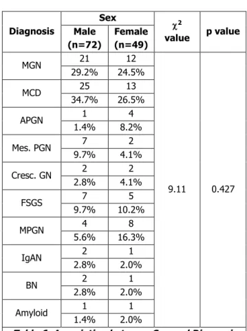
can conclude that there is a statistical significant association between sex and diagnosis (Table 5). LN was diagnosed in 15 (23.4%) females. MCD (34.7%), MGN (29.2%), Mes. PGN (9.7%), FSGS (9.7%), IgAN (2.8%), BN (2.8%) were diagnosed more in males as compared to females. For females these values are as MCD (20.3%), MGN (18.8%), Mes. PGN (3.1%), FSGS (7.8%), IgAN (1.6%), BN (1.6%). Like this MPGN (12.5%), Cresc. GN (3.1%), APGN (6.3%), Amyloid (1.6%) were more frequent in females than males. In males, MPGN was diagnosed as (5.6%), Cresc. GN (2.8%), APGN (1.4%) and Amyloid as (1.4%). (Fig. 5). As per the literature SLE (Systemic Lupus Erythematosus) has been commonly found in females so we did an analysis of the biopsy diagnoses excluding patients with lupus nephritis (LN) to see the association between sex and biopsy findings. Here, the 2 value was 9.11 and p =0.427 which is >0.05 so
J. Evid. Based Med. Healthc., pISSN- 2349-2562, eISSN- 2349-2570/ Vol. 3/Issue 54/July 07, 2016 Page 2758
since 2 value is 13.92 and p =0.125 which is >0.05. (Table
7). MCD (40.6%), MPGN (14.1%), APGN (4.7%), IgAN (3.1%), were more in the age group <40 years as compared
to the age group ≥40 years where MCD (21.1%), MPGN
(5.3%), APGN (3.5%), IgAN (1.8%), while in the age group
≥40 years; MGN (35.1%), FSGS (10.5%), Mes. PGN
(12.3%), Cres. GN (5.3%), BN (3.5%), AMYLOID (1.8%) were highly diagnosed as compared to the patients of the age group <40 years where the incidence was as: MGN (20.3%), FSGS (9.4%), Mes. PGN (3.1%), Cres. GN (1.6%), BN (1.6%), AMYLOID (1.6%).(Fig. 7).
20 of our patients (14.70%) had creatinine levels > 1.5mg/dl.
Patients
AGE (years) (MeanS.D.) 3715.7
Table 1: Mean Age of the Patients
Mean age of the patients was found to be 37±15.7 years.
Fig. 1: Age Distribution of Patients
From the pie diagram, it was observed that the
maximum number of patients were in the ≥40 years age
group (57.4%), followed by <40 years age group (42.6%).
Sex Number of cases
Percentage (%)
Ratio (Male: female)
Male 72 52.9
1.13: 1
Female 64 47.1
Total 136 100.0
Table 2: Sex Distribution of the Patients
It was observed that there was an overall male preponderance, 72 cases (52.9%) and female were 64 (47.1%). Male to female ratio is 1.13:1.
Fig. 2: Sex Distribution of the Patients
Diagnosis Number
of Cases Percentage (%)
MCD 38 27.9
MGN 33 24.3
LN 15 11.0
FSGS 12 8.8
MPGN 12 8.8
Mes. PGN 9 6.6
APGN 5 3.7
Cresc. GN 4 2.9
IgAN 3 2.2
BN 3 2.2
Amyloid 2 1.5
Total 136 100.0
Table 3: Diagnosis of the Patients
Fig. 3: Graphical presentation of Diagnosis of the Patients
Diagnosis
Age Group
(Age In Years) 2
value p value < 40
Years (n=78)
≥40
years (n=58)
LN 14 1
23.11 0.010
17.9% 1.7%
MGN 13 20
16.7% 34.5%
MCD 26 12
33.3% 20.7%
APGN 3 2
3.8% 3.4%
Mes. PGN 2 7
2.6% 12.1%
Cresc. GN 1 3
1.3% 5.2%
FSGS 6 6
7.7% 10.3%
MPGN 9 3
11.5% 5.2%
IgAN 2 1
2.6% 1.7%
B N 1 2
1.3% 3.4%
AMYLOID 1 1
1.3% 1.7%
J. Evid. Based Med. Healthc., pISSN- 2349-2562, eISSN- 2349-2570/ Vol. 3/Issue 54/July 07, 2016 Page 2759
Fig. 4: Age wise Distribution of Different Diagnosis types of the Patients
Diagnosis
Sex
2
value p value Male
(n=72)
Female (n=72)
LN 0 15
27.78 0.002
0.0% 23.4%
MGN 21 12
29.2% 18.8%
MCD 25 13
34.7% 20.3%
APGN 1 4
1.4% 6.3%
Mes. PGN 7 2
9.7% 3.1%
Cresc. GN 2 2
2.8% 3.1%
FSGS 7 5
9.7% 7.8%
MPGN 4 8
5.6% 12.5%
IgAN 2 1
2.8% 1.6%
BN 2 1
2.8% 1.6%
Amyloid 1 1
1.4% 1.6%
Table 5: Association between Sex and Diagnosis
Fig. 5:Sex wise Distribution of Different Types of Diagnosis of the Patients
Analysis Excluding LN:
Diagnosis
Sex
2
value p value Male
(n=72)
Female (n=49)
MGN 21 12
9.11 0.427
29.2% 24.5%
MCD 25 13
34.7% 26.5%
APGN 1 4
1.4% 8.2%
Mes. PGN 7 2
9.7% 4.1%
Cresc. GN 2 2
2.8% 4.1%
FSGS 7 5
9.7% 10.2%
MPGN 4 8
5.6% 16.3%
IgAN 2 1
2.8% 2.0%
BN 2 1
2.8% 2.0%
Amyloid 1 1
1.4% 2.0%
Table 6: Association between Sex and Diagnosis
Here, 2 value is 9.11 and p =0.427 which is >0.05, so
we can conclude that there is no statistical significant association seen between sex and diagnosis.
J. Evid. Based Med. Healthc., pISSN- 2349-2562, eISSN- 2349-2570/ Vol. 3/Issue 54/July 07, 2016 Page 2760 Diagnosis
Age group (age in years)
2
value p value < 40
years (n=64)
≥40
years (n=57)
MGN 13 20
13.92 0.125
20.3% 35.1%
MCD 26 12
40.6% 21.1%
APGN 3 2
4.7% 3.5%
Mes. PGN 2 7
3.1% 12.3%
Cresc. GN 1 3
1.6% 5.3%
FSGS 6 6
9.4% 10.5%
MPGN 9 3
14.1% 5.3%
IgAN 2 1
3.1% 1.8%
BN 1 2
1.6% 3.5%
Amyloid 1 1
1.6% 1.8%
Table 7: Association between Age and Diagnosis
Here, 2 value is 13.92 and p =0.125 which is >0.05, so
we can conclude that there is no statistical significant association seen between age and diagnosis.
Fig. 7: Age wise Distribution of Different Types of Diagnosis of the Patients
DISCUSSION: This study is an observational and retrospective single-Center experience in India which is restricted to the last 3 years only and is the latest data of GD from upper part of Assam, a North-Eastern state. We were unable to analyse the data for the period before this due to inadequate data and poor standardisation of the biopsy reporting. Another shortcoming of our study is the inability to perform EM in all cases which would have helped in better diagnosis. However, we feel that a relatively
accurate diagnosis could be achieved in a majority of cases. The analysis was done mainly on the type of pathological diagnosis with respect to frequency, age and gender. Regarding the other clinical parameters, all patients had
significant proteinuria of ≥ 2 g in 24 hours. The most common aetiology (pathological diagnosis) of GD in our study was MCD (27.9%) which correlated well with some studies such as Das U et al,[6] Reshi A R et al[3] where
diagnosis of MCD was 21.8% and 43.79% respectively. The study by Reshi A R et al also included children along with adults and adolescents which could account for the high incidence of MCD in Kashmir. However, this was in contrast to the large study by Rathi M et al [2], and a smaller study of 50 patients by Mundi I et al [4] from North India who found FSGS as the most common pathological lesion. Similarly, Golay V et al[7] from the Eastern part of our country
(West Bengal) also found FSGS as the predominant lesion. Narasimhan B et al[5] in their large series from CMCH, Vellore
found mesangioproliferative glomerulonephritis (Mes. PGN) (20.2%) to be the predominant pathological lesion whereas MCD was found in only 11.6% of their adult patients. Data from our neighbouring country Pakistan, Kazi J I et al[8] has
shown FSGS (39.87%) as the single most common pathological lesion, whereas a small study of 74 patients from Bangladesh by Huq N et al[9] has shown Mes. PGN
(36.48%) as the main morphological pattern.
In our study, MGN (24.3%) was the second commonest pathological lesion, but in the age group ≥ 40 years it was the predominant finding (34.5%). In contrast to the above-mentioned studies, incidence of FSGS was low (8.8%) in our study, even strikingly different from the study by Golay V et al [7] from Kolkata, West Bengal representing the Eastern part of India where FSGS (24.63%) was the predominant pathology. However, the incidence of MGN (22.44%) was similar with our study. The incidence of IgA Nephropathy was low at 2.2% in our study which is in contrast to the studies from Asia, Europe, America.
Lupus nephritis was found in 11% of the biopsies which was comparatively higher than the data from Eastern India[7]
and Kashmir.[3] It was the most common secondary
glomerular disease (75%) in our data, followed by Benign Nephrosclerosis (BN) (15%) and Amyloidosis (10%). This finding is quite similar to the data from the rest of the country[2,5,6] and all patients were females. The incidence of
amyloidosis (1.5%) was similar to the study from Kolkata by Golay V et al.[7]
There were limitations of our study due to lack of data, we did not have subcategorisation of the FSGS pathological lesions into NOS, collapsing variant, etc. ANCA levels could not be done in some patients with Cresc. GN due to affordability issues. EM (electron microscopy) was not done in any of the biopsies.
J. Evid. Based Med. Healthc., pISSN- 2349-2562, eISSN- 2349-2570/ Vol. 3/Issue 54/July 07, 2016 Page 2761
found in other parts of the country and western population. Lupus nephritis (LN) was significantly found in the pathological diagnosis of our study population from the Northeast. Larger data from our part of the country is definitely required to confirm the above trend and so that it can be integrated into a National biopsy registry to cover the heterogeneous population of India.
REFERENCES
1. Nachman PH, Jennette JC, Falk RJ. Primary glomerular disease. In: Brenner BM, ed. Brenner and
Rector’s the kidney. 8th edn. Philadelphia: Saunders
2008:1101–1191.
2. Rathi M, Bhagat RL, Mukhopadhyay P, et al. Changing histologic spectrum of adult nephritic syndrome over five decades in North India: a single center experience. Indian J Nephrol 2014;24(2):86-91. 3. Reshi AR, Bhat MA, Najar MS, et al. Etiological profile
of nephrotic syndrome in Kashmir. Indian J Nephrol 2008;18(1):9-12.
4. Mundi I, D’Cruz S, Punia RPS, et al. Clinico-pathological study of glomerular diseases in patients with significant proteinuria in north India. Saudi J Kidney Dis Transpl 2014;25(2):443-449.
5. Narasimhan B, Chacko B, John GT, et al. Characterization of kidney lesions in Indian adults: towards a renal biopsy registry. J Nephrol 2006;19(2):205-210.
6. Das U, Dakshinamurty KV, Prayaga A. Pattern of biopsy-proven renal disease in a single center of south India: 19 years experience. Indian J Nephrol 2011;21(4):250-257.
7. Golay V, Trivedi M, Kurien AA, et al. Spectrum of nephrotic syndrome in adults: clinicopathological study from a single center in India. Ren Fail 2013;35(4):487-491.
8. Kazi JI, Mubarak M, Ahmed E, et al. Spectrum of glomerulonephritides in adults with nephrotic syndrome in Pakistan. Clin Exp Nephrol 2009;13(1):38-43.
9. Huq N, Khatun M, Jinnah SA. Morphological pattern of glomerular diseases in adult nephrotic syndrome. Mymensingh Med J 2011;20(4):652-657.
10. Zhou FD, Zhao MH, Zou WZ, et al. The changing spectrum of primary glomerular diseases within 15 years: a survey of 3331 patients in a single Chinese centre. Nephrol Dial Transplant 2009;24(3):870-876. 11. Chang JH, Kim DK, Kim HW, et al. Changing
prevalence of glomerular diseases in Korean adults: a review of 20 years of experience. Nephrol Dial Transplant 2009;24(8):2406-2410.
12. Utsunomiya Y, Koda T, Kado T, et al. Incidence of pediatric IgA nephropathy. Pediatr Nephrol 2003;18(6):511–515.
13. Kurnatowska I, Jędrzejka D, Małyska A, et al. Trends in the incidence of biopsy-proven glomerular diseases in the adult population in central Poland in the years 1990-2010. Kidney Blood Press Res 2012;35(4):254-258.
14. Swaminathan S, Leung N, Lager DJ, et al. Changing incidence of glomerular disease in Olmsted County, Minnesota: a 30-year renal biopsy study. Clin J Am Soc Nephrol 2006;1(3):483-487.
15. Gesualdo L, Di Palma AM, Morrone LF, et al. The Italian experience of the national registry of renal biopsies. Kidney Int 2004;66(3):890-894.
16. Rychlík I, Jancová E, Tesar V, et al. The Czech registry of renal biopsies. Occurrence of renal diseases in the years 1994-2000. Nephrol Dial Transplant 2004;19(12):3040-3049.
17. Hanko JB, Mullan RN, O’Rourke DM, et al. The changing pattern of adult primary glomerular disease. Nephrol Dial Transplant 2009;24(10):3050-3054. 18. Braden GL, Mulhern JG, O’Shea MH, et al. Changing
incidence of glomerular diseases in adults. Am J Kidney Dis 2000;35(5):878-883.
19. Polito MG, de Moura LA, Kirsztajn GM. An overview on frequency of renal biopsy diagnosis in Brazil: clinical and pathological patterns based on 9,617 native kidney biopsies. Nephrol Dial Transplant 2010;25(2):490-496.