Journal of Evolution of Medical and Dental Sciences/ Volume 2/ Issue 46/ November 18, 2013 Page 9002
CLINICOPATHOLOGICAL STUDY OF UTERINE LEIOMYOMAS IN
HYSTERECTOMY SPECIMENS
Mangala Gowri1, Geetha Mala2, Srinivasa Murthy3, Vedavathy Nayak4
HOW TO CITE THIS ARTICLE:
Mangala Gowri, Geetha Mala, Srinivasa Murthy, Vedavathy Nayak. Clinicopathological study of uterine leiomyomas
in hysterectomy specimens . Journal of Evolution of Medical and Dental Sciences 2013; Vol. 2, Issue 46, November 18; Page: 9002-9009.
ABSTRACT: OBJECTIVE: Leiomyomas are the most common benign uterine neoplasms in women of reproductive age group. This study is undertaken to analyse various gamut of clinical and histopathological changes in hysterectomy specimens with uterine leiomyomas. MATERIAL & METHODS: A 3 year retrospective study conducted in the Dept. of Pathology and Obstetrics & Gynecology ESIC Medical College & PGIMSR, where in 259 hysterectomy specimens clinically diagnosed as uterine leiomyomas were subjected to histopathological examination and relevant clinical data analyzed. RESULTS: Leiomyomas occurred mostly in multiparous women aged 31-50 years (90.3%). Menorrhagia (49.03%) and pain abdomen (30.5%) were the chief clinical manifestations. Endometrial pattern commonly seen were proliferative and hyperplastic endometrium together accounting for 69.1% and associated adenomyosis (29%). One case each of tuberculosis and granulosa cell tumor of ovary was noted. CONCLUSION: Though hysterectomy is a routine procedure in the management of uterine leiomyomas, occasional cases of tumor or infective pathology may be missed. Therefore histopathology is mandatory for confirmed diagnosis and ensuring optimal management.
KEYWORDS: Leiomyomas, endometrial changes, hysterectomy.
INTRODUCTION: Leiomyomas synonymously called as fibromyomas, fibroids or myomas are the commonly encountered benign uterine neoplasms in women of reproductive age group accounting for 5-20% [1,2,3]. Leiomyomas need hormonal milieu for their growth and maintenanceas evidenced by the molecular studies that leiomyomas exhibit more estrogen receptors than normal myometrium [3,4,5]. Unopposed estrogenic stimulation manifests commonly as endometrial proliferative phase or
hyperplasia [3, 4,5]. Leiomyomas are usually asymptomatic, however depending on their size, location and hormonal effects, the commonest clinical manifestations are menorrhagia, dysmenorrhoea, pain abdomen, mass abdomen and mass effects [6]. Symptomatic leiomyomas need urgent attention either by myomectomy in younger women desirous of retaining the childbearing function. In elderly women hysterectomy still remains the traditional modality of treatment [7,8]. Leiomyomas undergo secondary changes so also adjacent tissue due to estrogenic stimulation [3,4,5]. However there are very few studies to elaborate on these pathological changes, hence in this context the present study was taken up.
OBJECTIVE: This study is undertaken to analyse various gamut of clinical and histopathological changes in hysterectomy specimens with uterine leiomyomas.
Journal of Evolution of Medical and Dental Sciences/ Volume 2/ Issue 46/ November 18, 2013 Page 9003 of three years from Jan 2010 to Jan 2013. A total of 259 hysterectomy specimens with or without salphingo-oophorectomy diagnosed clinical and radiologically as uterine leiomyomas were subjected to examination. Brief patients clinical data was retrieved with respect to age, parity, clinical manifestation, sonographic findings and basis of diagnosis. On receipt of surgical specimen, they were fixed in 10% neutral buffered formalin for 24-48 hours.A detailed gross examination of uterus, cervix with or without bilateral adnexae were carried out. Well circumscribed grey to tan lesions with whorled appearance was considered as leiomyoma and details related to its location, number and secondary changes noted. A minimum of two sections from cervix, endomyometrium and one section each of fallopian tubes and ovaries were taken. And representative additional sections from leiomyomas and other abnormal areas were also taken, processed and paraffin embedded. The blocks were sectioned and stained with hematoxylin eosin (H&E).A detailed microscopic histopathological examination pertaining to endometrial glandular and stromal changes, myometrial and leiomyomatous secondary changes, tubal and ovarian findings were noted to arrive at final diagnosis .Diagnosis of adenomyosis was considered when endometrial gland and stroma was noted within one low power field from endomyometrial junction. Specimens having more than one pathological change, all findings were cumulatively considered and included for further appropriate diagnosis.
RESULTS: 259 hysterectomy specimens with uterine leiomyomas were studied. Of which 221(85.3%) were abdominal hysterectomy with bilateral salphingo-opharectomy specimens remaining 38(14.7%) were only hysterectomy specimens (Table 1). Patients with leiomyomas were aged between 2nd and 5th decade of life. The youngest was 26 years and oldest was 59 years. The majority were multiparous women (246 cases 94.9 %) in 3rd and 4th decade of life (234 cases 90.3%). 1.3% were nulliparous women (Table 2& 3). Menorrhagia was the commonest clinical manifestation accounting to 49.03% followed by pain abdomen (30.5%), dysmenorrhea (20.07%), and retention of urine (0.4%). Diagnosis of uterine leiomyomas was made exclusively on clinical examination (54.1%).In the remaining 45.9% cases both clinical and ultrasonogram (USG) findings were needed for diagnosis (Table 5). However USG was done on all cases and diagnosis on clinical findings was confirmed.
Type of Hysterectomy Number Percentage Abdominal hysterectomy with bilateral
salphingo-oophorectomy specimens 221 85.3 Only hysterectomy specimens 38 14.7
Total 259 100
Table 1: Type of hysterectomy
Age in years Number Percentage
20-30 12 4.6
31-40 107 41.3
41-50 127 49
51-60 13 5.1
Total 259 100
Journal of Evolution of Medical and Dental Sciences/ Volume 2/ Issue 46/ November 18, 2013 Page 9004 Parity Number Percentage
Nulliparous 3 1.3
Primipara 10 3.8
Multipara 246 94.9
Total 259 100
Table 3: Parity of patients with leiomyoma
Clinical manifestations Number Percentage
Menorrhagia 127 49.03
Pain abdomen 79 30.50
dysmenorrhea 52 20.07
Retention of urine 01 0.4
Total 259 100
Table 4: Clinical manifestations in patients with leiomyomas
Basis for diagnosis Number Percentage Clinical diagnosis only 140 54.1
Clinical + USG 119 45.9
Total 259 100
Table 5: Basis of diagnosis in patients with leiomyoma
Most of the uteri showed 71% of unitary leiomyomas accounting for (184 cases) in the remaining 29% (75) the number varied from 2-10.In the present study 48%(124) of cases had intramural fibroid whereas subserosal (41 cases 16%) submucosal (8 cases 3%) and 33%(86) had leiomyomas in more than one location(Table 6).
Location of leiomyomas Number Percentage
Intramural 124 48
Subserosal 41 16
Submucosal 08 03
More than one location 86 33
Total 259 100
Table 6: Location leiomyomas in hysterectomy specimens
Journal of Evolution of Medical and Dental Sciences/ Volume 2/ Issue 46/ November 18, 2013 Page 9005 Secondary changes Number Percentage
Absent 198 76.4
Present 61 23.6
Hyalinisation 44 16.9
Cystic change 09 3.5
Myxoid change 04 1.6
Haemorrhage 02 0.8
Red degeneration 01 0.4
Calcification 01 0.4
Total 259 100
Table 7: Secondary changes within leiomyomas
Microscopic examination of endometrium revealed 46.3% (120) of proliferative phase and 22.8% (59) of endometrial hyperplasia. Other endometrial stromal changes were haemorrhage, chronic endometritis and tubercular endometritis (Table 8&9). Dual pathology of leiomyoma and adenomyosis was noted in 29 % (75 cases).
Endometrial changes Number Percentage
Proliferative phase 120 46.3
Simple hyperplasia 59 22.8
Secretory phase 36 13.9
Senile cystic atrophy 20 7.7
Atrophic endometrium 13 5.1
Proliferative with adenomyomatous polyp 11 4.2
Total 259 100
Table 8: Endometrial changes with uterine leiomyomas
Endometrial stromal changes Number Percentage
Haemorrhage 23 8.8
Chronic endometritis 04 1.5
Tubercular endometritis 01 0.4
Absent 231 89.43
Total 259 100
Table 9: Endometrial stromal changes in association with uterine leiomyomas
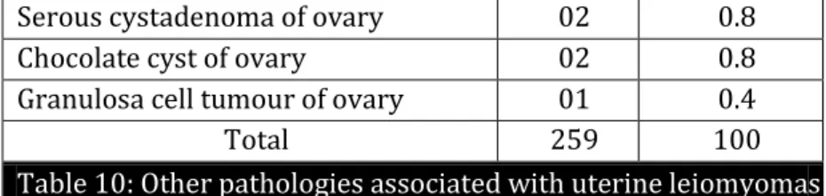
Other coincidental pathologies with uterine leiomyomas are depicted in the table 10 below.
Other pathologies Number Percentage
Absent 238 91.6
Cervical fibroid 05 2
Broad ligament fibroid 03 1.2
Journal of Evolution of Medical and Dental Sciences/ Volume 2/ Issue 46/ November 18, 2013 Page 9006
Serous cystadenoma of ovary 02 0.8
Chocolate cyst of ovary 02 0.8
Granulosa cell tumour of ovary 01 0.4
Total 259 100
Table 10: Other pathologies associated with uterine leiomyomas
DISCUSSION: The major gynaecological surgery done throughout the world is hysterectomy. Charles Clay was the first to perform subtotal and total hysterectomy in Manchester, England in 1843 and 1929 respectively [9,10]. It is a successful procedure done in terms of symptom relief, patient satisfaction and definitive cure in many disease .Benign conditions like leiomyoma, dysfunction uterine bleeding, adenomyosis, pelvis inflammatory disease, endometriosis, pelvic organ prolapse which account for major hysterectomies and rest for malignancy[11,12]. Of these benign lesions, leiomyoma followed by adenomyosis are the commonest indication for hysterectomy [13].
Leiomyomas are benign neoplasm composed of smooth muscle with variable amount of connective tissue [1,2,3]. Leiomyomas are commonly seen in the women of reproductive age [1-3, 7]. Present study had greater frequency between 31-50 years (90.3%) age group similar to studies by Ashraf T et al[14], and Begum S et al[7] whereas in contrast Hafiz R et al[15] observed that affected females were a decade lesser 20-40 years of age possibly since they included only menorrhagic patients with fibroid. Multiparous women (94.9%) were found to have leiomyomas more frequently then nulliparous (1.3%) analogous to study by Begum S et al [7]., in contrast to a study by Derek LJ et al[16] who observed fibroids are more common in nulliparous or infertile patients since he included more of asymptomatic infertile patients with fibroids.
Most of the leiomyomas are asymptomatic but if symptomatic, commonest clinical manifestation is menorrhagia due to increased vascularity, endometrial surface and altered uterine contractility which was 49.03% in present study followed by pain abdomen 30.5% possibly due to degenerative changes in leiomyomas similar to study by Begum S et al [7]., and Jaiswal C et al [17], 54.1% of patients were diagnosis with uterine leiomyomas only on clinical findings alone whereas USG was needed as additional examination equally (45.9%) similar to a study by Begum R et al[7] and also concluded that USG is confirmatory with 80% accuracy. Abraham et al in his study stated that diagnosis of fibroid is usually done on clinical findings but USG is helpful in ruling out that these tumours are not extrauterine masses or they have an extrauterine extension [18]. In the present study number of leiomyomas in uterus varied from 1-10 of which 71% of patients had unitary leiomyomas in concordance with Rosario et al [19].., in contrast study by Begum S et al[6], had majority of multiple fibroids. Most of the leiomyomas were intramural leiomyomas 48% similar to a study by Chhabra S et al [20], Begum S et al [7], and Rosario et al [19].
Journal of Evolution of Medical and Dental Sciences/ Volume 2/ Issue 46/ November 18, 2013 Page 9007 elective caesarean section. Also these secondary changes usually occur in old mature lesions and hence careful conscientious histopathological sampling should be done to rule out malignant changes [21,22].
In the present study proliferative phase and simple hyperplastic endometrium together accounted for 69.1% were the commonest endometrial changes seen in association with uterine leiomyomas possibly due to hyper-estrogenic status in accordance with the study by Rosario et al[19], Purandare et al[23], Sanyal et al[24],Chethana M et al[25].In the present study atrophic endometrium were 5.1% similar to studies by Denligdish et al[26] , Chethana M et al[25]and Rosario YP[19] and described these endometrial changes of normal, hyperplasia and atrophy may be possible due to irregular secretion of estrogens and mechanical effects of fibroid on endometrium.
Dual pathology of adenomyosis and leiomyomas were noted in 29% of patients in present study similar to studies by Denligdish et al[26], Rizvi et al[27]., and Rani S et al[13].., coexistence of these lesions are also due to unopposed estrogen and entrapment of glands within hypertrophied myometrium. Diagnosis of adenomyosis remains an incidental histopathological finding in uterine tissues examined for other clinically suspected pathology.
Extensive literature search showed no studies who reported on the various associated pathologies with uterine leiomyomas. In the present study, though the causative factor for hysterectomy was leiomyoma, there were varied incidental concurrent preoperatively undiagnosed lesions like granulosa cell tumour of ovary (0.4%), tubercular endometritis (0.4%), dermoid and chocolate cyst of ovary (0.8%), mucinous and serous cystadenoma of ovary (1.6%).
CONCLUSION: Leiomyomas are found frequently in multiparous women in reproductive and perimenopausal age group .Though hysterectomy is a routine procedure in the management of uterine leiomyomas, occasional cases of tumor or infective pathology may be missed. Therefore histopathology is mandatory for confirmed diagnosis and ensuring optimal management.
REFERENCES:
1. Crum C P. Body of uterus and Endometrium. In: Kumar V, Abbas A K, Fausto N, Eds. Robbins and Cotran Pathologic Basis of Disease. 7th ed. Philadelphia: Saunders, 2004:1089-90.
2. Silverberg S G, Tabbara S O. The uterine corpus. In: Silverberg S G, Delellis R A, Frable W J, Eds. Principles and Practice of Surgical Pathology and Cytopathology. Vol 3 (3rd edition) .New York: Churchill Livingstone, 1997: 2459-516.
3. Ackerman, Gull B, Karlsson B, Milsom I, Granberg S. Factors associated with endometrial thickness and uterine size in random sample of postmenopausal women. Am J Obstet Gynecol 2001 Aug ; 185(2): 386-91.
4. Witherspoon T J. The interrelationship between ovarian follicle cysts, hyperplasia of the endometrium and fibromyomata. Surg Gynecol Obstet 1933; 56: 1026-35.
5. Rein MS, Barbieri RL, Friedman AJ. Progesterone: A critical role in pathogenesis of uterine myomas. Am J Obst Gynecol 1995;172(1)14-8.
6. Begum S, Khan S. Audit of leiomyoma uterus at Khyber Teaching Hospital, Peshawar. J Ayub Med Coll 2004;16(2):46–9.
Journal of Evolution of Medical and Dental Sciences/ Volume 2/ Issue 46/ November 18, 2013 Page 9008 8. G. Gupta, D.S. Kotasthane, V.D. Kotasthane: Hysterectomy: A Clinico-Pathological Correlation Of
500 Cases. The Internet Journal of Gynecology and Obstetrics. 2010 Volume 14 Number 1. DOI: 10.5580/141b.
9. John A, Rock MD, Jhon D, Thompson MD; Telinds’s Operative Gynaecology. 1st Edition
Lippincott- Raven place.
10.Nausheen F, Iqbal J, Bhatti FA, Khan AT, Sheikh S. Hysterectomy: The patient’s perspective.
Annals Gynecol 2004; 10:339-41.
11.Gupta S, Manyonda I. Hysterectomy for benign gynecological diseases. Current Obstet Gynaecol 2006;16:147- 53.
12.Rani S. V. R, Thomas S. Leiomyoma, a major cause of abnormal uterine bleeding. J of Evolution of Medical and Dental Sciences.2013;2:2626-30.
13.Ashraf T. Management of uterine leiomyomas. J Coll Physicians Surg Pak 1997;7: 160–2.
14.Hafiz R, Ali M, Ahmed M. Fibroid as a causative factor in mennorhagia and its management.DHQ Hospital Rajan Pur, Nishtar Hospital Multan. J Med Res 2003;42(3):90–6.\
15.Derek LJ. Benign enlargement of uterus. In: Fundamentals of Obstetrics and Gynaecology. 5th Ed. London: Mosby; 1990. p. 193.
16.Jaiswal CJ.Vaginal management of uterocervical myomas .J obstet & Gynacol of India. 1996;46:260-63.
17.Abraham R. Uterine fibroids. Manual of clinical problems in Obstet Gynaecol 4th ed. 1994;227-29.
18.Rosario Y P. Uterine Leiomyomas. J of Obstet and Gynecol of India 1968: 101-107.
19.Chhabra S, Ohri N. Leiomyomas of uterus-A clinical study. J obstet & Gynacol of India. 1993;43:436-39.
20.Persaud V, Arjoon PD. Uterine leiomyoma. Incidence of degenerative change and correlation of associated symptoms. Obstet Gynecol. 1970;35:432– 436.
21.Prayson RA, Hart WR. Pathologic considerations of uterine smooth muscles tumors. Clin N America 1995;22(4):637-57.
22.Purandare. S, Jhalam L. Pathological picture in hysterectomy done for abnormal uterine bleeding. J obstet & Gynacol of India .1993;43:418-21.
23.Sanyal MK, Sanyal S, Bhattacharjee KK, Choudari NNK. Clinicopathological study of endometrium. a review of three hundred and twenty cases in different gynaecological abnormalities. J obstet & Gynacol of India .1981;31:816-21.
24.Chethana M,Kumar HML, Munikrishna M. Endometrial Changes in Uterine Leiomyomas. J Clin Biomed Sci 2013 ; 3 (2).72-79.
25.Deligdish L, Loewenthel M. Endometrial changes associated with myomata of the uterus. J Clin Pathol 1970; 23: 676-80.
Journal of Evolution of Medical and Dental Sciences/ Volume 2/ Issue 46/ November 18, 2013 Page 9009
AUTHORS: 1. Mangala Gowri 2. Geetha Mala 3. Srinivasa Murthy 4. Vedavathy Nayak
PARTICULARS OF CONTRIBUTORS:
1. Assistant Professor, Department of Obstetrics & Gynaecology, ESIC MC & PGIMSR, Rajajinagar, Bangalore.
2. Junior Resident, Department of Pathology, ESIC MC & PGIMSR, Rajajinagar, Bangalore. 3. Professor & Head, Department of Pathology,
ESIC MC & PGIMSR, Rajajinagar, Bangalore.
4. Assistant Professor, Department of Obstetrics & Gynaecology, ESIC MC & PGIMSR, Rajajinagar, Bangalore.
NAME ADDRESS EMAIL ID OF THE CORRESPONDING AUTHOR: Dr. Mangala Gowri,
172/A, 8th Main, 4th Block, Rajajinagar, Bangalore – 10. Email – vedavathynayak@yahoo.in
Date of Submission: 29/10/2013. Date of Peer Review: 30/10/2013. Date of Acceptance: 06/11/2013. Date of Publishing: 14/11/2013