The
agr
Inhibitors Solonamide B and
Analogues Alter Immune Responses to
Staphylococccus aureus
but Do Not Exhibit
Adverse Effects on Immune Cell Functions
Mara Baldry1, Betül Kitir2,3, Hanne Frøkiær1, Simon B. Christensen2, Nico Taverne4, Marjolein Meijerink4, Henrik Franzyk2, Christian A. Olsen2,3, Jerry M. Wells4, Hanne Ingmer1*
1Department of Veterinary Disease Biology, Faculty of Health and Medical Sciences, University of Copenhagen, Frederiksberg C, Denmark,2Department of Drug Design and Pharmacology, University of Copenhagen, Copenhagen, Denmark,3Center for Biopharmaceuticals, University of Copenhagen, Copenhagen, Denmark,4Host Microbes Interactomics Group, Wageningen University, Wageningen, The Netherlands
Abstract
Staphylococcus aureusinfections are becoming increasingly difficult to treat due to antibi-otic resistance with the community-associated methicillin-resistantS.aureus(CA-MRSA) strains such as USA300 being of particular concern. The inhibition of bacterial virulence has been proposed as an alternative approach to treat multi-drug resistant pathogens. One inter-esting anti-virulence target is theagrquorum-sensing system, which regulates virulence of CA-MRSA in response toagr-encoded autoinducing peptides. Agr regulation confines exo-toxin production to the stationary growth phase with concomitant repression of surface-expressed adhesins. Solonamide B, a non-ribosomal depsipeptide of marine bacterial origin, was recently identified as a putative anti-virulence compound that markedly reduced expres-sion ofα-hemolysin and phenol-soluble modulins. To further strengthen solonamide anti-viru-lence candidacy, we report the chemical synthesis of solonamide analogues, investigation of structure–function relationships, and assessment of their potential to modulate immune cell functions. We found that structural differences between solonamide analogues confer signifi-cant differences in interference withagr, while immune cell activity and integrity is generally not affected. Furthermore, treatment ofS.aureuswith selected solonamides was found to only marginally influence the interaction with fibronectin and biofilm formation, thus address-ing the concern that application of compounds inducaddress-ing anagr-negative state may have adverse interactions with host factors in favor of host colonization.
Introduction
Staphylococcus aureusis a colonizer of the human nasal cavity and skin in around 20–30% of the healthy human population and yet, it is a notorious opportunistic pathogen, causing severe OPEN ACCESS
Citation:Baldry M, Kitir B, Frøkiær H, Christensen SB, Taverne N, Meijerink M, et al. (2016) Theagr
Inhibitors Solonamide B and Analogues Alter Immune Responses toStaphylococccus aureusbut Do Not Exhibit Adverse Effects on Immune Cell Functions. PLoS ONE 11(1): e0145618. doi:10.1371/journal. pone.0145618
Editor:Holger Rohde, Universitätsklinikum Hamburg-Eppendorf, GERMANY
Received:September 19, 2015
Accepted:December 7, 2015
Published:January 5, 2016
Copyright:© 2016 Baldry et al. This is an open access article distributed under the terms of the
Creative Commons Attribution License, which permits unrestricted use, distribution, and reproduction in any medium, provided the original author and source are credited.
Data Availability Statement:All relevant data are within the paper and its Supporting Information files.
Funding:This work was supported by the European Union’s Seventh Framework Program for research, technological development and demonstration under grant agreement N° 289285, the Lundbeck Foundation’s Group Leader Fellowship (C.A.O.) and the Carlsberg Foundation (C.A.O.).
community-associated and nosocomial infections [1,2]. Infections caused byS.aureusrange from mild superficial skin infections, to toxinosis or toxic shock syndrome, and severe systemic
life-threatening conditions such as endocarditis or meningitis [2].S.aureuscan possess a wide
repertoire of acquired resistance genes, including methicillin resistance (i.e MRSA) which
lim-its treatment options [3]. A key regulator of virulence gene expression is the accessory gene
regu-lator (agr) quorum-sensing system, which is highly active in the CA-MRSA USA300 strain, one
of the most prevalent and virulent culprits of community-associated infections [3,4]. Activation
of theagrtwo-component system occurs in response to the accumulation of self-produced cyclic
thiolactone peptides also known as autoinducing peptides (AIPs). AIPs bind to the AgrC
histi-dine kinase of theagrtwo-component system and stimulate the expression of a regulatory RNA
designated RNAIII, the effector molecule ofagrsignaling [4]. At high cell densities AIP
accumu-lation is responsible for up-regulating expression of exoproteins including thehla-encodedα
-hemolysin, a prominent virulence factor inS.aureus, and down-regulating expression of
surface-associated proteins (such asspa-encoded protein A) [5,6]. Conversely, at low cell densities
exo-protein production is repressed and surface exo-proteins involved in interactions with host factors are
highly expressed [7,8]. There are at least four classes of AIPs inS.aureuseach differing slightly in
their chemical signaling through a cognate AgrC receptor and displaying antagonism in strains
harboring other classes of AIPs [7]. Synthetic analogues of AIPs can also inhibit MRSA virulence,
and naturally occurring antagonists of this system have been identified as well [8,9]. Recently,
two novel cyclodepsipeptides, named solonamide A (1) and B (2), were isolated from a marine
bacterium (Photobacteriumspp. strain S2753) with structures remarkably similar to those of the
AIPs [9,10]. They competitively inhibitagrby interfering with the binding of AIPs to theagr
sen-sor kinase, AgrC [10]. Interference with bacterial virulence and/or cell-to-cell signaling pathways
by solonamides may be a useful strategy for therapy againstS.aureusinfections. Such
anti-viru-lence approaches will inherently exert less selective pressure towards development of bacterial resistance as compared to antibiotics, and importantly they rely on a robust host immune
response for the ultimate clearance of the infection [11]. In this study, we investigated the
impor-tance of solonamide B and solonamide analogue structure in fine tuning theagrresponse.
Fur-thermore, we addressed the concern that these anti-virulence compounds might influence factors that promote host colonization, or have adverse effects on host immune responses.
Materials and Methods
Bacterial strains and growth conditions
Strains used in this study, and their sources are listed inTable 1. For preparations of live or UV
inactivated S. aureus samples specifically, overnight cultures were diluted 1/100 in fresh warm
TSB, incubated at 37°C while shaking at 200 rpm, and upon reaching OD6000.5 test
com-pounds in vehicle or pure vehicle were added to give a final concentration of 10μg/mL.
Cul-tures were grown to an OD6001.7 and spun down. The supernatants were collected and frozen
in 1 mL aliquots and the bacterial pellets were washed twice in sterile phosphate buffered saline
solution (PBS). Washed bacteria were adjusted to OD6000.5 in 20 mL PBS and 10 mL were
fro-zen directly in 1 mL aliquots and the remaining 10 mL were subjected to UV radiation (λ= 254
nm; CL-1000 cross-linker; UVP, Cambridge, United Kingdom) by pulsed UV radiation of 6 sec per pulse for a total of 90 sec. Samples prior to UV and after UV were plated on TSA for number of colony-forming unit (CFU) analysis and multiplicity of infection (MOI) calculation, as well as for checking the viability after UV-irradiation. The Gram-positive bacteria Lactoba-cillus acidophilus NCFM (Danisco, Copenhagen, Denmark), and the Gram-negative bacteria Escherichia coli Nissle 1917 O6:K5:H1 (Statens Serum Institut, Copenhagen, Denmark) were
Synthesis of modified solonamide analogues
For the synthesis of analogues SolB-NaI (5) polystyrene 2-chlorotrityl chloride resin was added
to a fritted syringe and swelled in dry CH2Cl2. A solution of Fmoc-L-Leu-OH (128 mg, 0.35
mmol, 2.5 equiv) andi-PrNEt2(0.12 ml, 0.7 mmol, 5 equiv) in dry CH2Cl2was added and the
resin loading was allowed to proceed on a rocking table for 1h. After washing with CH2Cl2
(×3) the resin was capped with CH2Cl2–MeOH–i-PrNEt2(7:2:1) for 30 min. The resin was
then washed with DMF (×3), MeOH (×3), and CH2Cl2(×3). The Fmoc group was removed
with piperidine–DMF (1:4, 4 ml, 2 × 30 min) and DBU–piperidine–DMF (2:2:96, 4 ml, 30
min), and the resin was then washed as described above. Fmoc-D-Ala-OH (139 mg, 0.42
mmol, 3 equiv) in DMF (3 mL) was pre-incubated with 2, 6-lutidine (98μl, 0.84 mmol, 6
equiv) and HATU (157 mg, 0.41 mmol, 2.95 equiv) for 10 min before addition to the resin and the reaction was allowed to proceed on a rocking table for 24 h. After this washing procedure
the Fmoc group was removed with piperidine–DMF (1:4, 4 ml, 2 × 30 min) and DBU–
piperi-dine–DMF (2:2:96, 4 ml, 30 min), and the resin was washed with DMF (×3), MeOH (×3) and
CH2Cl2(×3). The next two amino acids in the sequence (Fmoc-D-Leu-OH and
Fmoc-L-Nal-OH) were introduced according to the same procedure. A portion of the polystyrene 2-chloro-trityl-bound Fmoc-L-NaI-D-Leu-D-Ala-L-Leu (50 mg, 0.027 mmol) was transferred to fresh
fritted syringe and swelled with CH2Cl2before the standard Fmoc group removal and washing
as described above. Thenβ-hydroxyoctanoic acid (7 mg, 0.054 mmol, 2 equiv) was
pre-incu-bated for 10 min with DIC (8μl, 0.053 mmol, 1.95 equiv) and HOBt (7 mg, 0.053 mmol, 1.95
equiv) and added to the resin. The reaction was allowed to proceed on a rocking table for 24 h
and the resin was washed with DMF (×3), MeOH (×3) and CH2Cl2(×3), and then it was
treated with TFA–CH2Cl2(1:1, 2 mL, 2 × 30 min) followed by washing with CH2Cl2(3 ×2 mL)
and all fractions were pooled in a round-bottomed flask and concentrated under reduced
pres-sure. Co-evaporation with toluene (×2), toluene–CH2Cl2(1:1, ×2), and hexane–CH2Cl2(1:1,
×2) afforded the crude pentamer (18 mg), which was used directly for macrolactonization in
solution under high dilution. Thus,i-PrNEt2(21μl, 0.11 mmol, 4 equiv), DMAP (5.5 mg, 0.042
mmol, 1.5 equiv), and HATU (22.6 mg, 0.042 mmol, 1.5 equiv) dissolved in DMF (2 mL) were added to a stirred solution of linear crude (18 mg, 0.028 mmol) in DMF (60 mL). After stirring at ambient temperature for 16 h, the reaction mixture was diluted with EtOAc (50 mL), washed
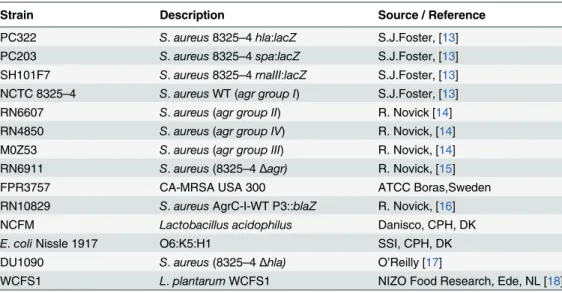
Table 1. Strains and their sources.
Strain Description Source / Reference
PC322 S.aureus8325–4hla:lacZ S.J.Foster, [13] PC203 S.aureus8325–4spa:lacZ S.J.Foster, [13] SH101F7 S.aureus8325–4rnaIII:lacZ S.J.Foster, [13] NCTC 8325–4 S.aureusWT (agr group I) S.J.Foster, [13]
RN6607 S.aureus(agr group II) R. Novick [14]
RN4850 S.aureus(agr group IV) R. Novick, [14] M0Z53 S.aureus(agr group III) R. Novick, [14] RN6911 S.aureus(8325–4Δagr) R. Novick, [15]
FPR3757 CA-MRSA USA 300 ATCC Boras,Sweden
RN10829 S.aureusAgrC-I-WT P3::blaZ R. Novick, [16] NCFM Lactobacillus acidophilus Danisco, CPH, DK
E.coliNissle 1917 O6:K5:H1 SSI, CPH, DK
DU1090 S.aureus(8325–4Δhla) O’Reilly [17]
WCFS1 L.plantarumWCFS1 NIZO Food Research, Ede, NL [18]
with 1 M HCl (50 mL), dried (NaSO4), filtered, and concentratedin vacuo. The residue was purified with complete separation of the two diastereosiomers by preparative HPLC.
Com-pound (5)was obtained as white fluffy solid (2.4 mg, 13%).1H NMR (400 MHz, DMSO-d6)δ
8.23 (d,J= 8.3, 1H), 8.21 (d,J= 7.2, 1H), 8.13 (d,J= 8.0, 1H), 7.99 (d,J= 7, 1H), 7.98 (d,J= 7,
1H), 7.89 (d,J= 8, 1H), 7.77 (m, 1H), 7.57 (dt,J= 6.6, 13.6, 1H), 7.51 (dt,J= 6.6, 13.6, 1H),
7.39 (m, 2H), 4.70 (m, 1H), 4.66 (m, 1H), 4.24 (t,J= 7.4, 1H), 4.20 (m, 1H), 3.68 (m, 1H), 3.43
(m, 1H), 3.23 (m, 1H), 2.20 (dd,J =16.5, 7.8, 1H), 2.13 (dd,J= 14.1 6.4), 1.53–1.17 (m, 17H),
0.86 (m, 6H), 0.83 (m, 3H), 0.72, (d,J= 6.9, 3H), 0.68 (d,J= 6.9, 3H);13C NMR (100 MHz,
DMSO-d6)δ171.6 (2C), 171.1 (2C), 171.0, 133.4, 131.8 (2C), 128.4, 127.4, 127.0, 125.8, 125.3,
125.2, 123.9, 88.4, 67.5, 53.9, 50.9, 48.3, 46.3, 43.3, 40.8, 40.5, 36.6, 31.3(2C), 23.9, 23.6, 23.4,
23.3, 22.3, 21.7, 21.5, 18.3, 14.1; HRMS: calcd for [M + H]+637.3887, found 637.3892;
ΔM = 0.8 ppm.
For the synthesis of the lactam analogues Am15-D (6), Am15-L (7), Am16-D (8), and
Am16-L (9); 2-Chlorotrityl resin (1.6 mmol/g; 0.250 g, 0.4 mmol) was transferred to a Teflon
reactor (10 mL) in which it was swelled in dry DCM (5 mL) and then treated with 10% DIPEA in dry DCM (5 mL) for 2 min, and then washed twice with dry DCM (each 5 mL for 2 min). The appropriate Fmoc-aa-OH building block (2 equiv, 0.8 mmol) in dry DCM (4.5 mL) con-taining DIPEA (4 equiv, 0.28 mL) was added to the resin and then shaken for 1 h. Then the
resin was capped with DCM–MeOH–DIPEA (80:15:5, 3 mL, 2 × 5 min). The Fmoc group was
removed with 20% piperidine in DMF (5 mL, 2 × 10 min), and then the resin was washed sequentially with DMF, MeOH and DCM (each 3 × 3 min with 5 mL). The following amino acid building block (4 equiv) was coupled with PyBOP (4 equiv) and DIPEA (8 equiv) in dry
DMF (4.5 mL) for>2 hours. The pentameric linear intermediates were assembled by repetition
of this Fmoc de-protection and coupling cycle, and were then cleaved from the resin with 50% TFA-DCM (3 × 30 min each with 3 mL). The filtrate was co-evaporated with toluene (3 ×), and the resulting residue purified by preparative HPLC (column: Phenomenex Luna C18(2),
5μm, 21.2 × 250 mm) using a gradient of 20%!50% eluent B during 20 min (A: H2O–
MeCN–TFA 95:5:0.1; B: H2O–MeCN–TFA 95:95:0.1) to give the linear peptides in yields of
25–50%. Cyclization of the linear intermediates (0.02–0.03 mmol) was perfomed by dissolution
in DMF (1–2 mL), and then this solution was added dropwise to a solution of TBTU (6 equiv),
HOAt (6 equiv) and DIPEA (12 equiv) in DMF–DCM (1:6; 10 mL). The mixture was stirred
for 16 h, after which the DCM was removedin vacuo, and then the residue was purified by
pre-parative HPLC (as above) with a gradient of 30%!95% B during 20 min to give the pure
ana-logues in 35–65% yield. The target compounds were characterized by analytical HPLC
(column: Phenomenex Luna C18(2), 3μm, 4.6 × 250 mm) using the same eluents A and B as
for preparative HPLC, as well asby HRMS for which spectra were obtained by using a Bruker
MicroTOF-Q II MS detector. The analyses were performed as ESI-MS (m/z): [M+H]+.
Am15-D(6). Analytical HPLC (20%!100% B during 30 min):tR= 26.60 min. HRMS:
calcd for [M + H]+586.3968, found 586.3939;ΔM = 4.9 ppm.
Am15-L(7). Analytical HPLC (20%!100% B during 30 min):tR= 28.65 min. HRMS:
calcd for [M + H]+586.3968, found 586.3969;ΔM = 0.1 ppm.
Synthesis of solonamides A and B (SolA (1) and SolB (2)), as well as epi-solonamides A and
B (ESA (3) and ESB (4)) was carried out as described by Kitir et al [19].
Agar Diffusion Reporter Assay
The reporter assay was conducted as described by Nielsen et al. [20] Test compounds in
DMSO, supernatants of strains 8325–4 (AIP-I) and M0Z53 (AIP-III), as well as H2O were
Activity of solonamides in WT and AgrC reporter strains using the
β
-lactamase assay
The method used is described by Nielsen et al. 2014 [10]. Briefly, 10μg/mL of the solonamides
and their analogues (final concentration), or DMSO (solvent), and 1/10 volume of spent
medium containing or free from AIP-I were used. Theβ-lactamase activity of the samples was
subsequently determined by using the nitrocefin hydrolysis method as described by Ji et al.
[14]. Statistical analysis was performed using the Student’s t-test (2-tailed).
Fibronectin-binding assay
Untreated 96-well plates (Nunc 265301) were incubated with 100μL per well of 10μg/mL
fibronectin from human plasma for 24 h while shaking at 4°C. The plates were then washed
three times with 1% bovine serum albumin in phosphate-buffered saline (PBS).S.aureus
strains 8325–4 (WT) and USA300 were grown with 10μg/mL of ESB(4),Am16-L (9), or
vehi-cle (DMSO) from OD6000.5 after inoculation into fresh TSB from an overnight culture.
RN6911 (Δagr) was also included. Samples were harvested at OD6001.7 and added to the
Fibro-nectin-coated wells and incubated for 1 h statically at 37°C. After washing, the attached cells were fixed with 2.5% glutaraldehyde in PBS for 1 h statically at 37°C and stained with 0.1% crystal violet for 30 min at room temperature, washed three times with water, and quantified by resuspension in acidified ethanol and measured at 570 nm. Significance between samples
was calculated using the Student’s t-test (2-tailed).
Static biofilm assay
A starter culture of each strain was grown in TSB to an OD6000.5. From this culture 50μL was
withdrawn and diluted 10-fold (from 10−1to 10−5) in 0.9% NaCl solution. 5
μL of each dilution
was inoculated into 200μL TSB. Compound in DMSO was added to each respective well to a
final concentration range of 5, 10, 20, 40 and 80μg/mL. DMSO and no cells were used as
con-trols. The microtiter plates were incubated for approximately 20 h at 37°C without shaking.
The biofilm was then washed twice with 0.9% NaCl (200μL), dried in a LAF bench, stained
with 125μL crystal violet (0.1%) for 30 min, followed by a 3× final wash with 200μL 0.9%
NaCl. To quantify the biofilm formation the stained biofilm was solubilized in 200μL 95%
eth-anol, of which 100μL was transferred to a new microtiter plate and the absorbance measured
at 590 nm.
Murine dendritic cell (DC) isolation and stimulation
All animals used as a source of bone marrow cells were housed under conditions approved by the Danish Animal Experiments Inspectorate (Forsøgdyrstilsynet) according to The Danish Animal Experimentation Act; LBK no. 474 from 15/05/2014, and experiments were carried out
in accordance with the guidelines of‘The Council of Europe Convention European Treaty
Series (ETS) 123 on the Protection of Vertebrate Animals used for Experimental and other
Sci-entific Purposes’. The source of bone marrow cells was female 4–6 month old C57/Black6–Jtac
mice (Taconic, Ejby, Denmark). All mice were sacrificed by cervical dislocation prior to bone marrow extraction for dendritic cell isolation. Dendritic cells were isolated and prepared as
previously described by Christensen et al. [21] with no modifications. Naïve DCs (2 × 10⁶cells/
mL) were resuspended in fresh medium and 500μL/well were seeded in 48-well tissue culture
plates (Nunc, Roskilde, Denmark). The stimuli were prepared to give a final volume of 100μL/
well at the following concentrations: 10μg/mLL.acidophilusNCFM, and/or 5, 10, and 20μg/
vehicle (0.1% DMSO). DCs were stimulated alone withL.acidophilus, solonamides or vehicle, or co-stimulated with bacteria and solonamides. For stimulation with pre-treated and
UV-inactivatedS.aureusstrains 8325–4, and RN6911 cultures were prepared to a final volume of
100μL/well at an MOI of 5 (5×10⁶CFU/mL per well) and added to the DCs.
Human peripheral blood mononuclear cell (PBMC) isolation and
stimulation
The human PBMC assays were approved by Wageningen University Ethical Committee and performed according to the principles of the Declaration of Helsinki. PBMCs were isolated and
prepared as previously described [22] with modifications. Peripheral blood of 3 healthy donors,
whose written informed consent had been provided, was obtained from the Sanquin Blood
Bank, Nijmegen, The Netherlands. Isolated PBMCs were washed and resuspended in Iscove’s
Modified Dulbecco’s Medium (IMDM) + glutamax supplemented with 10% heat inactivated
Fetal Bovine Serum (FBS), 100 U/mL penicillin and 100μg/mL streptomycin (Invitrogen,
Breda, The Netherlands) to a final concentration of 1×10⁶cells/mL and 500μl/well were seeded
in 48-well tissue culture plates. For the PBMC stimulation experiment, either compound or
vehicle alone were added at a final concentration of 10μg/mL, or thawed aliquots of theS.
aureussamples adjusted to an MOI of 10 in IMDM without added antibiotics and allowed to
adjust for 2 h before adding 50μL of each sample to the seeded PBMCs. TSB, PBS, IMDM and
L.plantarumWCFS1 were included as controls. The PBMCs were stimulated at 37°C and 5%
CO2for 24 h or 4 days. After incubation, the PBMC culture supernatants were collected and
frozen in at–20°C until cytokine analysis, and then the cells were harvested and tested for cell
viability with Annexin V/PI staining.
T-cell proliferation assay
Isolated PBMCs were counted and adjusted to a concentration of 1×10⁶cells/mL. They were
then spun down at 300×g for 5 min and the pellet was resuspended in 1 mL sterile PBS + 0.1%
BSA containing 50μg/mL CFDA/SE (Carboxyfluorescein diacetate succinimidyl ester, Cayman
Chemicals) and allowed to incubate for 10 min at 37°C. 5 mL IMDM + 10% FBS was added to the cells which were then placed on ice for a further 5 min prior to spinning down and washing 3x with IMDM + 10% FBS. Washed PBMCs were resuspended in complete culture medium to
a final concentration of 1×10⁶cells/mL and 500μl/well were seeded in 48-well tissue culture
plates. PBMCs were then stimulated with the lymphocyte proliferation inducers aCD3 and aCD28 (BD Pharmingen) at concentrations inducing 100% or 25% T-cell proliferation (10 ng/ mL and 0.4 ng/mL respectively for aCD3 and 0.6 ng/mL and 0.024 ng/mL respectively for aCD28). PBMCs were then further co-stimulated with either compound or vehicle alone to a
final concentration of 10μg/mL, or thawed aliquots of theS.aureussamples adjusted to an
MOI of 10 in IMDM without added antibiotics and allowed to adjust for 2 h before adding
50μL of each sample to the seeded PBMCs. TSB, PBS, and IMDM were included as controls.
The PBMCs were incubated at 37°C and 5% CO2for 4 days to allow for cell proliferation.
T-cell proliferation was measured by staining of harvested floating T-cells with PE-labeled aCD4
according to manufacturer’s instructions and evaluated by flow cytometry using FACS Diva
software. Lymphocytes were gated based on the expression of CD4, and the number of cell divi-sions was gated according to FITC excitation. Significance was tested using one-way ANOVA.
Cell viability assay
DC, PBMC and T-cell viability was assessed by using the commercially available Annexin V: PI
Viability was assessed by flow cytometry and analyzed using FACS Diva software. Significance was tested using one-way ANOVA.
Cytokine level detection assays
For DCs levels of IL-12, TNF-α, IL-6 and IL-10 (all purchased from R&D Systems,
Minneapo-lis, MN, USA) were detected in culture supernatants by commercially available enzyme-linked
immunosorbent assay (ELISA) kits according to the manufacturer’s instructions. For PBMC
and T-cell cytokine analysis, cytokines (IL-12, TNF-α, IL-6, IL-10, IL-8 and IL-1β) were
mea-sured by BD Cytometric Bead Array Flexset (BD Biosciences) using a FACS CantoII flow
cytometer, according to the manufacturer’s instructions and analyzed using the BD FCAP
soft-ware. Significance was tested using one-way ANOVA.
Ethical statement
All cells used for the generation DCs were generated from bone marrow cells isolated from mice sacrificed by cervical dislocation. The use of mice was approved by Danish Animal Exper-iments Inspectorate (Forsøgdyrstilsynet). This is the ethical committee, who approves all ani-mal experiments to be performed in Denmark as well as all the experimental aniani-mal facilities in Denmark. All animals used as a source of bone marrow cells were housed under conditions approved by the Danish Animal Experiments Inspectorate (Forsøgdyrstilsynet) according to The Danish Animal Experimentation Act; LBK no. 474 from 15/05/2014, and experiments
were carried out in accordance with the guidelines of‘The Council of Europe Convention
European Treaty Series (ETS)123 on the Protection of Vertebrate Animals used for Experimen-tal and other Scientific Purposes. The human PBMC assays were approved by Wageningen University Ethical Committee and performed according to the principles of the Declaration of
Helsinki. PBMCs were isolated and prepared as previously described [22] with modifications.
Peripheral blood of 3 healthy donors, whose written informed consent had been provided, was obtained from the Sanquin Blood Bank, Nijmegen, The Netherlands
Results and Discussion
Synthetic solonamides retain anti-virulence activity while
stereochemistry of analogues results in differential
agr
regulation
To explore the structure–activity relationships of solonamides, total syntheses of solonamides
A (1) and B (2) as well as theirβ-hydroxy acid epimers [ESA (3) and ESB (4)] were recently
reported [19], and the naphthylalanine analogue SolB-Nal (5) was prepared using the same
chemistry. The lactam analogues displaying variation in ring size as well as in chirality of selected amino acid residues were assembled by solid-phase peptide synthesis followed by cycli-zation in solution (vide infra). Structurally, solonamide B closely resembles the natural AIPs as
they both contain hydrophobic residues in a cyclic moiety of identical ring size (Fig 1) however;
the solonamides contain two D-amino acid residues. To investigate whether a difference in
chi-rality was crucial to obtain efficientagrinhibition, we synthesized solonamide-mimicking
lac-tam analogues (6and8) as well as all-L lactam analogues (7and9). In analogues6and7, the
ring size was reduced by a single atom to give a 15-membered macrocycle, while the side chain in both types of analogues was similar in length to that of solonamide B.
The ability of synthetic solonamides and lactam analogues to repressagractivity was
exam-ined by an agar diffusion assay [20] where reporter strains containing thelacZreporter fused
toS.aureusvirulence geneshla(encodingα-hemolysin),spa(encoding Protein A) and theagr
confirmed that synthetic solonamide B retained ability to modulateagractivity as monitored
by increased expression ofspaand decreased expression ofhlaandrnaIII(Fig 2). Furthermore,
activity of solonamides is clearly dependent on stereochemical features as the analogues of
solonamide B with opposite configuration of the stereocenter in theβ-hydroxy acid residue,
ESA (3) and ESB (4), showed markedly larger interference zones than the naturally occurring
solonamides, with ESB being the most effective. The lactam analogues also exhibited potent activity, with the most active compounds being those displaying the all-L stereochemical
con-figuration [i.e., Am15-L (7) and Am16-L (9)] also present in the AIPs.
Interference with theagrsensor system was further examined and quantified using the
RNAIII-reporter strain RN10829 [10,16]. In accordance with the agar diffusion assay, we
observed that all the solonamides showed varying degrees ofagrinhibition with the most
pro-nounced effect displayed by ESB(4), resulting in a three-fold reduction in RNAIII expression in
comparison to the control culture induced by AIP (Fig 3A). We found that the lactam
ana-logues Am15-L (7) and Am16-L (9), which have all-L amino acid configuration, were capable
ofagrinhibition with Am16-L (9) almost matching that of ESB (4), but with a shorter window
of activity (Fig 3A and 3B). Interestingly, the analogues [Am16-D (8) and Am15-D (6)]
con-taining two D-amino acids as also found in the solonamides, initially displayed marginal
acti-vation ofagrabove the level of AIP induction, which overtime reverted back to marginal
inhibition. These data highlight the importance of ring size and stereochemistry in fine-tuning the interactions of AIPs, solonamides, and their analogues with AgrC. Based on these
investiga-tions solonamide B (2), ESB (4), and Am16-L (9) were selected for further studies.
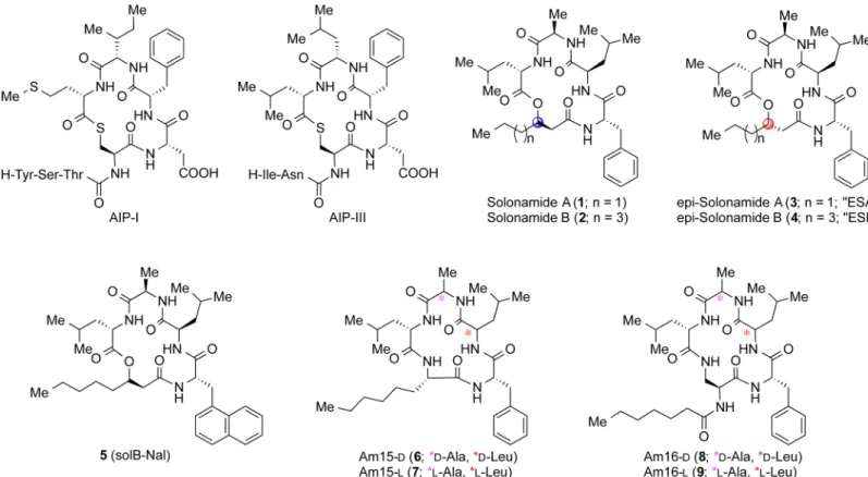
Fig 1. Compound and AIP Structures.Structures of autoinducing peptides (AIP-I and III) used in the study as well as tested depsipeptides and their modified lactam analogues.
Solonamide treatment of
S.
aureus
does not substantially promote
adhesion to host factors or biofilm formation
Asagrbalances the expression of exotoxins in the stationary growth phase with the repression
of surface-associated adhesins, a key question is whether repression ofagrby solonamides
leads to enhanced stationary phase expression of surface-associated proteins involved in
bio-film formation, adhesion and immune evasion such as thespaencoded Protein A or the
fibro-necting-binding protein [23–26]. To address this question we treatedS.aureusstrains with our
selected solonamides and studied the strains’capacities to adhere to human fibronectin or to
form biofilm. When investigating treatedS.aureuscells we observed that ESB (4) (P<0.0001)
and Am16-L (9) (P<0.02) significantly increased the fibronectin-binding capacity of the 8325–
4 WT strain, in comparison to the untreated and vehicle (DMSO)-treated controls, but not to
the extent observed in the RN6911agrdeletion mutant. This indicates that although an
increase in fibronectin binding is observed, it does not mirror anagr-negative phenotype. For
the CA-MRSA USA300 clinical isolate we observed a slight non-significant and
compound-independent decrease in fibronectin-binding capacity (Fig 4). These data suggest that
solona-mide treatment increases fibronectin binding, which is associated with inactivation ofagr
sig-naling, but in a strain-dependent fashion. With regards to the influence of treatment on
biofilm formation, we observed that solonamide B (2), ESB (4), and Am16-L (9) increased
static biofilm in a dose-independent manner; however, the overall increase was marginal (less
than 2-fold) as compared to non-treated and vehicle controls (Fig 5A, 5B and 5C). From these
results on fibronectin-binding and biofilm formation, we conclude that inhibition ofagrby the
selected anti-virulence compounds only marginally increases binding ofS.aureusto host
com-ponents without modulating static biofilm formation. These findings alleviate some of the
con-cerns that by interfering withagrthrough the application of our anti-virulence candidates, we
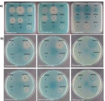
Fig 2. Biological validation of the synthetic solonamides (a) and lactam analogues (b).Agar plates containing thehla–lacZ(PC322), thernaIII-lacZ(SH101F7) orspa–lacZ(PC203) reporter strains ofS.aureus
were exposed to DMSO (20μL) containing the test compound (0.5 mg/mL). Vehicle (DMSO), H2O, AIP-I (autologous) and AIP-III (heterologous) were used as controls. Virulence gene down-regulation is represented by a white zone and up-regulation by a darker than background blue zone.
significantly promote complications often associated withagrnegative strains such as: host persistence, colonization and immune evasion.
Solonamide B (2), ESB (4), and lactam analogue Am16-L (9) are not
toxic to immune cells
Solonamide B was previously shown to display undetectable toxicity to both human and bovine
neutrophils [10]. To further support these data, we examined the viability of human PBMCs
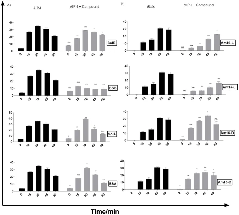
Fig 3. Solonamides (A) and lactam analogues (B) differentially interfere with AgrC activation as monitored by direct RNAIII expression.Cultures of RN10829 (P2-agrA:P3-blaZ) containing the pagrC-I-WT, were grown to an OD600of 0.4–0.5 where a 1/10 volume of AIP-I containing supernatant was added and solonamides and analogues in DMSO to a final concentration of 10μg/mL. Samples obtained at 15 min time intervals after addition of test solutions were analysed forβ-lactamase activity. Each bar represents the average of 3 replicates and the error bars represent the standard deviation. Comparisons were made for each individual time point between AIP and AIP + Compound samples. ns (no significance);*, p<0.05;**, p<0.01;***, p<0.001.
and T-cells as well as murine bone marrow-derived DCs, exposed to our selected compounds, by using the Annexin V/PI staining method. Toxicity testing of our compounds on a wider range of immune cells is imperative given the fact that for any anti-virulence compound to be
effective, a healthy immune response towards the disarmed pathogen is needed [6]. We found
Fig 4. Solonamides marginally enhance adhesion to the extracellular matrix component fibronectin.Upon exposure ofS.aureusstrains to 10μg/mL anti-virulence compounds or vehicle control, attachment to fibronectin was measured in 96-well plates coated with 10μg/mL fibronectin and staining with 0.1% crystal violet. Absorbance values were corrected for cell density and represented as arbitrary binding units. For statistical significance, comparisons were made between untreated versus vehicle and treated (black bracket), and between vehicle control versus compound treated (red square). ns (no significance);*, p<0.05;**, p<0.01;***, p<0.001.
doi:10.1371/journal.pone.0145618.g004
Fig 5. Solonamides marginally enhance biofilm formation of WT strain 8325–4.A dilution series of an 8325–4 culture were inoculated (5μL) into wells of a 96-well microtiter plate containing 200μL TSB. SolB (A), ESB (C) or Am16-L (C) was added to final concentrations of 5, 10, 20, 40, and 80μg/mL. Inoculum alone, DMSO and no inoculum were used as controls. Biofilm formation was assessed by crystal violet staining and OD590 nm measurement. Each bar represents the average of 3 experiments, and the error bars represent the standard deviation. For statistical significance, comparisons were made between untreated versus vehicle and treated (black bracket), and between vehicle control versus compound treated (red square). ns (no significance);*, p<0.05;**,
p<0.01;***, p<0.001.
that none of the tested solonamides nor the lactam analogue at the effective concentration of
10μg/mL displayed toxicity towards human PBMCs and T-cells, as viability remained above
80% irrespective of incubation period (24 h or 4 days), and was equal to the non-exposed and
DMSO-exposed PBMC samples (S1 Fig). Increasing the concentration through a range of 5,
10, 20 and 40μg/mL over a 4 day incubation period only showed a slight
concentration-depen-dent decrease in viability for solonamide B (2) and ESB (4) that nevertheless did not drop
below 73% at 40μg/mL (S2 Fig). When examining murine dendritic cells (mDCs) for viability,
we observed that after a 20 h incubation period viability ranged from 65% to 71% irrespective
of compound exposure (S3 Fig). Collectively, these data imply that our potential anti-virulence
compounds are not toxic to immune cells at the concentration that effectively antagonizesagr
signaling.
Solonamide-treated
S.
aureus, but not solonamides alone, influence
immune cell responses
As the selected anti-virulence compounds did not affect viability of human PBMCs, prolifer-ated T-cells or murine DCs, we investigprolifer-ated whether they would modulate cytokine and che-mokine responses in unstimulated and microbially stimulated antigen-presenting cells as well
as proliferating T cells. Cytokines IL-6, TNF-α, IL-12, and IL-10 were measured for DCs, and
for the PBMCs and aCD3 and aCD28 stimulated T-cells, IL-1βand the chemokine IL-8 were
also measured. The tested compounds had no effect on the cytokine and chemokine secretion in non-stimulated DCs, PBMCs or T-cells activated with aCD3 and aCD28 compared to untreated controls, indicating that the compounds alone do not stimulate an immune response. Furthermore, the selected solonamides and analogues exerted no immune-modulating effect
on cytokine secretion byL.acidophilus-activated murine dendritic cells, and neither hampered
nor enhanced DC function (S4 Fig).
AsS.aureusharbors a wide repertoire of immune evasion mechanisms [27] we
hypothe-sized that treatment with ouragr-inhibiting compounds would interfere with some of these
mechanisms, and thus allow for a more robust immune response against the treated target
pathogen. To investigate this, we exposed DC and PBMCs to compound-treatedS.aureusand
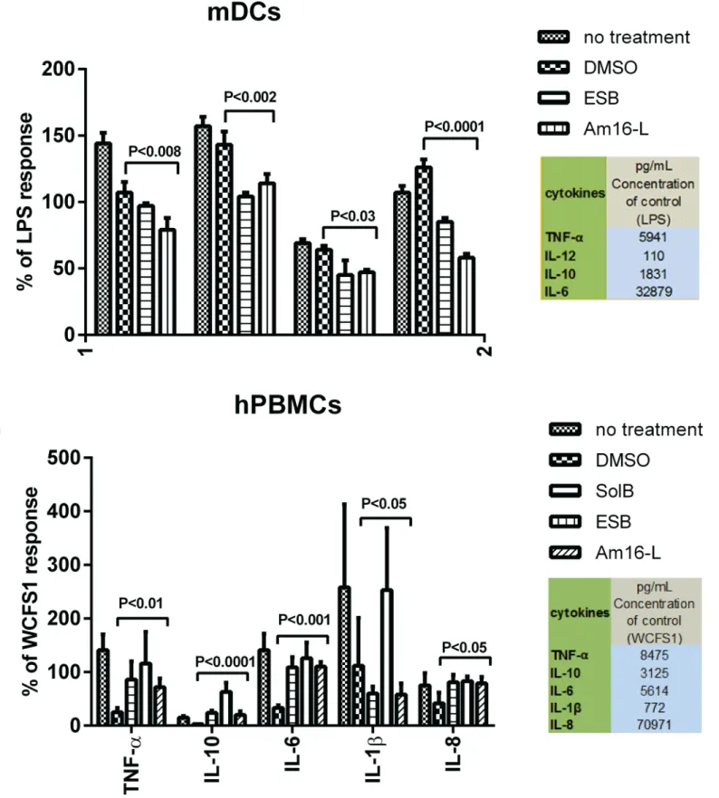
measured cytokine responses as indicators of immune modulation and response. We found
that the amounts of cytokines secreted by DCs in response toS.aureustreated with theagr
inhibitors were significantly lower than those measured with untreated or DMSO treatedS.
aureuscontrols (Fig 6A). In contrast PBMCs incubated withS.aureustreated with these
com-pounds secreted significantly higher amounts of cytokines compared to DMSO-treatedS.
aureusalthough this was not consistent for IL-1β(Fig 6B). These findings reveal that treatment ofS.aureuswith ouragr-interfering compounds significantly alters the cytokine release pattern depending on the immune cell type.
One reported mechanism ofS.aureusimmune evasion is via the inhibition of T-cell
prolif-eration. A reported mediator of such inhibition is theS.aureussecretion of the MHC class II
analog protein Map, whose expression is regulated by theagr-controlled RNAIII regulatory
RNA [28,29]. To investigate the possible effects of compound-treatedS.aureuson host
immune modulation or evasion, we also examined whether treated bacteria were capable of interfering with T-cell proliferation. We hypothesized that as Map expression is partially
con-trolled by RNAIII, the inhibition of RNAIII viaagrinterference with our compounds could
interfere with some of the T-cell proliferation-inhibition properties ofS.aureus. In accordance
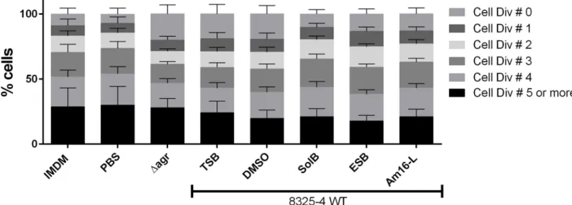
presence of compound-treatedS.aureusand untreated control strains, we observed that the
Δagr, non-treated WT strain and the DMSO-treated samples showed a marked increase in
non-dividing T cells compared to the PBS, medium control, and compound-treatedS.aureus
samples (Fig 7). This observation suggests that the selected solonamides are capable of
reduc-ing the partial inhibition of T cell proliferation exerted by WTS.aureus, thus displaying some
degree of modulation. The data also suggests surprisingly that this effect may beagr
-indepen-dent, thus rendering our hypothesis partially void. When the compound-treatedS.aureus
strains were UV-inactivated the effect was not observed (S5 Fig) suggesting that inhibition of T
cell proliferation by WTS.aureusmight depend on a secreted metabolite or protein, which
may not be produced when WTS.aureusis treated with solonamides.
Conclusions
In summary our investigations show that solonamide B and analogues harbor no toxicity towards immune cells, and they suggest that no adverse effects are anticipated when using
these compounds to targetS.aureusinfections. These findings provide important background
information for the futurein vivocharacterizations of these compounds as potential safe
candi-dates for anti-virulence therapy targetingS.aureus.
Supporting Information
S1 Fig. Solonamides and the selected analogue Am16-L do not influence the viability of human PBMCs or proliferated T-cells.Human PBMCS were stimulated with 10μg/mL solB
(2), ESB (4) or the analogue Am16-L (9) for 24 h (A) and 4 days (B) or stimulated for T-Cell proliferation with aCD3/aCD28 and co-stimulated with the selected compounds for 4 days (C) Culture media and DMSO were used as controls. PBMCs and T-cells were harvested; stained with Annexin V/PI, and cell viability measured by flow cytometry. The results are representa-tive of 3 different donors.
(TIF)
Fig 7. SolB, ESB and Am16-L- reduce interference of T-cell proliferation byS.aureus.CFDA/SE (25μg/mL) stained human PBMCs (1x10⁶/ml) were co-stimulated with aCD3/aCD28 (0.4ng and 0.024ng/mL respectively) and S. aureus either treated or not with 10μg/mL SolB, ESB or Am16-L at an MOI of 10 for 4 days. Harvested cells were stained with aCD4-PE and analyzed by Flow Cytometry using FACS Diva software. Lymphocytes were gated based on the expression of CD4, and the number of cell divisions was gated according to the CFDA/SE FITC excitation. The data represent the average of 3 donors, and the error bars represent the standard deviation.
S2 Fig. Increasing concentrations of solonamides and the selected analogue Am16-L do not influence the viability of human PBMCs.Human PBMCS were stimulated with a
concentra-tion gradient of 5, 10, 20 and 40μg/mL of solB (2), ESB (4) or the analogue Am16-L (9) for 4
days. Culture media and DMSO were used as controls. PBMCs were harvested; stained with Annexin V/PI, and cell viability measured by flow cytometry. The results are representative of 3 different donors.
(TIF)
S3 Fig. Solonamides and selected analogues do not influence the viability of naïve or stimu-lated murine dendritic cells.Bone-marrow-derived DCs were stimulated withL.acidophilus
NCFM (10μg/mL), solB (2), the ESB (4) or the analogue Am16-L (9) at 20μg/mL, either alone
or in combination withL.acidophilusNCFM for 20 hrs. DCs were harvested; stained with
Annexin V/PI, and then cell viability was measured by flow cytometry. The results are repre-sentative of one of 3 reproducible independent experiments.
(TIF)
S4 Fig. Solonamides do not exert immunomodulating effects on NCFM-stimulated murine dendritic cells. (a)SolB,(b)ESB, and(c)analogue Am16-L. (Columns 1 = NCFM; 2 = 0.1%
DMSO; 3, 4 and 5 = 5, 10 and 20μg/mL of each compound; 6 = un-stimulated DCs).
Bone-marrow-derived dendritic cells (DCs) were co-stimulatedwith L.acidophilusNCFM (10μg/
mL) and increasing concentrations of compounds at 5, 10 and 20μg/mL. Concentrations of
IL-6, TNF-α, IL-12 and IL-10 in the supernatants after 20 h were measured by enzyme-linked
immunosorbent assay (ELISA). The results are based on at least 3 independent experiments. (TIF)
S5 Fig. SolB, ESB and Am16-L do not reduce interference of T-cell proliferation by UV-inactivated and treatedS.aureus.CFDA/SE (25μg/mL) stained human PBMCs (1x10⁶/ml)
were co-stimulated with aCD3/aCD28 (0.4ng and 0.024ng/mL respectively) and either treated
or not treated with 10μg/mL SolB, ESB or Am16-L and UV-inactivatedS.aureusat an MOI of
10 for 4 days. 200μL floating cells were harvested, washed and stained with aCD4-PE for 30
min at 4°C. Cells were further washed and resuspended in FACS buffer. Cells were analyzed by Flow Cytometry using FACS Diva software. Lymphocytes were gated based on the expression of CD4, and number of cell divisions was gated according to the FITC excitation. The data rep-resent the average of 3 donors, and the error bars reprep-resent the standard deviation.
(TIF)
Acknowledgments
The authors thank Anni Mehlsen for skilled technical assistance as well as Lisbeth D. Lund and Anita Nielsen for expert guidance.
Author Contributions
Conceived and designed the experiments: MB H. Frøkiær H. Franzyk CO JMW HI. Performed the experiments: MB BK SBC NT MM. Analyzed the data: MB H. Frøkiær NT JMW HI. Con-tributed reagents/materials/analysis tools: BK H. Frøkiær H. Franzyk CO. Wrote the paper: MB H. Frøkiær H. Franzyk CO JMW HI.
References
1. Plata K, Rosato AE, Wegrzyn G.Staphylococcus aureusas an infectious agent: overview of biochemis-try and molecular genetics of its pathogenicity. Acta Biochim Pol 2009 Nov 12; 56(4):597–612. PMID:
2. Bien J, Sokolova O, Bozko P. Characterization of Virulence Factors ofStaphylococcus aureus: Novel Function of Known Virulence Factors That Are Implicated in Activation of Airway Epithelial Proinflam-matory Response. Journal of Pathogens 2011 Sep 14; ( 2011: 601905.). doi:10.4061/2011/601905
PMID:22567334
3. Gordon RJ, Lowy FD. Pathogenesis of methicillin-resistantStaphylococcus aureusinfection. Clin Infect Dis 2008 Jan 6;(46: ).
4. Wang B, Zhao A, Novick RP, Muir TW. Activation and inhibition of the receptor histidine kinase AgrC occurs through opposite helical transduction motions. Mol Cell 14 A.D. Mar 20; 53(6):929–40. 5. Queck SY, Jameson-Lee M, Villaruz AE, Bach TH, Khan BA, Sturdevant DE, et al. RNAIII-independent
target gene control by theagrquorum-sensing system: insight into the evolution of virulence regulation inStaphylococcus aureus. Mol Cell 2008 Oct 10; 32(1):150–8. doi:10.1016/j.molcel.2008.08.005
PMID:18851841
6. Shoham M. Antivirulence agents against MRSA. Future Med Chem 2011 May; 3(7):775–7. doi:10. 4155/fmc.11.43PMID:21644821
7. George EA, Muir TW. Molecular mechanisms of agr quorum sensing in virulent staphylococci. Chem-biochem 2007 May 25; 8(8):847–55. PMID:17457814
8. George EA, Novick RP, Muir TW. Cyclic peptide inhibitors of staphylococcal virulence prepared by Fmoc-based thiolactone peptide synthesis. J Am Chem Soc 2008 Apr 9; 130(14):4914–24. doi:10. 1021/ja711126ePMID:18335939
9. Mansson M, Nielsen A, Kjaerulff L, Gotfredsen CH, Wietz M, Ingmer H, et al. Inhibition of virulence gene expression inStaphylococcus aureusby novel depsipeptides from a marine photobacterium. Mar Drugs 2011 Dec; 9(12):2537–52. doi:10.3390/md9122537PMID:22363239
10. Nielsen A, Mansson M, Bojer MS, Gram L, Larsen TO, Novick RP, et al. Solonamide B inhibits quorum sensing and reducesStaphylococcus aureusmediated killing of human neutrophils. PLoS One 2014; 9 (1):e84992. doi:10.1371/journal.pone.0084992PMID:24416329
11. Rasko DA, Sperandio V. Anti-virulence strategies to combat bacteria-mediated disease. Nat Rev Drug Discov 2010 Feb; 9(2):117–28. doi:10.1038/nrd3013PMID:20081869
12. Weiss G, Rasmussen S, Zeuthen LH, Nielsen BN, Jarmer H, Jespersen LF, et al.Lactobacillus aci-dophilusinduces virus immune defence genes in murine dendritic cells by a Toll-like receptor-2-depen-dent mechanism. Immunology 2010 Oct 1; 131(2):268–81. doi:10.1111/j.1365-2567.2010.03301.x
PMID:20545783
13. Chan PF, Foster SJ. The role of environmental factors in the regulation of virulence-determinant expression inStaphylococcus aureus8325–4. Microbiology 1998 Sep 1; 144(9):2469–79.
14. Ji G, Beavis RF, Novick RP. Bacterial interference caused by autoinducing peptide variants. Science 1997 Jun 27; 276(5321):2027–30. PMID:9197262
15. Novick RP, Ross HF, Projan SJ, Kornblum J, Kreiswirth B, Moghazeh S. Synthesis of staphylococcal virulence factors is controlled by a regulatory RNA molecule. EMBO J 1993 Oct; 12(10):3967–75. PMID:7691599
16. Novick RP, Morse SI. In vivo transmission of drug resistance factors between strains of Staphylococ-cus aureus. J Exp Med 1967 Jan 1; 125(1):45–59 1967 Jan 1;125(1):45–59. PMID:6016896
17. O'Reilly M, de Azavedo JC, Kennedy S, Foster TJ. Inactivation of the alpha-haemolysin gene of Staph-ylococcus aureus8325–4 by site-directed mutagenesis and studies on the expression of its haemoly-sins. Microbial Pathogenesis 1986 Apr 1; 1(2):125–38. PMID:3508485
18. Kleerebezem M, Boekhorst J, van Kranenburg R, Molenaar D, Kuipers OP., Leer R, et al. Complete genome sequence ofLactobacillus plantarumWCFS1. Proc Natl Acad Sci U S A 2003 Feb 3; 100(4). 19. Kitir B, Baldry M, Ingmer H, Olsen CA. Total synthesis and structural validation of cyclodepsipeptides
solonamide A and B. Tetrahedron 2014 Oct 21; 70(42):7721–32.
20. Nielsen A, Nielsen KF, Frees D, Larsen TO, Ingmer H. Method for screening compounds that influence virulence gene expression inStaphylococcus aureus. Antimicrob Agents Chemother 2010 Jan; 54 (1):509–12. doi:10.1128/AAC.00940-09PMID:19917763
21. Christensen HR, Frokiaer H, Pestka JJ. Lactobacilli differentially modulate expression of cytokines and maturation surface markers in murine dendritic cells. J Immunol 2002 Jan 1; 168(1):171–8. PMID:
11751960
22. van, Hemert S, Meijerink M, Molenaar D, Bron PA, de Vos P, Kleerebezem M, et al. Identification of Lactobacillus plantarumgenes modulating the cytokine response of human peripheral blood mononu-clear cells. BMC Microbiology 2010 Nov 16; 10(293).
24. Otto M. Staphylococcal infections: mechanisms of biofilm maturation and detachment as critical deter-minants of pathogenicity. Annual Review of Medicine 2012 Aug 16; 64:175–88. doi: 10.1146/annurev-med-042711-140023PMID:22906361
25. Yarwood JM, Bartels DJ, Volper EM, Greenberg EP. Quorum sensing inStaphylococcus aureus bio-films. J Bacteriol 2004 Mar 1; 186(6):1838–50. PMID:14996815
26. Saravia-Otten P, Muller HP, Arvidson S. Transcription ofStaphylococcus aureusfibronectin binding protein genes is negatively regulated byagrand anagr-independent mechanism. J Bacteriol 1997 Sep 1; 179(17):5259–63. PMID:9286974
27. Foster TJ. Immune evasion by staphylococci. Nat Rev Microbiol 2005 Dec 1; 3(12):948–58. PMID:
16322743
28. Lee LY, Miyamoto YJ, McIntyre BW, Hook M, McCrea KW, McDevitt D, et al. TheStaphylococcus aureusMap protein is an immunomodulator that interferes with T cell-mediated responses. J Clin Invest 2002 Nov 15; 110(10):1461–71. PMID:12438444