Natural Variants of the KPC-2
Carbapenemase have Evolved Increased
Catalytic Efficiency for Ceftazidime
Hydrolysis at the Cost of Enzyme Stability
Shrenik C. Mehta, Kacie Rice, Timothy Palzkill*
Department of Pharmacology, Baylor College of Medicine, Houston, Texas, United States of America
Abstract
The spread ofβ-lactamases that hydrolyze penicillins, cephalosporins and carbapenems among Gram-negative bacteria has limited options for treating bacterial infections. Initially,
Klebsiella pneumoniaecarbapenemase-2 (KPC-2) emerged as a widespread carbapenem hydrolyzingβ-lactamase that also hydrolyzes penicillins and cephalosporins but not cepha-mycins and ceftazidime. In recent years, single and double amino acid substitution variants of KPC-2 have emerged among clinical isolates that show increased resistance to ceftazi-dime. Because it confers multi-drug resistance, KPCβ-lactamase is a threat to public health. In this study, the evolution of KPC-2 function was determined in nine clinically isolated vari-ants by examining the effects of the substitutions on enzyme kinetic parameters, protein sta-bility and antibiotic resistance profile. The results indicate that the amino acid substitutions associated with KPC-2 natural variants lead to increased catalytic efficiency for ceftazidime hydrolysis and a consequent increase in ceftazidime resistance. Single substitutions lead to modest increases in catalytic activity while the double mutants exhibit significantly increased ceftazidime hydrolysis and resistance levels. The P104R, V240G and H274Y substitutions in single and double mutant combinations lead to the largest increases in ceftazidime hydro-lysis and resistance. Molecular modeling suggests that the P104R and H274Y mutations could facilitate ceftazidime hydrolysis through increased hydrogen bonding interactions with the substrate while the V240G substitution may enhance backbone flexibility so that larger substrates might be accommodated in the active site. Additionally, we observed a strong correlation between gain of catalytic function for ceftazidime hydrolysis and loss of enzyme stability, which is in agreement with the‘stability-function tradeoff’phenomenon. The high Tmof KPC-2 (66.5°C) provides an evolutionary advantage as compared to other class A
en-zymes such as TEM (51.5°C) and CTX-M (51°C) in that it can acquire multiple destabilizing substitutions without losing the ability to fold into a functional enzyme.
OPEN ACCESS
Citation:Mehta SC, Rice K, Palzkill T (2015) Natural Variants of the KPC-2 Carbapenemase have Evolved Increased Catalytic Efficiency for Ceftazidime Hydrolysis at the Cost of Enzyme Stability. PLoS Pathog 11(6): e1004949. doi:10.1371/journal. ppat.1004949
Editor:Robert Bonomo, Case Western Reserve University School of Medicine, UNITED STATES
Received:January 21, 2015
Accepted:May 11, 2015
Published:June 1, 2015
Copyright:© 2015 Mehta et al. This is an open access article distributed under the terms of the
Creative Commons Attribution License, which permits unrestricted use, distribution, and reproduction in any medium, provided the original author and source are credited.
Data Availability Statement:All relevant data are within the paper.
Funding:The work was funded by National Institutes of Health grant AI32956 to TP. The funders had no role in study design, data collection and analysis, decision to publish, or preparation of the manuscript.
Author Summary
The absence of new antibiotics combined with the emergence of antibiotic-resistance en-zymes like KPC-2 that can inactivate mostβ-lactam antibiotics has resulted in a longer du-ration of medical treatment, higher costs of medical care, and increased mortality. In recent years, a number of amino acid sequence variants of KPC-2 have been identified in clinical isolates worldwide suggesting continued evolution of resistance in KPC-2. In this study we have characterized nine clinically isolated variants of KPC-2 (KPC-3 to -11) that differ from the initial KPC-2 isolate by one to two amino acids. The KPC variants confer increased resistance to the antibiotic ceftazidime as compared to KPC-2. This increase in resistance is correlated with improved ability of the variant enzymes to hydrolyze the anti-biotic. Additionally, the changes associated with increased ceftazidime hydrolysis also re-duce the thermal stability of the enzyme, indicating the mutations that assist catalysis come with a cost on the overall stability of the enzyme. The high thermal stability of KPC-2 allows destabilizing mutations that enhance catalysis to accumulate while the enzyme re-tains a folded, functional structure. Thus, the high stability of KPC-2 provides an evolu-tionary advantage to acquire multiple mutations and retain function as compared to other β-lactamase enzymes.
Introduction
Because of their broad-spectrum activity, safety and favorable pharmacokinetic properties [1], β-lactam antibiotics have been the drugs of choice to treat bacterial infections. While antibiot-ics have helped save millions of lives, the extensive use of these drugs has resulted in the emer-gence of antibiotic resistant bacterial strains. This problem is compounded by the ability of these organisms to acquire mutations or obtain genes encoding antibiotic-inactivating enzymes from other bacteria, thereby reducing the efficacy of drugs. Thus, treating antibiotic resistant bacterial infections is a complex clinical challenge [2].
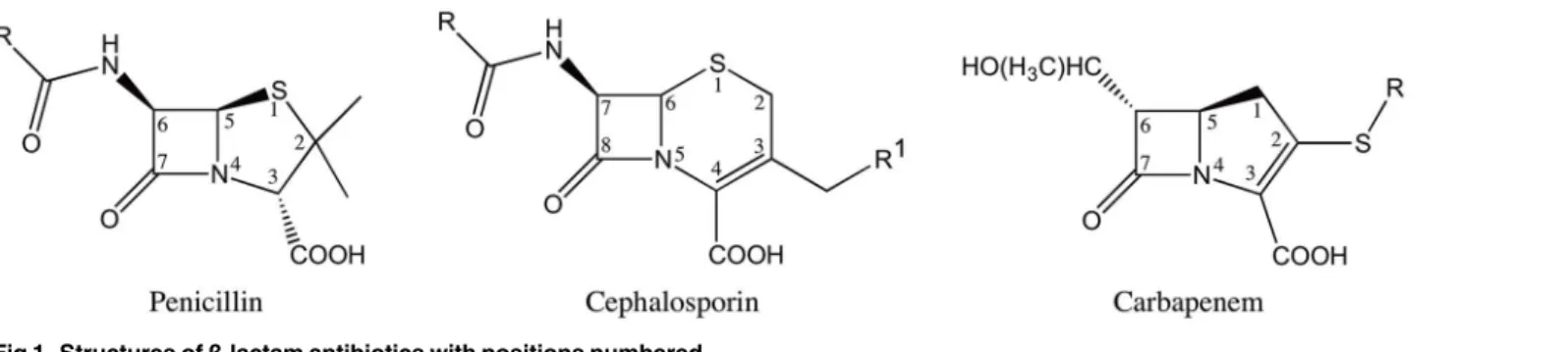
Theβ-lactam antibiotics contain a characteristic four-memberedβ-lactam ring and act as covalent inhibitors of the essential transpeptidase enzymes known as penicillin binding pro-teins (PBP’s). Theβ-lactam antibiotics are classified into different groups based on their chemi-cal structure [3]. The most clinically relevant classes are penicillins, cephalosporins and carbapenems (Fig 1). The penicillins and cephalosporins contain theβ-lactam ring fused to a five or six-membered ring, respectively. The carbapenems consist of theβ-lactam ring fused to a five-membered ring with a carbon atom replacing the sulfur at the C-1 position along with an unsaturated C2-C3 bond [4] (Fig 1). The presence of a6-α-hydroxyethyl side-chain at the C-6 position of theβ-lactam nucleus is a feature that distinguishes the carbapenems from the peni-cillins and cephalosporins that have a6-β- or7-β-acylamino side-chain in the same position, respectively [5] (Fig 1). In addition to being a structural distinguishing factor for the carbape-nems, the6-α-hydroxyethyl side-chain is also responsible for the broad-spectrum activity of the carbapenem antibiotics [6–8].
stable acyl-enzyme intermediate [6–8]. However, the increasing use of carbapenems has led to the emergence of carbapenem hydrolyzingβ-lactamases [11]. Carbapenemase activity has been reported in class A, B and Dβ-lactamases [12]. In particular, resistance to carbapenems medi-ated by class A enzymes such as KPC-2, SME-1-3, IMI-1-2, SFC-1, NmcA, GES-2 and GES-4 to 6 poses a serious clinical threat [13]. Among these class A carbapenemases, KPC-2 is the most clinically important enzyme due to its prevalence in enteric bacteria [14]. Additionally, the presence ofblaKPC-2gene on the mobile transposon Tn4401has facilitated its dissemination among Gram-negative bacteria [15].
Biochemical data have shown that KPC-2 is effective in hydrolyzing penicillins, cephalo-sporins and carbapenems. However, KPC-2 hydrolyzes cephamycins and ceftazidime poorly [16]. This broad-spectrum activity has resulted in severely limited treatment options leading to high fatality rates [14]. The problem of antibiotic resistance due to KPC enzymes has been compounded by the recent identification of a number of clinical variants of KPC-2. Current-ly, a total of 22 KPC variants have been annotated by Genbank and listed on the Lahey Clinic website (http://www.lahey.org/studies/other.asp#table1). In this study, we have character-ized the variants KPC-3 to KPC-11 that possess 1 to 2 amino acid substitutions as compared to KPC-2. KPC-2 was first isolated in North Carolina, however the KPC variants have been isolated from Columbia, Italy, Spain, Puerto Rico, Scotland and Israel, indicating that this enzyme has rapidly disseminated throughout the world [17–22] (Fig 2). While KPC-2 was named afterKlebsiella pneumoniae, these variants have been isolated from a variety of or-ganisms includingE.coli,Enterobacter cloacae,Actinobacter calcoaceticus-baumanniiand Pseudomonas aeroginosa. [17–22] (Table 1).
Previous studies on KPC 3–6 and 9–11 have indicated that the amino acid substitutions as-sociated with these variants result in increased resistance to ceftazidime while not affecting car-bapenem resistance [17–22]. However, there is a need for more detailed biochemical analysis of the effect of these substitutions on the enzyme kinetics, stability and substrate profile of KPC-2. Additionally, no information about the substrate profiles is available for the variants KPC-7 and KPC-8. This information is essential to determine treatment regimens and for the design of new inhibitors for these enzymes. The results presented here indicate that the ac-quired mutations in KPC-2 expand its substrate profile by increasing catalytic efficiency for ceftazidime hydrolysis as much as 80-fold compared to wild-type KPC-2. This study also high-lights the evolutionary advantage conferred to KPC-2 as compared to other class Aβ -lacta-mases due to its high structural stability and consequent ability to acquire destabilizing mutations while still maintaining a folded, active structure.
Fig 1. Structures ofβ-lactam antibiotics with positions numbered.
Results
Resistance profiles of KPC-2 variants expressed in
E
.
coli
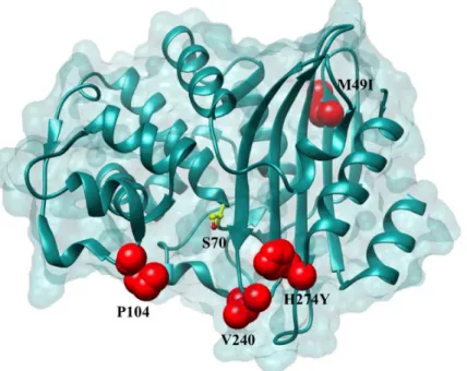
A total of nine KPC-2 variants, four variants differing from KPC-2 by a single amino acid and five variants differing from KPC-2 by two amino acids, were constructed by site-directed muta-genesis and introduced intoE.coliRB791 cells (Materials and Methods). The same strain con-taining the expression plasmid that did not encode KPC-2 was used as a negative control. The effect of the mutations on the resistance profiles inE.coliwas evaluated by determining MIC’s Fig 2. Positions of the variants residues included in this study on the KPC-2 enzyme.Positions that are substituted in variant enzymes are highlighted in red. The catalytic Ser70 is represented in yellow as a ball and stick model.
doi:10.1371/journal.ppat.1004949.g002
Table 1. Nucleotide polymorphisms and amino acid changes in variants as compared to KPC-2.
Variant Nucleotide Change Amino Acid Change GenBank ID
KPC-3 814 C–>T H274Y AF395881
KPC-4 308 C–>G P104R AY700571
716 T–>G V240G
KPC-5 308 C–>G P104R EU400222
KPC-6 716 T–>G V240G EU555534
KPC-7 147 G–>A M49I EU729727
814 C–>T H274Y
KPC-8 716 T–>G V240G FJ234412
814 C–>T H274Y
KPC-9 716 T–>C V240A FJ624872
814 C–>T H274Y
KPC-10 308 C–>G P104R GQ140348
814 C–>T H274Y
KPC-11 308 C–>T P104L HM066995
for each variant for a penicillin (ampicillin), an oxyimino-cephalosporin (ceftazidime) and the carbapenems, imipenem and meropenem (Table 2). Comparing the resistance profiles of the variant enzymes in an identical genetic background allows assignment of any changes in resis-tance to the corresponding single or double amino acid change, thus highlighting the role of specific residues in resistance to specific substrates (Table 2).
The MIC values of the four single amino acid variants, H274Y (KPC-3), P104R (KPC-5), V240G (KPC-6) and P104L (KPC-11) for ampicillin, imipenem and meropenem were within 2-fold of the KPC-2 MIC’s for these substrates. Interestingly, the H274Y (KPC-3) and V240G (KPC-6) substitutions resulted in a 4-fold increase in resistance to ceftazidime, while the P104R (KPC-5) substitution resulted in a 5-fold increase in resistance to ceftazidime. While the P104L (KPC-11) substitution did not display any change in resistance to ceftazidime, the other clinically observed single amino acid changes in KPC-2 result in an increase in resistance to ceftazidime while maintaining the resistance levels to penicillin and carbapenem antibiotics.
Similar to the single amino acid variants, the two amino acid variants did not display any significant differences for ampicillin, imipenem and meropenem MIC’s. The only exception was P104R:H274Y (KPC-10) that displayed a 4-fold and 3-fold decrease in MIC for ampicillin and meropenem, respectively. However, consistent with the observation for the single amino acid variants, each of the double amino acid variants resulted in an increase in resistance to tazidime. While M49I:H274Y (KPC-7) resulted in a modest 4-fold increase in resistance to cef-tazidime, V240A:H274Y (KPC-9), P104R:V240G (KPC-4), P104R:H274Y (KPC-10) and V240G:H274Y (KPC-8) resulted in 10-, 30-, 40- and 80-fold increases in ceftazidime MIC, re-spectively. The observation that these substitutions do not affect resistance to penicillin and carbapenem antibiotics while increasing resistance to ceftazidime implicates ceftazidime as the selective pressure for the acquisition of these variants in clinical isolates. Also, the dramatic in-crease in ceftazidime resistance in the two amino acid variants as compared to the single amino acid variants indicates a step-wise evolution with the acquisition increasing resistance with each amino acid variation.
Steady-state enzyme kinetics
In order to have a biochemical correlate to the MIC data, each KPC variant was purified and steady-state kinetic parameters were determined for ampicillin, imipenem, meropenem and cef-tazidime (Table 3). Consistent with the MIC data, the single amino acid changes did not result Table 2. Minimum inhibitory concentrations (MIC’s) of antibiotics for KPC variants.
MIC (μg / mL)
AMP CAZ IMI MERO
pTP123-empty 16 0.125 0.38 0.064
pTP123-blaKPC-2 (KPC-2) 128 0.38 1 0.38
P104R (KPC-5) 64 2.0 0.75 0.25
P104L (KPC-11) 64 0.5 0.75 0.25
V240G (KPC-6) 128 1.5 1.0 0.25
H274Y (KPC-3) 64 1.5 1 0.25
P104R:V240G (KPC-4) 64 12 1 0.25
P104R:H274Y (KPC-10) 32 16 1 0.125
V240A:H274Y (KPC-9) 128 4 1 0.19
V240G:H274Y (KPC-8) 128 32 2 0.25
M49I:H274Y (KPC-7) 64 1.5 1 0.25
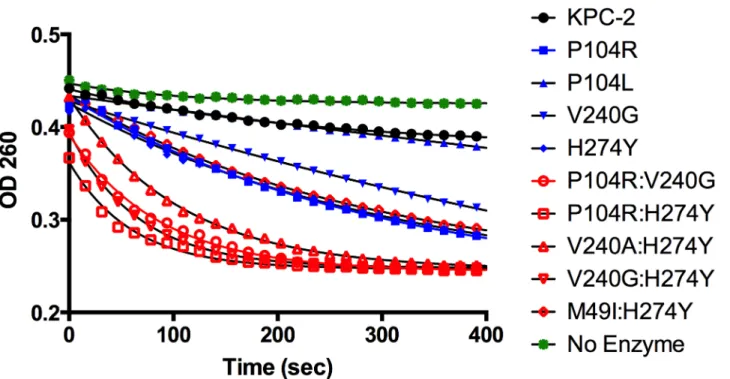
in greater than 2-fold changes in catalytic efficiencies (kcat/Km) for ampicillin, imipenem and meropenem. The KPC variants, as well as the parental KPC-2, have highKmvalues for ceftazi-dime, and saturating levels of substrate cannot be obtained. However, thekcat/Kmvalue was de-termined under conditions where [S]<<Km. Although the individualkcatandKmvalues could not be determined for ceftazidime hydrolysis by KPC-2 and the variants, a progress curve of the reaction with identical amounts of enzyme and substrate clearly shows the differences in activity of the enzymes (Fig 3). The KPC-2 enzyme hydrolyzes ceftazidime poorly with a catalytic effi-ciency of 8 x 10⁻⁴μM-1sec-1. Consistent with the ceftazidime MIC results, the P104L mutant ex-hibited only a modest 2-fold increase in catalytic efficiency for ceftazidime hydrolysis. In contrast, 5-fold, 9-fold and 11-fold increases were observed for the V240G, H274Y and P104R mutants, respectively. Thus, while both the P104R and P104L substitutions are found in clinical isolates, arginine at this position seems to be preferred as compared to leucine for ceftazidime hydrolysis. Both the MIC and enzymatic data suggest that mutation of Pro104 to Arg results in the highest resistance levels to ceftazidime among the single amino acid variants due to the in-creased ability of this variant enzyme to hydrolyze ceftazidime as compared to KPC-2. Table 3. Kinetic parameters of KPC variants.
AMP IMI MERO CAZ
KPC-2 kcat(sec-1) 50±7 48±3 3.8±0.7
Km(μM) 226±68 252±30 36±16
kcat/Km(μM-1.sec-1) 0.23±0.04 0.19±0.03 0.11±0.036 0.0008±0.0002
P104R kcat(sec-1) 314±15 28±1 3.0±0.4
(KPC-5) Km(μM) 1407±123 247±22 53±10
kcat/Km(μM-1.sec-1) 0.22±0.01 0.12±0.01 0.06±0.004 0.009±0.001
P104L kcat(sec-1) 12.8±0.2 54±4 2.5±0.1
(KPC-11) Km(μM) 136±18 244±31 35±7
kcat/Km(μM-1.sec-1) 0.1±0.01 0.22±0.01 0.07±0.01 0.002±0.0002
V240G kcat(sec-1) 146±4 27±1 2.3±0.07
(KPC-6) Km(μM) 318±11 172±23 30±4
kcat/Km(μM-1.sec-1) 0.46±0.01 0.16±0.02 0.08±0.008 0.004±0.0005
H274Y kcat(sec-1) 224±16 34±4 1.6±0.1
(KPC-3) Km(μM) 432±48 108±25 31±8
kcat/Km(μM-1.sec-1) 0.52±0.02 0.32±0.04 0.05±0.019 0.007±0.0003
P104R:V240G kcat(sec-1) 61±5 21±1 2.3±0.04
(KPC-4) Km(μM) 538±84 157±18 30±3
kcat/Km(μM-1.sec-1) 0.12±0.01 0.14±0.01 0.08±0.006 0.04±0.002
P104R:H274Y kcat(sec-1) 190±3 31±1 3.1±0.07
(KPC-10) Km(μM) 633±42 211±23 36±1
kcat/Km(μM-1.sec-1) 0.3±0.02 0.15±0.01 0.08±0.003 0.06±0.01
V240A:H274Y kcat(sec-1) 49±4 27±3 2.2±0.2
(KPC-9) Km(μM) 148±24 113±28 21±2
kcat/Km(μM-1.sec-1) 0.33±0.03 0.24±0.04 0.1±0.001 0.02±0.004
V240G:H274Y kcat(sec-1) 24±0.3 24±2 2.6±0.2
(KPC-8) Km(μM) 132±23 110±22 17±2
kcat/Km(μM-1.sec-1) 0.18±0.03 0.22±0.02 0.16±0.007 0.03±0.003
M49I:H274Y kcat(sec-1) 225±26 32±1 3.0±0.1
(KPC-7) Km(μM) 411±108 120±1 31±3
kcat/Km(μM-1.sec-1) 0.56±0.08 0.26±0.01 0.1±0.006 0.006±0.0002
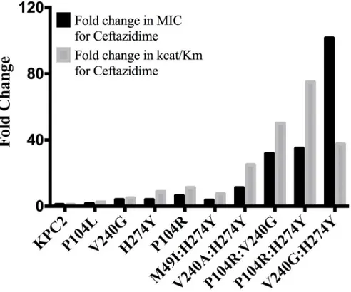
The catalytic efficiencies of the double mutants for ampicillin, imipenem and meropenem hydrolysis remained within 2-fold of the KPC-2 catalytic efficiencies for the same substrates. Except for P104R:H274Y (KPC-10), which displayed 4-fold and 3-fold decreases in MIC for ampicillin and meropenem, respectively, the enzyme kinetic data is in agreement with the MIC values for these substrates. For ceftazidime hydrolysis, M49I:H274Y (KPC-7) had the least im-pact with an 8-fold increase in catalytic efficiency as compared to KPC-2. The V240A:H274Y (KPC-9) and V240G:H274Y (KPC-8) variants exhibited 25- and 40-fold increases in catalytic efficiency while the P104R:V240G (KPC-4) and P104R:H274Y (KPC-10) double mutants had the largest impact with 50- and 75-fold increases, respectively, in catalytic efficiency for ceftazi-dime hydrolysis (Table 3). V240G:H274Y (KPC-8) exhibited the highest MIC for ceftazidime amongst all the mutants but did not display the highest catalytic efficiency. Thus, while the V240G:H274Y (KPC-8) mutant follows the overall trend of increasing activity, its catalytic effi-ciency does not directly correlate with the MIC value (Fig 4). This may reflect the fact that the MIC value is influenced by a number of variables including protein expression, stability and solubility in addition to catalytic efficiency. In summary, acquisition of the single and double substitutions associated with the variants allows KPC-2 to hydrolyze ceftazidime more effi-ciently and broadens the substrate profiles of the enzymes (Fig 3).
Additivity relationships between single and double amino-acid
substitutions
The MIC and enzyme kinetics data indicate that the double mutants P104R:V240G (KPC-4), P104R:H274Y (KPC-10) and V240G:H274Y (KPC-8) show higher ceftazidime resistance and hydrolysis rates as compared to the constituting single mutants. When two substitutions are in-troduced into the enzyme together, their combined effect on catalysis may be additive or coop-erative. For additive interactions, the fold change in the double mutant is expected to be the Fig 3. Progress curves of KPC-2 (black), single mutants (blue) and double mutants (red) and no enzyme control (green) for ceftazidime hydrolysis. All reactions were performed with 500 nM enzyme and 50μM ceftazidime. Hydrolysis of ceftazidime results in a loss of absorbance at 260 nm.
product of the fold changes for the individual mutants. However, if the two substitutions inter-act (directly or indirectly), the fold change in the double mutant may be much higher (or lower) than that expected from the additive effects of the two single substitutions. To determine whether the interactions between P104R, V240G and H274Y are additive or cooperative for ceftazidime hydrolysis, the free energies (ΔΔG) of the single and double mutants were calculat-ed as describcalculat-ed previously [23]. Briefly, the free energies associated withkcat/Kmvalues for KPC-2 and the single and double variants were calculated usingEq 2:
DDG¼ RTln ðkcat=KMÞmutant ðk
cat=KMÞwild type
ð2Þ
Table 4. Free energy values and additivity relationships between substituents for ceftazidime hydrolysis.
ΔΔG ΔGi
KPC2 0
-P104R -0.63
-V240G -0.42
-H274Y -0.57
-P104R:V240G -1.02 0.03
P104R:H274Y -1.13 0.07
V240G:H274Y -0.95 0.04
Calculated fromkcat/Kmof ceftazidime hydrolysis.
doi:10.1371/journal.ppat.1004949.t004
Fig 4. Bar graph comparing the MIC for ceftazidime (black) and catalytic efficiency for ceftazidime hydrolysis (gray).Both values are represented as fold changes compared to KPC-2.
Subsequently, the coupling free energy (ΔGI) was calculated usingEq 3:
DDG
ðX;YÞ ¼ DDGðXÞþDDGðYÞþDGI ð3Þ
HereΔΔG(x,y)represents the free energy difference between the wild-type and double mutant;
ΔΔG(x)andΔΔG(y)represent the differences in free energy between the wild-type and each sin-gle mutant, respectively, andΔGIrepresents the coupling free energy [24]. The KPC-2 enzyme is considered wild-type for the purposes of these comparisons. If the interactions between the single mutations are purely additive, then the coupling free energy is zero,ΔGI= 0; however, if the substitutions are non-additive (positive or negative cooperativity), thenΔGI6¼0. The re-sultingΔΔG values andΔGIvalues are summarized inTable 4. TheΔGIvalues for P104R: V240G (KPC-4), P104R:H274Y (KPC-10) and V240G:H274Y (KPC-8) are 0.03, 0.07 and 0.04 respectively. These values are small compared to theΔΔG values of the individual mutants and, therefore, the P104R, V240G and H274Y residues interact additively to facilitate ceftazidime hydrolysis. This means that the individual substitutions act independently and do not influence each other’s function when present in the double mutants. It also indicates that the order in which the individual mutations that make up a double mutant occur is not important.
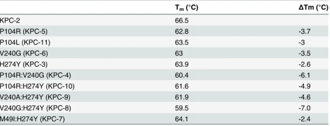
Determination of protein stability
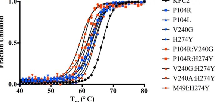
Substitutions close to the active site that alter enzyme function are often associated with a cost in terms of loss of protein stability [25–27]. Creating new, exposed hydrophobic surfaces or polar interactions that are satisfied only when substrate binds will be destabilizing to the en-zyme in the absence of substrate. In order to determine any cost associated with the substitu-tions in the KPC variants, the thermal stability of each purified variant enzyme was determined using circular dichroism spectroscopy by monitoringα-helix content at 222 nm with increas-ing temperature. The fit of the data and theTmvalues of the variants are summarized inFig 5 andTable 5. Single substitutions close to the active site result in a 2.6 to 3.7°C loss in protein stability as compared to KPC-2. With the exception of M49I:H274Y (KPC-7), the double
Fig 5. Thermal unfolding curves of KPC variants as measured by circular dichroism at 222 nm.The identity of each variant is indicated by the symbol shape and color shown in the inset.
mutants exhibited an even more dramatic reduction inTm. The P104R:V240G (KPC-4) and P104R:H274Y (KPC-10) double mutants displayed 6°C and 5°C reductions in Tm, respectively. The V240G:H274Y (KPC-8) mutant exhibited the largest effect among all variants with a de-crease in Tmof 7°C as compared to KPC-2. Interestingly, the V240A:H274Y (KPC-9) variant displayed a decrease in Tmof 5°C as compared to KPC-2. Thus, an alanine substitution at posi-tion 240 in combinaposi-tion with H274Y provides KPC-9 with 2°C increased stability compared to V240G:H274Y (KPC-8). Overall, the results clearly indicate that the substitutions found in the KPC variants decrease enzyme stability. Thus, the increase in ceftazidime hydrolysis resulting from the substitutions in the variants is associated with a cost in terms of stability.
Effect of mutations on protein expression levels
In addition to thermal stability and hydrolytic activity, protein expression levelsin vivoalso contribute to the overall resistance levels. Thus, to assess the effect of the single and double mu-tations on protein expression and the resulting effect on resistance levels, the steady-state ex-pression levels of KPC-2 and the variant enzymes were measured (Fig 6). As expected, KPC-2, which has the highest Tm, also exhibits the highest expression level. The single mutants P104R, P104L and V240G showed a marginal decrease in expression while H274Y showed a 2-fold crease. Among the double mutants, V240:H274Y and M49I:H274Y displayed the largest de-crease in expression levels (3-and 4-fold respectively) while P104R:V240G and P104R:H274Y displayed a modest 2-fold decrease. The V240G:H274Y variant displayed the highest expres-sion levels amongst all the double mutants. This provides an explanation for why this mutant showed the highest resistance to ceftazidime but not the highest catalytic efficiency (Fig 4). Taken together, the overall trends in expression levels are similar to the thermal stability results wherein the single and double mutants show a decrease in expression level as compared to KPC-2. The small magnitude of differences amongst mutants is not surprising considering that even the lowest Tmobserved among the KPC variants is 59.5°C, which is higher as compared to other class Aβ-lactamases such as TEM-1β-lactamase [28].
In silico binding studies
Due to the absence of any structural data for the variants, molecular modeling was used to ex-amine potential mechanisms by which the mutations increase the catalytic efficiencies for cef-tazidime hydrolysis. Autodock Vina [29] was used to predict the binding conformation and interactions of ceftazidime with the wild-type and variant enzymes. The P104R:H274Y (KPC-10) variant was selected for study as it exhibited the largest increase in catalytic efficiency for Table 5. Melting temperatures of KPC-2 and variants.
Tm(°C) ΔTm (°C)
KPC-2 66.5
P104R (KPC-5) 62.8 -3.7
P104L (KPC-11) 63.5 -3
V240G (KPC-6) 63 -3.5
H274Y (KPC-3) 63.9 -2.6
P104R:V240G (KPC-4) 60.4 -6.1
P104R:H274Y (KPC-10) 61.6 -4.9
V240A:H274Y (KPC-9) 61.9 -4.6
V240G:H274Y (KPC-8) 59.5 -7.0
M49I:H274Y (KPC-7) 64.1 -2.4
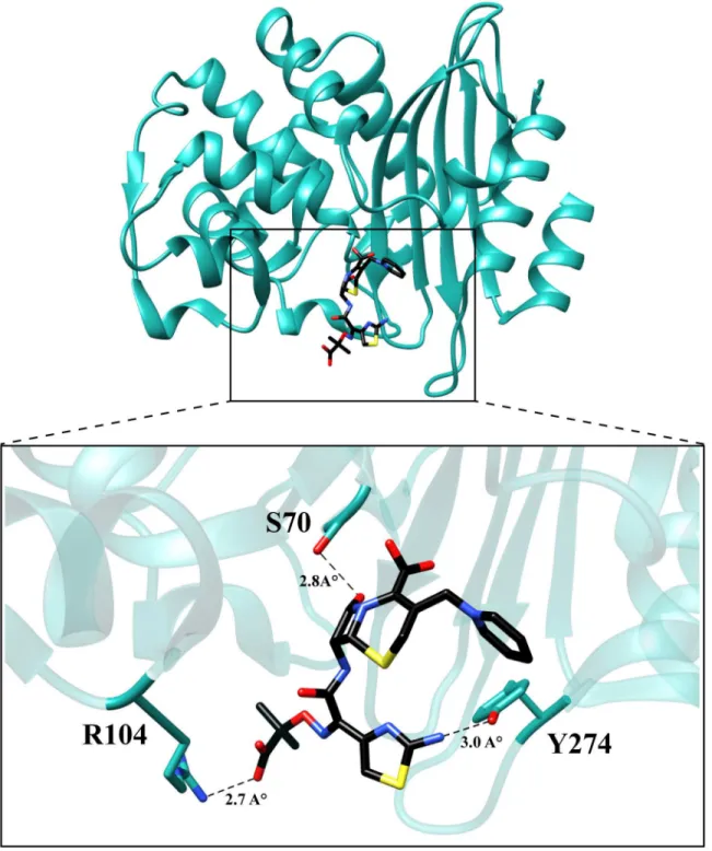
ceftazidime hydrolysis. The KPC-2 structure was used as a starting point and the P104R and H274Y substitutions were modeled based on predicted low energy conformations (Materials and Methods) [30]. Ceftazidime was then docked into the mutant structure using Autodock Vina and the top five results were compared. The binding conformation that displayed theβ -lactam carbonyl oxygen positioned in the oxyanion hole and exhibited the highest number of hydrogen bonding interactions with ceftazidime was selected for further analysis. The analysis suggests that mutating residue 104 from proline to arginine promotes hydrolysis of ceftazidime by formation of an additional hydrogen bond between the guanidinium nitrogen of the argi-nine and the carboxyl functionality of the oxyimino group on ceftazidime. The docking results further suggest that substitution of histidine with tyrosine at position 274 results in the forma-tion of a hydrogen bond between the tyrosine hydroxyl side chain and the amine funcforma-tionality of the aminothiazole ring. (Fig 7). These interactions could result in increased catalytic efficien-cy through improved substrate binding or via transition state stabilization.
Discussion
Infections caused by bacteria producing KPC-2β-lactamase have been associated with high rates of morbidity and mortality [14]. The presence of the KPC-2 enzyme and the consequent Fig 6. Protein expression levels of KPC-2β-lactamase and variant enzymes.KPC-2 is represented in black, single mutants in blue and double mutants in red. Band intensities from two independent experiments were used to plot the bar graph.
carbapenem-resistance reduces treatment options [14]. In recent years, a number of variants of KPCβ-lactamase have been identified from patient samples covering a wide geographic distri-bution [17–22]. In this study, amino acid substitutions associated with several clinically isolat-ed KPC variants were examinisolat-ed in an identical plasmid and genetic background to evaluate if the substitutions result in altered patterns of hydrolysis and resistance toβ-lactam antibiotics. Fig 7. Molecular model of ceftazidime binding to the variant P104R:H274Y (KPC-10).The residues are represented in cyan and ceftazidime is represented in black. The dotted lines represent hydrogen bonds with the distances labeled.
The clinically-identified single and double mutants of KPC-2 represent evolved versions of KPC-2 with an expanded substrate profile that includes the oxyimino-cephalosporin ceftazi-dime. Importantly, these variants do not exhibit substantially reduced activity towards carbape-nems, creating a further threat to antibiotic therapy.
A broadening substrate profile through the acquisition of mutations has previously been ob-served for the TEM and CTX-Mβ-lactamases [27,31,32]. Interestingly, residue 104 plays a cru-cial role in conferring ceftazidime hydrolyzing ability to TEMβ-lactamase [31,33]. The E104K mutation in TEM-1 results in a 4-fold increase in MIC for ceftazidime and a 50-fold increase in catalytic efficiency for ceftazidime hydrolysis [33]. Similarly, in the case of CTX-Mβ -lacta-mases, a D240G substitution increases the ceftazidime MIC by 8-fold due to a 10-fold increase in catalytic efficiency of ceftazidime hydrolysis [34]. The improved ceftazidime hydrolyzing ability of KPC-2 variants containing substitutions at residues 104 and 240 reveals that this is a common strategy among class A enzymes for expanding the substrate spectrum to ceftazidime. Interestingly, the H274Y substitution and the combinations such as P104R:V240G (KPC-4), P104R:H274Y (KPC-10), and V240G:H274Y (KPC-8) have not been associated with increased ceftazidime hydrolysis in other class Aβ-lactamases, suggesting these mutational pathways to ceftazidime resistance are unique to KPC-2. Modeling studies suggest that hydrogen bonds with amino acid residues at position 104 and 274 and substrate improve the catalytic efficiency of ceftazidime hydrolysis. Since glycine does not have a side-chain, modeling was not per-formed; however, substitution of residue 240 to glycine or alanine may expand the active site or increase flexibility in the region to accommodate ceftazidime.
A number of studies on class Aβ-lactamases as well as other enzymes indicate that muta-tions that alter function often lead to decreased stability [25–27,35,36]. This function-stability trade-off is attributed to the increased active site strain resulting from the increased activity as-sociated with a gain-of-function substitution [27]. The observed decrease in stability of the sin-gle and double mutants of KPC-2 is consistent with a function-stability trade-off. This is illustrated inFig 8by a plot of the log of the catalytic efficiency for ceftazidime hydrolysis ver-sus the thermal stability of the KPC mutants, which reveals a strong correlation between the gain of function and loss of stability (R2= 0.8). Thus, there is a clear inverse relationship be-tween function (catalytic efficiency) and stability for the KPC group of enzymes studied here.
The evolutionary advantage of a highly stable enzyme as a starting point for the selection of variants with altered function has been demonstrated for several systems in directed evolution experiments [39]. For example, the presence of the M182T stabilizing substitution in TEM-1 reduces the number of random single amino acid substitutions that inactivate the enzyme by one-third [40]. Similar observations have been made with a P450 enzyme as well as a thermo-stable chorismate mutase [41,42]. Thus, excess stability provides a buffer for an enzyme to ab-sorb mutations that are catalytically beneficial but are associated with a stability cost, such as the KPC mutations characterized in this study.
To date 22 KPC variants have been identified. This study provides a detailed analysis of 9 var-iants. Besides these variants, a recent study characterized KPC-15, which exhibits increased cef-tazidime activity and has the P104R, V240G, H274Y mutations as well as A120L and G147K substitutions [43]. Our results suggest that the P104R, V240G and H274Y substitutions contrib-ute strongly to the ceftazidime hydrolysis activity of this variant. Of the remaining uncharacter-ized variants, KPC-13 (GenBank: HQ342889) and KPC-19 (GenBank: KJ775801) have the H274Y mutation in conjunction with D92G and N293T substitutions, respectively. Based on the presence of the H274Y mutation, we would speculate that these variants have increased ceftazi-dime hydrolysis activity. However, it is difficult to speculate on the roles of D92G and N293T considering their position is far away from the active site. 14 (GenBank: JX524191), KPC-16 (GenBank: KC465199), KPC-17 (GenBank: KC465200) and KPC-22 (GenBank: KM379100), each possess unique mutations that have not been characterized. Additionally, while KPC-18, KPC-20 and KPC-21 have been identified and annotated a Genbank ID, their sequences are not yet available for analysis (http://www.lahey.org/studies/other.asp#table1).
As described here, the KPC enzymes have evolved substitutions that result in increased ceftazi-dime hydrolysis. Yet, the number of KPC variant enzymes identified to date is an order of magni-tude less than the number of TEM variant extended spectrumβ-lactamases (ESBLs) known (~220). One reason for this is that TEM was identified in 1963 and TEM ESBLs were first identi-fied in 1983 while the KPC-2 was identiidenti-fied in the late 1990s and the first variants in 2001 Fig 8. Correlation plot of log catalytic efficiency for ceftazidime (y-axis) as compared to thermal stability of the variants (x-axis).KPC-2 (black circle), Single mutants (blue circle), Double mutants (red circle).
[1,16,44]. Thus, there has been less time and opportunity for the evolution of KPC variants. It is also possible that the increased stability of the KPC variants relative to TEM variants results in less selective pressure for second site substitutions that stabilize KPC such as are commonly observed among TEM ESBLs; for example M182T [38]. This would lead to less diversification of the KPC enzymes versus the TEM enzymes due to the accumulation of fewer stabilizing substitutions.
The KPC enzymes represent a versatile and adaptable group. This study highlights how KPC-2 is evolving under the pressure of current antibiotic therapy towards increased resistance to the oxyimino-cephalosporin, ceftazidime. The V240G:H274Y (KPC-8) double mutant pro-vides high-level resistance to allβ-lactam classes including penicillins, cephalosporins and car-bapenems antibiotics. The stability of this double mutant is 8°C lower than KPC-2, however, it is still substantially more stable than other class A enzymes. Therefore, one can expect the evo-lution of more KPC-variants with altered substrate profiles.
Materials and Methods
Bacterial strains and plasmids
E.coliK12 XL1-Blue strain (recA1endA1gyrA96thi-1hsdR17supE44relA1lac[F’proAB lacIqZΔM15 Tn10 (Tetr)] was obtained from Stratagene (La Jolla, CA) and used in site-directed mutagenesis experiments. TheE.coliRB791 strain was used for protein expression, purifica-tion and MIC determinapurifica-tions [45]. TheblaKPC-2gene was inserted in the previously described pTP123 plasmid [46]. The resulting plasmid was used to express and purify the KPC-2 enzyme and also used as a template for site-directed mutagenesis and subsequent expression of mutant enzymes inE.coliRB791 [47].
Site-directed mutagenesis
All KPC-2 mutants were created using the QuikChange kit (Stratagene, La Jolla, CA). Oligonu-cleotides were obtained from Integrated DNA Technologies (Coralville, IA). The following is the list of primers used to introduce mutations (underlined) into pTP123 KPC-2:
P104R: CAAAAATGCGCTGGTTCGCTGGTCACCCATCTC P104L: CAAAAATGCGCTGGTTCTGTGGTCACCCATCTC V240A: CGGAACCTGCGGAGCGTATGGCACGGCAAATG V240G: CGGAACCTGCGGAGGGTATGGCACGGCAAATG H274Y: CAAGGATGACAAGTACAGCGAGGCCGTCATC M49I: CGGTGTGTACGCGATAGATACCGGCTCAG
Minimum inhibitory concentration (MIC) determinations
Minimum inhibitory concentrations (MIC’s) forE.colistrain RB791 containing the KPC mu-tants was determined for imipenem, meropenem and ceftazidime using Etest strips (Ab Bio-disk, Sweden) according to the manufacturers recommendations. The MIC’s of the variants for ampicillin were determined using the broth dilution method in a 100-well microtiter format. Overnight cultures of the variants were diluted into wells containing two-fold dilutions of am-picillin in a total volume of 300μl LB broth. The plate was allowed to incubate overnight at 37°C with continuous shaking and scored for visible growth to determine the MIC.
Protein purification
overnight at 37°C. The overnight culture was added to 1 L LB broth containing 12.5μg/mL chloramphenicol at a final dilution of 1:100 and subsequently allowed to grow to OD6000.7 at 37°C. Protein expression was induced by addition of 1 M IPTG to a final concentration of 0.2 mM and the cultures were grown at 23°C overnight. The cells were harvested by centrifugation at 4000 xgfor 20 minutes and the pellet frozen for at least 1 hour at -80°C. To release the peri-plasmic contents, the pellet was resuspended in 50 mL of 10 mM Tris-HCl buffer, pH 8.0 con-taining 1 tablet of Complete Protease Inhibitor Cocktail (Roche Diagnostics Corporation, Indianapolis, IN) and incubated on ice for 1 hour. Subsequently, osmotic shock was initiated by addition of 50 mL of cold, sterile water. The insoluble material was pelleted by centrifugation at 10,000gfor 1 hour. The supernatant was filtered and passed through a HiTrap SP column (GE Healthcare, Piscataway, NJ). The P104R, P104R:V240G and P104R:H274Y mutants bound the column at pH 8.0 and were eluted using a NaCl gradient. The remaining enzyme variants were bound to the column by adjusting the buffer to pH 5.5 using MES acid and subsequently they were eluted using a NaCl gradient. The purity of theβ-lactamase containing fractions was deter-mined using SDS-PAGE and the pooled fractions were concentrated and subjected to size ex-clusion chromatography using a HiLoad Superdex 75 column (GE Healthcare, Piscataway, NJ). Protein concentrations were determined by measuring the optical density at 280 nm and using the following extinction coefficients for respective proteins: 39,545 M-1cm-1was used for KPC-2, KPC-4, KPC-6, KPC-11; 41,035 M-1cm-1for KPC-3, KPC-7, KPC-8, KPC-9, KPC-10 and 39,420 M-1cm-1for KPC-5. All the extinction coefficients were calculated using the‘ProtParam’
tool from the Swiss Institute of Bioinformatics online resource portal [48].
Enzyme kinetics
Michaelis-Menten kinetic parameters for KPC-2 and the variant enzyme-substrate pairs were determined at 25°C in 50 mM sodium phosphate buffer, pH 7.0, containing 0.1 mg/mL BSA using variable amounts of enzyme depending on the enzyme-substrate pair. The initial veloci-ties ofβ-lactam hydrolysis were measured on a Beckman-Coulter spectrophotometer model DU-800 (Fullerton, CA) using the following extinction coefficients: imipenem,Δε295= -9000 M-1cm-1; meropenem,Δε295= -10,940 M-1cm-1; ceftazidimeΔε295= -7600 M-1cm-1; ampicillin, Δε235= -900 M-1cm-1. GraphPad Prism 5 was used to obtain the steady-state parameters by non-linear least squares fit of the data to the Michaelis-Menten equationv = kcat[S]/(Km+ [S]). The velocity of ceftazidime hydrolysis could not be saturated by measurable concentrations due to a highKm. Thus, the second order rate constant at steady-state,kcat/Km, was determined by
fitting the progress curves to the equationv = kcat/Km[E][S], where [S]<<Km(eq. 1).
Thermal denaturation
Thermal denaturation experiments were performed as described previously [28]. In short, the thermal stability of the KPC variants was measured on a Jasco J-815 circular dichroism spectro-polarimeter (Jasco, Essex, UK) coupled with a Peltier effect temperature controller. A total of 0.15 mg/mL of enzyme in 50 mM sodium phosphate buffer, pH 7.0, was placed in a 0.1 cm quartz cuvette and unfolding of the proteins was observed at 222 nm by heating the samples from 40°C to 80°C in 0.1°C increments at a rate of 1°C min-1. The melting temperature (Tm) is the temperature mid-point of protein unfolding and was determined by fitting the data to a sin-gle Boltzmann model.
Protein expression levels
diluted 1:100 into LB broth containing 12.5μg/mL chloramphenicol. Cells were grown to OD600= 0.6 at 37°C and 5 mL culture was pelleted by centrifugation at 13,000 rpm. The peri-plasmic contents were released by resuspending the cells in 100μl of 10 mM Tris pH 8.0 buffer, containing 20% sucrose and osmotic shock induced by adding 100μl of cold sterile water. The insoluble fraction was separated by centrifugation [23]. Total protein concentrations were de-termined using the Bradford protein assay. 1μg of total periplasmic proteins were loaded in each well. Purified KPC-2 enzyme was used as a positive control and periplasmic fraction from cells containing the empty vector was used as a negative control. The blot was probed using polyclonal anti-KPC antibody as the primary antibody and anti-rabbit, horseradish peroxidase (HRP) conjugated antibody as the secondary antibody. SuperSignal West Pico Chemilumines-cent Substrate (Thermo Scientific, Rockford, IL) was used as substrate for the secondary anti-body. Band intensities were quantified using ImageJ software.
Molecular modeling
In the absence of structural data, molecular modeling was performed to evaluate the effects of the mutations on KPC-2β-lactamase. A molecular model of the P104R:H274Y was created by mutating the KPC-2 structure [30] (PDB ID: 2OV5)in silicousing the Dunbrack rotamer li-brary as a part of the UCSF Chimera software [49,50]. The Dunbrack backbone-dependent rotamer library predicts the conformation of the amino acid side-chain based on the global en-ergy minimum of the protein. A PDB file for ceftazidime was created using the CORINA soft-ware, Molecular Networks Gmbh, Earlangen, Germany. Ceftazidime was then docked into this model to predict the Michaelis-Menten complex using the Autodock Vina docking method as described previously [29,51]. Briefly, the protein was prepared for docking by adding polar hy-drogen atoms using AutoDockTools. The grid box was centered on the catalytic Ser-70 residue and the dimensions of the docking space (26 x 30 x 22 A°) were adjusted to include the entire catalytic site. Of the 5 results, the model with ceftazidime positioned in the binding conforma-tion with theβ-lactam carbonyl directed into the oxyanion hole and displaying the largest number of hydrogen bond and hydrophobic interactions was chosen for further analysis.
Acknowledgments
The authors thank Dr. Paul Leonard, Dr. Todd Link and the Center for Biomolecular Structure and Function, University of Texas MD Anderson Cancer Center for the use of their Circular Dichroism instrument. The authors also thank Dr. Hardik I. Parikh for his insights for the mo-lecular modeling experiments and Dr. Hiram Gilbert for comments on the manuscript.
Author Contributions
Conceived and designed the experiments: SCM TP. Performed the experiments: SCM KR TP. Analyzed the data: SCM KR TP. Contributed reagents/materials/analysis tools: SCM KR. Wrote the paper: SCM TP.
References
1. Livermore DM, Woodford N. Theβ-lactamase threat in Enterobacteriaceae,Pseudomonasand Acine-tobacter. Trends Microbiol. 2006; 14: 413–420. PMID:16876996
2. Arias CA, Murray BE. Antibiotic-Resistant Bugs in the 21st Century—A Clinical Super-Challenge. N Engl J Med. 2009; 360: 439–443. doi:10.1056/NEJMp0804651PMID:19179312
3. Kong KF, Schneper L, Mathee K. Beta-lactam antibiotics: from antibiosis to resistance and bacteriolo-gy. APMIS. 2010; 118: 1–36. doi:10.1111/j.1600-0463.2009.02563.xPMID:20041868
5. Birnbaum J, Kahan FM, Kropp H, MacDonald JS. Carbapenems, a new class of beta-lactam antibiotics. Discovery and development of imipenem/cilastatin. Am J Med. 1985; 78: 3–21. PMID:3925777
6. Maveyraud L, Mourey L, Kotra LP, Pedelacq J-D, Guillet V, Mobashery S, et al. Structural Basis for Clinical Longevity of Carbapenem Antibiotics in the Face of Challenge by the Common Class Aβ -Lac-tamases from the Antibiotic-Resistant Bacteria. J Am Chem Soc. 1998; 120: 9748–9752.
7. Nukaga M, Bethel CR, Thomson JM, Hujer AM, Distler A, Anderson VE, et al. Inhibition of class Aβ -lac-tamases by carbapenems: crystallographic observation of two conformations of meropenem in SHV-1. J Am Chem Soc. 2008; 130: 12656–12662. doi:10.1021/ja7111146PMID:18761444
8. Fonseca F, Chudyk EI, van der Kamp MW, Correia A, Mulholland AJ, Spencer J. The basis for carbape-nem hydrolysis by class Aβ-lactamases: a combined investigation using crystallography and simula-tions. J Am Chem Soc. 2012; 134: 18275–18285. doi:10.1021/ja304460jPMID:23030300
9. Helfand MS, Bonomo RA.β-lactamases: a survey of protein diversity. Curr Drug Targets Infect Disord. 2003; 3: 9–23. PMID:12570729
10. Crowder MW, Spencer J, Vila AJ. Metallo-β-lactamases: novel weaponry for antibiotic resistance in bacteria. Acc Chem Res. 2006; 39: 721–728. PMID:17042472
11. Pfeifer Y, Cullik A, Witte W. Resistance to cephalosporins and carbapenems in Gram-negative bacterial pathogens. Int J Med Microbiol. 2010; 300: 371–379. doi:10.1016/j.ijmm.2010.04.005PMID:
20537585
12. Walsh TR. Emerging carbapenemases: a global perspective. Int J Antimicrob Agents. 2010; 36 Suppl 3: S8–14. doi:10.1016/S0924-8579(10)70004-2PMID:21129630
13. Bush K, Fisher JF. Epidemiological expansion, structural studies, and clinical challenges of newβ -lac-tamases from gram-negative bacteria. Annu Rev Microbiol. 2011; 65: 455–478. doi: 10.1146/annurev-micro-090110-102911PMID:21740228
14. Nordmann P, Cuzon G, Naas T. The real threat ofKlebsiella pneumoniaecarbapenemase-producing bacteria. Lancet Infect Dis. 2009; 9: 228–236. doi:10.1016/S1473-3099(09)70054-4PMID:19324295
15. Naas T, Cuzon G, Villegas MV, Lartigue MF, Quinn JP, Nordmann P. Genetic structures at the origin of acquisition of theβ-lactamaseblaKPCgene. Antimicrob Agents Chemother. 2008; 52: 1257–1263. doi:
10.1128/AAC.01451-07PMID:18227185
16. Yigit H, Queenan AM, Anderson GJ, Domenech-Sanchez A, Biddle JW, Steward CD, et al. Novel car-bapenem-hydrolyzingβ-lactamase, KPC-1, from a carbapenem-resistant strain ofKlebsiella pneumo-niae. Antimicrob Agents Chemother. 2001; 45: 1151–1161. PMID:11257029
17. Hidalgo-Grass C, Warburg G, Temper V, Benenson S, Moses AE, Block C, et al. KPC-9, a novel carba-penemase from clinical specimens in Israel. Antimicrob Agents Chemother. 2012; 56: 6057–6059. doi:
10.1128/AAC.01156-12PMID:22964247
18. Lamoureaux TL, Frase H, Antunes NT, Vakulenko SB. Antibiotic resistance and substrate profiles of the class A carbapenemase KPC-6. Antimicrob Agents Chemother. 2012; 56: 6006–6008. doi:10. 1128/AAC.01338-12PMID:22908150
19. Lascols C, Hackel M, Hujer AM, Marshall SH, Bouchillon SK, Hoban DJ, et al. Using nucleic acid micro-arrays to perform molecular epidemiology and detect novelβ-lactamases: a snapshot of extended-spectrumβ-lactamases throughout the world. J Clin Microbiol. 2012; 50: 1632–1639. doi:10.1128/ JCM.06115-11PMID:22322349
20. Robledo IE, Aquino EE, Sante MI, Santana JL, Otero DM, Leon CF, et al. Detection of KPC in Acineto-bacterspp. in Puerto Rico. Antimicrob Agents Chemother. 2010; 54: 1354–1357. doi:10.1128/AAC. 00899-09PMID:20038618
21. Wolter DJ, Kurpiel PM, Woodford N, Palepou MF, Goering RV, Hanson ND. Phenotypic and enzymatic comparative analysis of the novel KPC variant KPC-5 and its evolutionary variants, KPC-2 and KPC-4. Antimicrob Agents Chemother. 2009; 53: 557–562. doi:10.1128/AAC.00734-08PMID:19015357
22. Woodford N, Tierno PM Jr., Young K, Tysall L, Palepou MF, Ward E, et al. Outbreak ofKlebsiella pneu-moniaeproducing a new carbapenem-hydrolyzing class Aβ-lactamase, KPC-3, in a New York Medical Center. Antimicrob Agents Chemother. 2004; 48: 4793–4799. PMID:15561858
23. Adamski CJ, Cardenas AM, Brown NG, Horton LB, Sankaran B, Prasad BV, et al. Molecular Basis for the Catalytic Specificity of the CTX-M Extended-Spectrumβ-Lactamases. Biochemistry. 2014; 54: 447–458. doi:10.1021/bi501195gPMID:25489790
24. Wells JA. Additivity of mutational effects in proteins. Biochemistry. 1990; 29: 8509–8517. PMID:
2271534
25. Schreiber G, Buckle AM, Fersht AR. Stability and function: two constraints in the evolution of barstar and other proteins. Structure. 1994; 2: 945–951. PMID:7866746
27. Wang X, Minasov G, Shoichet BK. Evolution of an Antibiotic Resistance Enzyme Constrained by Stabil-ity and ActivStabil-ity Trade-offs. J Mol Biol. 2002; 320: 85–95. PMID:12079336
28. Brown NG, Pennington JM, Huang W, Ayvaz T, Palzkill T. Multiple global suppressors of protein stabili-ty defects facilitate the evolution of extended-spectrum TEMβ-lactamases. J Mol Biol. 2010; 404: 832– 846. doi:10.1016/j.jmb.2010.10.008PMID:20955714
29. Trott O, Olson AJ. AutoDock Vina: improving the speed and accuracy of docking with a new scoring function, efficient optimization, and multithreading. J Comput Chem. 2010; 31: 455–461. doi:10.1002/ jcc.21334PMID:19499576
30. Ke W, Bethel CR, Thomson JM, Bonomo RA, van den Akker F. Crystal structure of KPC-2: insights into carbapenemase activity in class Aβ-lactamases. Biochemistry. 2007; 46: 5732–5740. PMID:
17441734
31. Sowek JA, Singer SB, Ohringer S, Malley MF, Dougherty TJ, Gougoutas JZ, et al. Substitution of lysine at position 104 or 240 of TEM-1pTZ18R beta-lactamase enhances the effect of serine-164 substitution on hydrolysis or affinity for cephalosporins and the monobactam aztreonam. Biochemistry. 1991; 30: 3179–3188. PMID:1901218
32. Cantu C 3rd, Huang W, Palzkill T. Cephalosporin substrate specificity determinants of TEM-1β -lacta-mase. J Biol Chem. 1997; 272: 29144–29150. PMID:9360991
33. Petit A, Maveyraud L, Lenfant F, Samama JP, Labia R, Masson JM. Multiple substitutions at position 104 ofβ-lactamase TEM-1: assessing the role of this residue in substrate specificity. Biochem J. 1995; 305 (Pt 1): 33–40. PMID:7826350
34. Bonnet R, Recule C, Baraduc R, Chanal C, Sirot D, De Champs C, et al. Effect of D240G substitution in a novel ESBL CTX-M-27. J Antimicrob Chemother. 2003; 52: 29–35. PMID:12775683
35. Brown NG, Pennington JM, Huang W, Ayvaz T, Palzkill T. Multiple Global Suppressors of Protein Sta-bility Defects Facilitate the Evolution of Extended-Spectrum TEMβ-Lactamases. J Mol Biol. 2010; 404: 832–846. doi:10.1016/j.jmb.2010.10.008PMID:20955714
36. Meiering EM, Serrano L, Fersht AR. Effect of active site residues in barnase on activity and stability. J Mol Biol. 1992; 225: 585–589. PMID:1602471
37. Chen Y, Delmas J, Sirot J, Shoichet B, Bonnet R. Atomic resolution structures of CTX-Mβ-lactamases: extended spectrum activities from increased mobility and decreased stability. J Mol Biol. 2005; 348: 349–362. PMID:15811373
38. Huang W, Palzkill T. A natural polymorphism inβ-lactamase is a global suppressor. Proc Natl Acad Sci U S A. 1997; 94: 8801–8806. PMID:9238058
39. Bloom JD, Arnold FH. In the light of directed evolution: pathways of adaptive protein evolution. Proc Natl Acad Sci U S A. 2009; 106 Suppl 1: 9995–10000. doi:10.1073/pnas.0901522106PMID:
19528653
40. Bloom JD, Silberg JJ, Wilke CO, Drummond DA, Adami C, Arnold FH. Thermodynamic prediction of protein neutrality. Proc Natl Acad Sci U S A. 2005; 102: 606–611. PMID:15644440
41. Besenmatter W, Kast P, Hilvert D. Relative tolerance of mesostable and thermostable protein homo-logs to extensive mutation. Proteins. 2007; 66: 500–506. PMID:17096428
42. Bloom JD, Labthavikul ST, Otey CR, Arnold FH. Protein stability promotes evolvability. Proc Natl Acad Sci U S A. 2006; 103: 5869–5874. PMID:16581913
43. Wang D, Chen J, Yang L, Mou Y, Yang Y. Phenotypic and enzymatic comparative analysis of the KPC variants, KPC-2 and its recently discovered variant KPC-15. PLoS One. 2014; 9: e111491. doi:10. 1371/journal.pone.0111491PMID:25360633
44. Datta N, Kontomichalou P. Penicillinase synthesis controlled by infectious R factors in Enterobacteria-ceae. Nature. 1965; 208: 239–241. PMID:5326330
45. Amann E, Brosius J, Ptashne M. Vectors bearing a hybridtrp-lacpromoter useful for regulated expres-sion of cloned genes inEscherichia coli. Gene. 1983; 25: 167–178. PMID:6363212
46. Petrosino J, Rudgers G, Gilbert H, Palzkill T. Contributions of aspartate 49 and phenylalanine 142 resi-dues of a tight binding inhibitory protein ofβ-lactamases. J Biol Chem. 1999; 274: 2394–2400. PMID:
9891008
47. Brown NG, Chow DC, Palzkill T. BLIP-II is a highly potent inhibitor ofKlebsiella pneumoniae carbapene-mase (KPC-2). Antimicrob Agents Chemother. 2013; 57: 3398–3401. doi:10.1128/AAC.00215-13
PMID:23587951
48. Artimo P, Jonnalagedda M, Arnold K, Baratin D, Csardi G, de Castro E, et al. ExPASy: SIB bioinformat-ics resource portal. Nucleic Acids Res. 2012; 40: W597–603. doi:10.1093/nar/gks400PMID:
49. Dunbrack RL Jr. Rotamer libraries in the 21st century. Curr Opin Struct Biol. 2002; 12: 431–440. PMID:
12163064
50. Pettersen EF, Goddard TD, Huang CC, Couch GS, Greenblatt DM, Meng EC, et al. UCSF Chimera–a visualization system for exploratory research and analysis. J Comput Chem. 2004; 25: 1605–1612. PMID:15264254