D I A B E T E S & M E T A B O L I S M J O U R N A L
his is an Open Access article distributed under the terms of the Creative Commons At-tribution Non-Commercial License (http://creativecommons.org/licenses/by-nc/3.0/) which permits unrestricted non-commercial use, distribution, and reproduction in any medium, provided the original work is properly cited.
Exercise Treadmill Test in Detecting Asymptomatic
Coronary Artery Disease in Type 2 Diabetes Mellitus
Mee Kyoung Kim, Ki Hyun Baek, Ki Ho Song, Hyuk Sang Kwon, Jung Min Lee, Moo Il Kang, Kun Ho Yoon, Bong Yun Cha, Ho Young Son, Kwang Woo Lee
Department of Internal Medicine, he Catholic University of Korea School of Medicine, Seoul, Korea
Background: he present study was designed to develop criteria for screening patients with type 2 diabetes mellitus (T2DM) for asymptomatic coronary artery disease (CAD).
Methods: A total of 213 patients with T2DM without typical angina or chest pain were studied between 2002 and 2007. We also evaluated 53 patients with T2DM who had reported chest discomfort using an exercise treadmill test (ETT).
Results: hirty-one of the 213 asymptomatic patients had positive ETT results. We performed coronary angiography on 23 of the 31 patients with a positive ETT and found that 11 of them had signiicant coronary stenosis. he main diferences between the patients with signiicant stenosis and those with a negative ETT were age (63.1±9.4 vs. 53.7±10.1 years, P=0.008) and
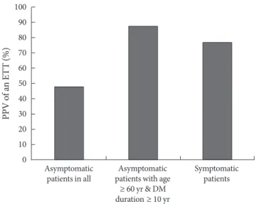
dura-tion of diabetes (16.0±7.5 vs. 5.5±5.7 years, P<0.001). he positive predictive value (PPV) of the ETT was calculated to be 47.8%.
he PPV of the ETT increased to 87.5% in elderly patients (≥60 years) with a long duration of diabetes (≥10 years). he latter value is similar to that of patients with T2DM who presented with chest discomfort or exertional dyspnea. he PPV of the ETT in symptomatic patients was 76.9%.
Conclusion: In the interest of cost-efectiveness, screening for asymptomatic CAD could be limited to elderly patients with a duration of diabetes ≥10 years.
Keywords: Diabetes mellitus; Duration of diabetes; Exercise treadmill test; Silent myocardial ischemia
Corresponding author: Ki Hyun Baek
Division of Endocrinology and Metabolism, Department of Internal Medicine, Yeouido St. Mary’s Hospital, he Catholic University of Korea School of Medicine, 62 Yeouido-dong, Yeongdeungpo-gu, Seoul 150-713, Korea E-mail: [email protected]
INTRODUCTION
In patients with type 2 diabetes mellitus (T2DM), coronary artery disease (CAD) is generally detected at an advanced stage with extensive atherosclerosis and poor outcomes, whereas CAD is commonly missed in the asymptomatic stage [1]. Dia-betic patients with asymptomatic CAD have a higher cardiac mortality risk than those with symptomatic CAD [2]. In the largest autopsy study of diabetic patients without antemortem evidence of CAD, approximately 50% of patients under 65 years of age and 75% of those 65 years of age and older had high-grade coronary atherosclerosis [3].
Performing routine screening for asymptomatic CAD in all patients with T2DM is debatable for several reasons [4-8]; there-fore, it is important to adopt appropriate criteria for selecting high risk patients who would beneit most from routine CAD screening. he CAD screening results may inluence a physi-cian’s lifestyle advice for the patient, treatment targets for gly-cemia, blood pressure, or cholesterol, or decisions regarding the appropriateness of aspirin or angiotensin-converting en-zyme inhibitor therapy [9]. CAD screening might identify in-dividuals who could benefit from anti-ischemia therapy (β- blocker) or from revascularization, and speciically those with let main or severe multivessel disease [4].
In the present study, which involved asymptomatic patients with T2DM, we used the diagnostic approach of performing a coronary angiogram following a demonstration of myocardial ischemia during an exercise treadmill test (ETT). he present study was designed to develop criteria for screening patients with T2DM for asymptomatic coronary artery disease.
METHODS
Study patients
A total of 213 outpatients who underwent ETT were enrolled from St. Mary’s Hospital Diabetes Clinics (Seoul, Korea) be-tween March 2002 and November 2007. he exclusion criteria were type 1 or secondary diabetes, previous cardiovascular disease, typical angina or chest pain, resting electrocardiogram (ECG) signs of myocardial ischemia, and severe systemic dis-ease with a poor prognosis. he diagnosis of T2DM was based on American Diabetes Association (ADA) criteria [10]. Pati-ents with blood pressure over 140/90 mm Hg or those taking antihypertensive agents were recorded as being hypertensive (HBP). Diabetic nephropathy was deined as the appearance of abnormal urinary albumin levels: microalbuminuria (30-299 mg albumin/24 hr) or macroalbuminuria (>300 mg albu-min/24 hr). We also analyzed 53 patients with T2DM who un-derwent an ETT to evaluate their chest discomfort (typical or atypical) or exertional dyspnea during the same period.
Protocol for coronary artery disease
he ETT was performed following Bruce protocol. he study patients ceased taking beta blockers and calcium channel bloc-kers 72 hours before the exercise test [11]. A 12-lead ECG was used and continuously monitored during the test, and blood pressure was recorded at rest and every 2 minutes during exer-cise and recovery. he ETT was considered positive when a horizontal or downsloping ST-segment with a depression ≥1 mm occurred 0.08 seconds ater the J point. he test was con-sidered equivocal when a ST segment depression<1 mm was observed, or when a left bundle branch block or premature ventricular beats >6 beats/min were seen. he test was consid-ered inconclusive if the patient failed to reach 85% of the pre-dicted maximal heart rate for his or her age. All subjects with a positive screening test underwent cardiology consultation. Coronary angiography was performed only when the ETT was positive. We deined asymptomatic CAD as objective evi-dence of ischemia occurring in an asymptomatic patient
dur-ing the ETT. We also deined low-limitdur-ing CAD as a stenosis of more than 70% on coronary angiography. Coronary artery stenosis greater than 70% compared to pre-stenotic measure-ments was considered signiicant for CAD.
Assays
All blood samples were taken in the morning following a min-imum 8-hour fast. Glycated hemoglobin (A1C) levels were de-termined by high-performance liquid chromatography (HLC-723 G7; Tosoh Co., Tokyo, Japan). Total cholesterol, triglycer-ides, and high density lipoprotein cholesterol (HDL-C) were determined using commercial kits.
Statistical analysis
All data were analyzed using SPSS version 10.0 for Windows statistical sotware (SPSS Inc., Chicago, IL, USA). he data are presented as mean±standard deviation, unless otherwise stated. Patient characteristics were compared by presence or ab-sence of symptoms using independent sample Student’s t-tests
for continuous measures and χ2 tests for categorical measures. Com parisons of the clinical and biochemical characteristics among the groups were made using ANOVA and post hoc analysis (Tukey test). P values <0.05 were considered
statisti-cally signiicant.
RESULTS
Clinical characteristics
he baseline clinical characteristics of all asymptomatic and
Table 1. Baseline clinical and laboratory characteristics
Asymptomatic (n=213)
Symptomatic
(n=53) P value
Age, yr 55.3±10.2 56.7±10.0 0.300
Sex, M/F 167/46 28/25 < 0.001
Duration of diabetes, yr 6.4±6.5 10.3±8.9 0.004
BMI, kg/m2 24.7±2.6 24.7±2.8 0.873
A1C, % 6.7±1.1 7.2±1.1 0.010
Total cholesterol, mmol/L 4.6±0.9 4.8±1.0 0.130
Triglyceride, mmol/L 1.7±1.1 1.7±1.3 0.536
HDL-C, mmol/L 1.1±0.3 1.1±0.3 0.821
HBP, % 46.4 58.5 0.063
Nephropathy, % 22.5 42.5 0.005
symptomatic patients are summarized in Table 1. he mean duration of diabetes was signiicantly longer in symptomatic patients versus those without symptoms (10.3±8.9 years vs. 6.4±6.5 years, P=0.004). he symptomatic patients had higher
A1C levels (7.2±1.1% vs. 6.7±1.1%, P=0.010) and a greater
prevalence of HBP (58.5% vs. 46.4%, P=0.063) and
nephropa-thy (42.5% vs. 22.5%, P=0.005).
Flowchart of asymptomatic patients who underwent the ETT
Fig. 1 shows the lowchart for patients who underwent an ETT. Eighteen patients (8.5%) were unable to complete the ETT; nine due to leg discomfort, ive due to fatigue, three due to dys-pnea, and one due to dizziness. Nine of the 195 completed tests were not considered diagnostic because either the upsloping ST segment depression or the horizontal ST segment depres-sion was less than 1 mm. he remaining 186 completed tests provided data suitable for diagnosis and were subdivided into those having a positive ETT (n=31, 16.6%) and those having a
negative ETT (n=155, 83.3%). All subjects with a positive
screening test underwent cardiology consultation. Among the 31 patients with a positive treadmill test, two had no defects on the thallium scan and six patients refused further evalua-tion. he remaining 23 patients underwent coronary angiog-raphy. Coronary angiography revealed coronary stenosis ≥70% in eleven patients: three patients had one diseased vessel, sev-en had two diseased vessels, and one had three diseased vessels. Of the remaining twelve patients, one presented with a 50-70% lumen reduction and eleven had either a lumen reduction of <50% or no lumen reduction. The positive predictive value (PPV) for the ETT was calculated to be 47.8% (37.5% for men and 71% for women) (Fig. 2).
Comparison of patient characteristics according to the results of the ETT and coronary angiography
We divided the 213 patients into three groups. One group had negative ETT results (Group A). he second group had a posi-tive ETT, but negaposi-tive coronary angiography (Group B). his second group, therefore, had only a positive functional test. he third group had a positive ETT and positive coronary an-giography, indicative of low-limiting coronary artery disease (Group C).
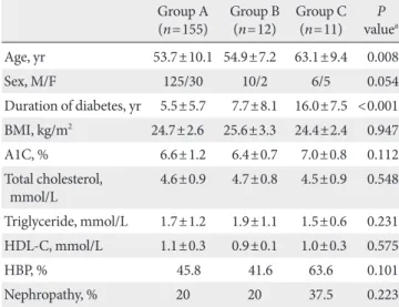
Table 2 shows a comparison of the diferences in variables for the three groups by ANOVA. he main diferences between the 11 patients who presented with coronary lesions (Group C) and the 155 patients with a negative ETT (Group A) were age (63.1±9.4 years vs. 53.7±10.1 years, P=0.008) and
dura-tion of diabetes (16.0±7.5 years vs. 5.5±5.7 years, P<0.001).
18 (8.5%) Were unable to complete the test
9 Were not diagnostic test
31 (16.6%) Were positive test 155 (83.3%) Were negative test 213 Underwent exercise treadmill test
195 (91.5%) Were able to complete the test
186 Were diagnostic test
Fig. 1. Flowchart of patients who underwent the exercise treadmill test.
Fig. 2. Positive predictive values (PPV) of an exercise treadmill test (ETT) for coronary artery stenosis. DM, diabetes mellitus.
Asymptomatic patients in all
Asymptomatic patients with age
≥ 60 yr & DM duration ≥ 10 yr
Symptomatic patients 100
90
80
70
60
50
40
30
20
10
0
P
P
V
o
f a
n
E
T
T
(
%
Serum A1C levels and lipid proiles were not signiicantly dif-ferent among the three groups.
ETT in elderly patients (≥60 years) with a long duration of diabetes (≥10 years)
We determined that the main factors inluencing the results of the ETT and coronary angiography were age and duration of
diabetes. We arbitrarily selected an age of 60 years and a 10-year duration of diabetes as cut-of points. We examined how the results might change if the elderly patients with a long du-ration of diabetes underwent only an ETT. Fig. 3 shows the lowchart for the 29 elderly study patients with a long duration of diabetes who underwent only the ETT. Of these 29 patients who underwent an ETT, 4 (13.7%) were unable to complete the ETT. In addition, 2 of the 25 completed tests were not con-sidered diagnostic, and the remaining 23 tests were concon-sidered suitable for diagnosis. Of the 23 suitable tests, almost half of them (n=10, 43.4%) had a positive ETT. Eight of the ten
pa-tients who had a positive ETT underwent coronary angiogra-phy, and 7 of the 8 had coronary stenosis greater than 70%. In this segment of our study population, the PPV using the ETT was 87.5% (Fig. 2).
ETT results for patients who presented with chest discomfort or exertional dyspnea
We analyzed 53 study patients who underwent the ETT to evaluate their (typical or atypical) chest discomfort or exer-tional dyspnea. Among them, 9 patients (16.9%) were unable to complete the ETT. Four of the 44 completed tests were not considered diagnostic and the remaining 40 patients’ tests were considered suitable for diagnosis. Seventeen patients (42.5%) had a positive ETT. hirteen of the seventeen patients under-went coronary angiography and ten of the thirteen patients (three patients with one diseased vessel, four with two diseased Table 2. Clinical and laboratory characteristics by exercise
tread mill test and coronary angiography results
Group A (n=155)
Group B (n=12)
Group C (n=11)
P valuea
Age, yr 53.7±10.1 54.9±7.2 63.1±9.4 0.008
Sex, M/F 125/30 10/2 6/5 0.054
Duration of diabetes, yr 5.5±5.7 7.7±8.1 16.0±7.5 <0.001
BMI, kg/m2 24.7±2.6 25.6±3.3 24.4±2.4 0.947
A1C, % 6.6±1.2 6.4±0.7 7.0±0.8 0.112
Total cholesterol, mmol/L
4.6±0.9 4.7±0.8 4.5±0.9 0.548
Triglyceride, mmol/L 1.7±1.2 1.9±1.1 1.5±0.6 0.231
HDL-C, mmol/L 1.1±0.3 0.9±0.1 1.0±0.3 0.575
HBP, % 45.8 41.6 63.6 0.101
Nephropathy, % 20 20 37.5 0.223
Group A, negative ETT; Group B, positive ETT & negative CAG; Group C, positive ETT & positive CAG.
ETT, exercise treadmill test; CAG, coronary angiography; BMI, body mass index; HDL-C, high density lipoprotein cholesterol; HBP, high blood pressure.
aGroup A vs. Group C.
Fig. 3. Flowchart of elderly patients (≥60 years) with a long duration of diabetes (≥10 years). CAG, coronary angiography.
2 Were not diagnostic test 4 (13.7%) Were unable to complete the test 29 Underwent exercise treadmill test
25 (86.2%) Were able to complete the test
23 Were diagnostic test
13 (56.5%) Were negative test 10 (43.4%) Were positive test
8 Underwent CAG
7 Were angiographic stenosis 1 Was normal CAG
vessels, and three with three diseased vessels) had coronary stenosis greater than 70%. In this study population, the PPV using the ETT was calculated to be 76.9% (Fig. 2).
DISCUSSION
We found the main diferences between the asymptomatic pa-tients with signiicant coronary stenosis and those with nega-tive ETT to be duration of diabetes and age. he PPV of the ETT for predicting angiographic coronary disease was 48%. his value was found to be higher when the patients were el-derly (≥60 years) and had a long duration of diabetes (≥10 years), or were symptomatic. To improve cost-effectiveness, screening for asymptomatic CAD could be limited to elderly patients with a duration of diabetes of 10 years or more. As previously reported, even when a satisfactory ETT test is completed, the PPV in detecting silent coronary stenosis is low, between 40 and 60% [8,12,13]. In our study, the PPV of the ETT increased to 88% in elderly patients with a long duration of diabetes. his value was similar to that of type 2 diabetes mellitus patients who presented with chest discomfort or exer-tional dyspnea: the PPV of the ETT in symptomatic patients was 76.9%.
he ETT is oten not feasible for diabetic patients because their exercise capability may be impaired by peripheral vascu-lar or neuropathic disease [14]. In our study, 18 patients (8.5%) were unable to complete the ETT, mostly due to leg discom-fort or dyspnea. A completely normal ETT has been reported to be a marker for a good prognosis in patients with diabetes [14]. Compared with such imaging procedures as coronary computed tomography (CT), echocardiography, and stress single photon emission computed tomography myocardial per-fusion imaging, the treadmill exercise test can be performed at a much lower cost. he ETT is preferable to a pharmacological stress test because it depicts the heart’s actual workload and better simulates everyday cardiac strain [15]. Furthermore, patients are not exposed to ionizing radiation and contrast. herefore, initially performing an ETT in patients who are able to exercise is recommended. he more recently developed non-invasive, multi-slice CT angiography is recommended for ex-clusion of coronary heart disease, but has the associated disad-vantages of high radiation exposure and cost [14]. An estimat-ed 1 in 270 women who underwent CT coronary angiography at age 40 will develop cancer from that CT scan, compared with an estimated 1 in 8,100 women who had a routine head CT scan
at the same age [16]. For these reasons, we chose the ETT as a screening tool for coronary artery disease in asymptomatic patients with T2DM. Further studies will guide us with respect to which modality is most appropriate.
he 1998 ADA Consensus Development Conference on the diagnosis of coronary artery disease in people with diabetes recommended performing stress screening for coronary ar-tery disease in asymptomatic patients with ≥2 cardiovascular risk factors (smoking, arterial hypertension, hypercholesterol-emia, family history of premature CAD, microalbuminuria) [17]. Unfortunately, recent studies have shown that the pres-ence of traditional categorical risk factors did not help to iden-tify asymptomatic patients with a higher prevalence of coro-nary artery disease [18]. In the absence of symptomatic CAD, clinical features that help to identify patients with an increased risk for myocardial infarction or cardiac death include evidence of other atherosclerotic vascular disease, microalbuminuria and chronic kidney disease, autonomic neuropathy, retinopa-thy, hyperglycemia, abnormal resting ECGs, age, sex, and both traditional and novel cardiac risk factors [5]. However, the rou-tine screening of asymptomatic patients with T2DM and nor-mal ECGs remains controversial [19]. In our study, patients with signiicant coronary stenosis tended to have higher A1C levels and to have a greater prevalence of HBP and nephropa-thy, however, the diference was not statistically signiicant (Hb-A1C, 7.0% vs. 6.6%; HBP, 63.6% vs. 45.8%; Nephropathy, 37.5% vs. 20%). he size of our study population may have been too small to draw conclusions about the other covariates studied. We found the main diferences between the asymptomatic patients with significant coronary stenosis and those with a negative ETT were the duration of diabetes and age. It is well known that age is a strong predictor of CAD [5], however, du-ration of diabetes is not included among CAD predictors in the ADA recommendations [5]. Debate on the relationship between duration of diabetes and CAD continues, and several recent studies have reported a positive correlation [8,20]. Our study has several limitations. First, it used a retrospec-tive design, which is prone to selection bias. Second, data on other traditional cardiovascular risk factors, such as smoking, family history of premature CAD, autonomic neuropathy and retinopathy were not available. hird, clinical features such as chronic kidney disease, hyperglycemia, and hypertension did not difer between the groups. he small number of subjects in our study may also be a limitation.
ar-tery disease in elderly patients (≥60 years) with a duration of diabetes ≥10 years. Further studies are needed to evaluate the efectiveness of routine screening for asymptomatic CAD in this patient subgroup.
ACKNOWLEDGMENT
here are no conlicts of interest.
REFERENCES
1. Alexander CM, Landsman PB, Teutsch SM. Diabetes mellitus, impaired fasting glucose, atherosclerotic risk factors, and prev-alence of coronary heart disease. Am J Cardiol 2000;86:897-902.
2. Choi EK, Koo BK, Kim HS, Cho YM, Kang HJ, Cho YS, Chung WY, Chae IH, Choi DJ, Oh BH, Park YB, Choi YS. Prognostic significance of asymptomatic coronary artery disease in pa-tients with diabetes and need for early revascularization thera-py. Diabet Med 2007;24:1003-11.
3. Goraya TY, Leibson CL, Palumbo PJ, Weston SA, Killian JM, Pfeifer EA, Jacobsen SJ, Frye RL, Roger VL. Coronary athero-sclerosis in diabetes mellitus: a population-based autopsy study. J Am Coll Cardiol 2002;40:946-53.
4. Rutter MK, Nesto RW. he changing costs and beneits of screen-ing for asymptomatic coronary heart disease in patients with diabetes. Nat Clin Pract Endocrinol Metab 2007;3:26-35. 5. Bax JJ, Young LH, Frye RL, Bonow RO, Steinberg HO, Barrett
EJ; ADA. Screening for coronary artery disease in patients with diabetes. Diabetes Care 2007;30:2729-36.
6. Bacci S, Villella M, Villella A, Langialonga T, Grilli M, Rauseo A, Mastroianno S, De Cosmo S, Fanelli R, Trischitta V. Screen-ing for silent myocardial ischaemia in type 2 diabetic patients with additional atherogenic risk factors: applicability and ac-curacy of the exercise stress test. Eur J Endocrinol 2002;147: 649-54.
7. Araz M, Celen Z, Akdemir I, Okan V. Frequency of silent myo-cardial ischemia in type 2 diabetic patients and the relation with poor glycemic control. Acta Diabetol 2004;41:38-43.
8. Janand-Delenne B, Savin B, Habib G, Bory M, Vague P, Lass-mann-Vague V. Silent myocardial ischemia in patients with di-abetes: who to screen. Diabetes Care 1999;22:1396-400. 9. Buse JB, Ginsberg HN, Bakris GL, Clark NG, Costa F, Eckel R,
Fonseca V, Gerstein HC, Grundy S, Nesto RW, Pignone MP, Plutzky J, Porte D, Redberg R, Stitzel KF, Stone NJ; American
Heart Association; American Diabetes Association. Primary prevention of cardiovascular diseases in people with diabetes mellitus: a scientiic statement from the American Heart Asso-ciation and the American Diabetes AssoAsso-ciation. Circulation 2007;115:114-26.
10. Expert Committee on the Diagnosis and Classiication of Dia-betes Mellitus. Report of the Expert Committee on the Diag-nosis and Classification of Diabetes Mellitus. Diabetes Care 2003;26 Suppl 1:S5-20.
11. Gibbons RJ, Balady GJ, Beasley JW, Bricker JT, Duvernoy WF, Froelicher VF, Mark DB, Marwick TH, McCallister BD, hom-pson PD Jr, Winters WL, Yanowitz FG, Ritchie JL, Gibbons RJ, Cheitlin MD, Eagle KA, Gardner TJ, Garson A Jr, Lewis RP, O’Rourke RA, Ryan TJ. ACC/AHA Guidelines for Exercise Testing. A report of the American College of Cardiology/Ame-rican Heart Association Task Force on Practice Guidelines (Committee on Exercise Testing). J Am Coll Cardiol 1997;30: 260-311.
12. Penfornis A, Zimmermann C, Boumal D, Sabbah A, Mene-veau N, Gaultier-Bourgeois S, Bassand JP, Bernard Y. Use of dobutamine stress echocardiography in detecting silent myo-cardial ischaemia in asymptomatic diabetic patients: a com-parison with thallium scintigraphy and exercise testing. Diabet Med 2001;18:900-5.
13. Valensi P, Sachs RN, Lormeau B, Taupin JM, Ouzan J, Blasco A, Nitenberg A, Metz D, Paries J, Talvard O, Leutenegger M, At-tali JR. Silent myocardial ischaemia and let ventricle hypertro-phy in diabetic patients. Diabetes Metab 1997;23:409-16. 14. Djaberi R, Beishuizen ED, Pereira AM, Rabelink TJ, Smit JW,
Tamsma JT, Huisman MV, Jukema JW. Non-invasive cardiac imaging techniques and vascular tools for the assessment of cardiovascular disease in type 2 diabetes mellitus. Diabetolo-gia 2008;51:1581-93.
15. Xanthos T, Ekmektzoglou KA, Papadimitriou L. Reviewing myocardial silent ischemia: specific patient subgroups. Int J Cardiol 2008;124:139-48.
16. Smith-Bindman R, Lipson J, Marcus R, Kim KP, Mahesh M, Gould R, Berrington de Gonzalez A, Miglioretti DL. Radiation dose associated with common computed tomography exami-nations and the associated lifetime attributable risk of cancer. Arch Intern Med 2009;169:2078-86.
18. Scognamiglio R, Negut C, Ramondo A, Tiengo A, Avogaro A. Detection of coronary artery disease in asymptomatic patients with type 2 diabetes mellitus. J Am Coll Cardiol 2006;47:65-71. 19. American Diabetes Association. Standards of medical care in
diabetes--2009. Diabetes Care 2009;32 Suppl 1:S13-61. 20. Yoo WS, Kim HJ, Kim D, Lee MY, Chung HK. Early detection