Silent Myocardial Ischemia in Patients with Stable Coronary Artery
Disease Receiving Conventional Antianginal Drug Therapy
João Fernando Monteiro Ferreira, Luiz Antonio Machado César, César J. Gruppi, Dante M. A. Giorgi, Whady A. Hueb,
Antonio P. Mansur, José A. F. Ramires
Instituto do Coração do Hospital das Clínicas da Faculdade de Medicina de São Paulo, São Paulo, SP - Brazil
Summary
Background: Few data are available on the behavior of myocardial ischemia during daily activities in patients with coronary artery disease receiving antianginal drug therapy.
Objectives: To study the mechanism generating myocardial ischemia by evaluating blood pressure and heart rate changes in patients with stable atherosclerotic disease receiving drug therapy and with evidence of myocardial ischemia.
Methods: Fifty non-hospitalized patients (40 males) underwent 24-hour electrocardiographic monitoring synchronized with blood pressured monitoring.
Results: Thirty five episodes of myocardial ischemia were detected in 17 patients, with a total duration of 146.3 minutes; angina was reported in five cases. Twenty nine episodes (100.3 minutes) occurred during wakefulness, with 11 episodes (35.3 + 3.7 min) in the period from 11 a.m. to 3 p.m. Blood pressure and heart rate evaluation in the three ten-minute intervals following the ischemic episodes showed a statistically significant difference (p< 0.05), unlike that shown for the three intervals preceding the episodes. However, during the ischemic episode, a higher than 10-mmHg elevation in blood pressure and 5 beats per minute in heart rate were observed when compared with the time interval between 20 and 10 minutes before the episode. The mean heart rate at the onset of ischemia during the exercise test performed before the study was 118.2 + 14.0, and 81.1 + 20.8 beats per minute on the 24-hour electrocardiogram (p < 0.001).
Conclusions: The incidence of silent myocardial ischemia is high in stable coronary artery disease and is related to alterations in blood pressure and heart rate, with different thresholds for ischemia for the same patient. (Arq Bras Cardiol 2007;88(5):283-288)
Key words: Coronary disease; ischemia; electrocardiography, ambulatory; blood pressure.
Mailing address: João Fernando Monteiro Ferreira •
Rua Madre Rita Amada de Jesus, 72 / 222B – Granja Julieta - 04721-050 - São Paulo, SP - Brazil E-mail: [email protected]
Manuscript received November 21, 2006; revised manuscript received April 15, 2007; accepted June 11, 2007.
Introduction
Silent myocardial ischemia is considered the most frequent presentation of chronic coronary artery disease and was defined by Cohn et al, in 1981, as the evidence of myocardial ischemia in the absence of symptoms1. Its presence is related
to a higher risk of death and cardiac events2-4, and up to
three-fold higher mortality rates are observed in relation to individuals without ischemia5.
However, the role and behavior of the factors determining the occurrence of silent myocardial ischemia have not been fully established. Coronary artery obstruction, endothelial alterations, alterations in oxygen consumption factors, mainly in patients receiving antianginal, lipid-lowering and antihypertensive drugs, are known to participate in the process6-9.
To explain the behavior of the episodes of myocardial ischemia during daily activities and their determining aspects, we evaluated patients with stable coronary artery disease and proven active myocardial ischemia even when receiving drug therapy.
Methods
After approval by the Hospital’s Ethics Commission, 50 patients aged from 40 to 81 years (mean of 61.9 ± 10.7years) were studied. Forty (80%) were males with coronary artery disease confirmed by coronary angiography (with > 50% stenosis in at least one major coronary artery), in addition to the following criteria:
Inclusion criteria - evidence of clinical myocardial ischemia (effort angina) or functional myocardial ischemia (exercise test with > 1.0-mm ST-segment depression for males and > 2.0 mm for females and/or myocardial perfusion scintigraphy showing transient perfusion defects considered at least moderate).
minimum, maximum, mean values and standard deviations were analyzed. For qualitative variables, absolute and relative frequencies were calculated.
The Student’s t test was used to analyze the hypothesis of equality of means between the two groups. The non-parametric Mann-Whitney test was used when the hypothesis of normality of data was rejected. The chi-square test or the Fisher’s exact test was used to compare the proportions between the two groups. To assess the behavior of the group considering the ischemic conditions studied, the Analysis of Variance was used with repetitive measurements, which consists of the adjustment of a multivariate linear model. The significance level used for the tests was 5%.
Results
Characterization of patients - The analysis of risk factors for coronary artery disease showed 36.0% of patients with diabetes mellitus, 72.0% with hypertension, 32.0% currently smoking, 82.0% with hypercholesterolemia, 36.0% with hypertriglyceridemia, and 24.0% with a positive family history for coronary artery disease. During the study, patients were kept on the usual medications as prescribed by their physicians, with the following distribution: 78.0% beta-blockers, 40.0% calcium channel beta-blockers, 76.0% nitrates, 96.0% acetylsalicylic acid, 34.0% angiotensin converting enzyme inhibitors, and 38.0% lipid-lowering drugs. The evaluation of the lipid profile and blood glucose showed the following mean serum levels: total cholesterol 209.1 + 41.3 mg/dL, HDL-cholesterol 39.1 + 7.9 mg/dL, LDL-cholesterol 138.2 + 34.8 mg/dL, triglycerides 166.6 + 96.6 mg/dL, and blood glucose 140.0 + 66.8 mg/dL.
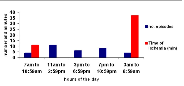
Characterization of the ischemic events - A total of 1112.4 hours of electrocardiographic monitoring was recorded, with an overall mean of 22.7 hours per patient. Thirty five episodes of myocardial ischemia with the duration of 146.3 minutes were detected in 17 patients (34.0%). Most of the ischemic episodes occurred during the active period, in a total of 29 (82.8%), whereas six (17.2%) episodes occurred during the passive period. Total ischemia time was 100.3 min in the active period, and 44.5 minutes in the passive period. The distribution of number and duration of ischemic episodes by 4-hour periods is shown in Graphic 1.
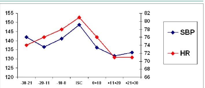
Of the 17 patients with ischemia, five (29.4%) reported angina during the study period, whereas four (12.1%) reported angina without ischemia on electrocardiography. Findings on SBP, DBP and HR during the ischemic episodes by time intervals are shown in Graphic 2.
The analysis of SBP, HR and DP variables between the time points prior to ischemia (-30-21), (-20-11) and (-10-0) in relation to the time point of ischemia (0) did not show any significant difference. The same variables following ischemia (0+10), (+11+20) and (+21+30), in turn, showed statistically significant differences (Table 1).
Comparison between the ischemic and non-ischemic groups - Once the characteristics of HR and blood pressure were determined at the time point of ischemia on dynamic electrocardiography, these variables could be compared between the ischemic and non-ischemic groups.
The analysis of the exercise test variables showed the following means for the ischemic and non-ischemic groups, respectively: exercise time to ischemia of 3.6 + 1.7 min vs.4.0 + 1.8 min (p=0.077), HR at the onset of electrocardiographic atrial flutter and fibrillation, tachycardias, artificial pacemaker,
left ventricular overload, or other ventricular repolarization abnormalities that affected the analysis of the ST-segment.
During the study, the patients continued receiving the antianginal drugs they had been using and underwent continuous 24-hour electrocardiographic and blood pressure monitoring with the Meditech CardioTens equipment (Medical Electronics, Budapest, Hungary). This system allows retrieval of simultaneous information on blood pressure and ischemic episodes.
The system was programmed to record the electrocardiogram automatically in two channels (CM5 and CM1), considering the basic ECG and ST-segment patterns. Patients were advised to report any type of symptom in an attached diary.
For the diagnosis of ischemic events, an isoelectric point was manually set (PQ interval and a point 60 ms from the J point) to determine the ST-segment baseline from which the presence of elevation or depression were considered. The ST-segment baseline was defined as the mean obtained during the 30 minutes preceding the ischemic episodes.
Diagnostic criteria of ischemic events - a > 2-mm elevation or a > 1-mm horizontal or downsloping ST-segment depression from the baseline that persisted for at least 1 min and reverted to baseline for at least one minute were considered diagnostic criteria of ischemic events10. A report on the ischemia time
of each event, as well as the time of the day when the events occurred, was prepared.
Patients were divided into two groups – ischemic and non-ischemic, according to the findings of myocardial ischemia on dynamic electrocardiography, and comparisons of different variables were made between the groups.
Assessment of the ischemic events - in the patients with ischemia, the total number of episodes and the total time in minutes (min) of all episodes during the 24 hours of observation, the total number of events, and the total ischemia time in minutes for each 4-hour period in the 24 hours, from 11:00p.m. to 2:59a.m., from 3:00a.m. to 6:59p.m., from 7:00a.m. to 10:59a.m., from 11:00a.m. to 2:59p.m., from 3:00p.m. to 6:59p.m., and from 7:00p.m. to 10:59p. m., respectively, were considered. The period from 7a.m. to 10:59p.m. was considered active, and that from 11p.m. to 6:59 a.m. was considered passive.
Blood pressure monitoring - this granted 24 consecutive hours of measurements, programmed to perform six systolic (SBP) and diastolic blood pressure (DBP) measurements/hour in mmHg, and heart rate (HR) in beats per minute (bpm). A measurement was started whenever the ST-segment level or the heart rate exceeded the limits set. Additional measurements could be taken manually at any time.
After each ischemic event was identified, we defined as (-30-21), (-20-11) and (-10-0) time points the measurements that occurred 30 to 20 min, 20 to 10 min, and up to 10 min before the onset of the ischemic episode, respectively. The same intervals [(0+10), (+11+20), and (+21+30)] were considered up to 30 minutes after the ischemic episode. Measurements taken when the (-30-21) time point was not more than 20 min from the last ischemic episode, and/or the (+21+30) time point was not less than 20 min from the next episode were disregarded. Thus, the mean SBP, DBP, HR and double product (DP) at each time point were obtained.
ischemia of 111.0 + 12.9 bpm vs. 121.3 + 13.6 bpm (p=0.085), maximum ST-segment depression of 2.7 + 0.9 mm vs.2.5 + 0.7 mm (p=0.760), peak SBP of 179.4 + 30.1 mmHg vs.190.6 + 28.8 mmHg (p=0.277), peak DBP of 87.0 + 14.7 mmHg vs.96.5 + 20.0 mmHg (p=0.248). The HR was determined both at the onset of myocardial ischemia on dynamic electrocardiography performed during the study, and on the exercise test performed at study admission: a significant difference was found with p<0.001 (Graphic 3).
No statistical difference was found between ischemic and non-ischemic patients as regards age (p = 0.098), as well as regards the different risk factors for coronary artery disease such as diabetes mellitus (p = 0.584), hypertension (p=0.746), smoking (p=0.357), hypercholesterolemia (p=0.467), hypertriglyceridemia (p=0.584) and positive family history for coronary artery disease (p=0.181).
Also, no difference was found in relation to the distribution of medication used by ischemic and non-ischemic patients: beta-blockers (p=1.000), calcium channel blockers (p=0.273), nitrates (p=0.728), acetylsalicylic acid (p=1.000), angiotensin converting enzyme inhibitors (p=0.442) and lipid lowering drugs (p=0.344).
Lipid and blood glucose values were similar between ischemic and non-ischemic patients: total cholesterol (p=0.186), HDL-cholesterol (p=0.258), LDL-cholesterol (p=0.072), triglycerides (p=0.743) and blood glucose (p=0.327).
As regards the results of coronary angiography, no difference was found in the pattern of coronary stenosis (equal to or higher than 70%) between ischemic and non-ischemic patients: ischemic single vessel 17.7% - non-ischemic single vessel: 27.3%; ischemic two-vessel 58.8% - non-ischemic two-vessel: 51.5% and ischemic three-vessel 23.5% - non-ischemic three-vessel: 21.2%.
Results of blood pressure monitoring according to the presence of ischemia on dynamic electrocardiography are shown in Table 2.
DISCUSSION
Our results show rates of patients with ischemia (34%) lower than those found by other researchers (43 to 89%)3,4,11. This
lower incidence of ischemia may possibly be explained by the fact that the medication was kept during the study and that the monitoring time was shorter (24 instead of 48 hours).
Most of the episodes occurred in the absence of symptoms, thus characterizing the diagnosis of silent ischemia, a result that is consistent with other studies7,11-13. The probable mechanisms
for the lack of perception of the ischemic phenomenon remain not fully understood. Maseri et al14 consider defects
in the system of pain perception, higher endorphin release, degeneration of nociceptive pathways, and baroceptor alterations as some of the mechanisms that explain the absence of angina. Even considering the greater number of patients with diabetes mellitus, a higher incidence of myocardial ischemia was not observed, which is consistent with Solimene and Oliveira’s15 as well as with Battlouni’s16 findings.
The evaluation of the variables of the exercise test performed before study admission showed that this method was not able to predict which patients would develop ischemia on daily activities. Paul et al17 also demonstrated
that the time for onset of ischemia, ST-segment depression, and total exercise time did not correlate with the presence of ischemia on the exercise test. Carboni et al18, Borzac et
al19 and other investigators also concluded that the exercise
test is not predictive of the development of ischemia during daily activities on electrocardiography and that the correlation between its severity indexes is low, except for the cases where prognostic signs of high risk were present. We also demonstrated a significant difference between the HR at the onset of the ischemic episode as measured by the exercise test and electrocardiography. Deedwania and Nelson13
demonstrated a variation in heart rate and blood pressure values at the onset of the ischemic episode, and that the numeric values of these parameters on the exercise test were
higher than those of the electrocardiography (110.6 vs. 91.7 beats per minute). Rehman et al20 found similar results for HR,
SBP and DP. Carboni et al18 also found similar results, although
only for patients who had presented a symptomatic ischemic episode. Deanfield et al21 used dynamic electrocardiography
to evaluate thirty patients with stable coronary artery disease and positive exercise test and also found a lower HR at the onset of ischemia on electrocardiography (98.0+20.5 vs. 124.0+17.0 bpm). Although factors determining oxygen consumption have an influence on the genesis of ischemic episodes during daily activities as presumed from the results of exercise test, their variation, and not only their absolute value, may determine ischemia, as observed on the 24-hour electrocardiography. That is, as we demonstrated, ischemia occurred even at a lower heart rate, thus showing the existence of different thresholds for ischemia.
As regards the distribution of the number and duration of ischemic episodes by periods and in the 24 hours, we observed predominance in the active period with a peak in the period going from 11a.m. to 2:59p.m., and a lower peak from 7p.m. to 10:59p.m. This circadian tendency of myocardial ischemia was reported in several studies that repeatedly demonstrated a higher number of episodes and longer time of ischemia during wakefulness, especially in the morning. Some investigators also reported a bimodal behavior with the presence of another ischemic peak at the beginning of the night, which is consistent with our findings8,13,22-24.
In relation to BP monitoring, we verified that both ischemic and non-ischemic patients maintained their SBP, DBP and
MBP controlled in the 24 hours and in the active and passive periods, like in Rehman et al’s study20 in a population similar
to ours. Therefore, the pressure load per se did not interfere with the occurrence of episodes of myocardial ischemia in our sample, thus suggesting that other mechanisms should participate in the genesis of silent ischemia.
Although we did not detect a relation between oxygen consumption factors (HR and SBP) and the ischemic episodes with the analysis chosen, heart rate means were observed to be 5 bpm higher at the moment of the ischemic episode, and systolic blood pressure means to be 10 mmHg higher than the measurements preceding ischemia. Deanfield et al21 evaluated
dynamic electrocardiography of patients with stable coronary artery disease and a positive exercise test and found only 23% of ischemic episodes preceded by a 10-beats-per-minute increase in heart rate. This additional assessment criterion, admitting slight variations of HR and SBP, was also used by other researchers. Panza et at25, in turn, evaluated 48 patients
with a similar profile and showed that 89.0% of the ischemic episodes were preceded by an increase equal to or higher than 10 bpm, and that this HR variation occurred up to 10 minutes before the onset of ischemia. Lupi et al26 conducted
an interesting study with 14 patients with stable coronary disease who had positive exercise tests. A second exercise test was performed 10 minutes after the first one, showing that heart rate and exercise time were significantly different, thus suggesting that the preconditioning phenomenon interferes with the genesis of myocardial ischemia in these patients. Rehman et al20 studied 38 patients with coronary artery
Table 1 – P values for SBP, HR and DP measurements in the intervals before and after ischemia compared to the time point of ischemia (0)
-30-21 -20-11 -10-0 0 0+10 +11+20 +21+30
SBP 0.195 0.081 0.265 - 0.019 0.046 0.040
HR 0.054 0.078 0.271 - 0.022 0.030 0.040
DP 0.151 0.081 0.302 - 0.028 0.044 0.047
SBP – systolic blood pressure, HR – heart rate, DP – double product
disease and positive exercise test for ischemia and performed combined electrocardiography and blood pressure monitoring for up to 48 hours in patients not taking medication. The authors did not find any significant difference in blood pressure and heart rate measurements in the 10 minutes before the ischemic event when compared to the measurements taken in the period of four hours before the ischemic event. However, DP, HR and SBP measurements during the ischemic episode were significantly higher than those in the other time points, thus showing that the changes in these variables are related to the genesis of ischemia, although occurring very close to the onset of ischemia. This result was also found in our study, since no statistically significant differences were observed between HR, SBP and DP measurements in the 30 minutes preceding the ischemic episode. However, when compared to the measurements obtained after the end of ischemia, the increase in these variables was demonstrated to participate in the onset of ischemia. Deedwania and Nelson13 evaluated 25
patients receiving medication and with proven silent ischemia for 24 hours with simultaneous electrocardiography and blood pressure monitoring and concluded that increases in SBP, HR and DP precede the ischemic event, reaching a peak at the moment of ischemia. However, the statistical analysis was
not performed so as to identify differences in the means, but considered increases of 5 beats per minute for heart rate and of 10 mmHg for blood pressure as significant; these variations were also found in our study. In addition to this analysis, a repetitive pattern is observed in the great majority of the studies, with progressive increases in HR, SBP and DP until their peak is reached during the ischemic episodes, followed by a decrease after the episodes. Therefore, patients with stable coronary artery disease had variations of HR and SBP preceding the ischemic episodes, thus showing that alterations in oxygen consumption participate in the genesis of myocardial ischemia. However, although variations in HR and SBP are present, they are not usually great, thus suggesting that an association of factors related to the coronary flow demand is necessary to the development of ischemic episodes, perhaps even with a common triggering factor.
Final considerations
During daily activities silent myocardial ischemia is present and frequent, even in patients with stable coronary artery disease receiving medication.. Paradoxically, it occurs at a lower HR than that observed in the exercise test, and is Table 2 – Blood pressure monitoring according to the presence of ischemia
Variables Ischemic Non-Ischemic p
SBP 7a.m.-10:59 p.m. (mmHg) 135.3 + 14.5 132.3 + 15.9 0.531
DBP 7a.m.-10:59p.m. (mmHg) 74.5 + 9.6 78.1 + 10.5 0.258
MBP 7a.m.-10:59p.m. (mmHg) 94.8 + 9.0 96.1 + 11.4 0.677
HR 7a.m.-10:59p.m. (bpm) 69.3 + 12.4 68.4 + 9.5 0.769
SBP 11p.m.-06:59a.m. (mmHg) 128.4 + 16.4 123.8 + 17.1 0.373
DBP 11p.m.-06:59a.m. (mmHg) 67.8 + 11.9 71.4 + 12.5 0.331
MBP 11p.m.-06:59a.m. (mmHg) 87.9 + 11.9 88.8 + 13.2 0.822
HR 11p.m.-06:59a.m. (bpm) 61.1 + 8.1 62.3 + 9.1 0.646
HR – heart rate; SBP – systolic blood pressure; DBP – diastolic blood pressure
Graph 3 - Heart rate at the onset of the ischemic episode on exercise test and on dynamic electrocardiography.
118.2*
81.1*
111.4
81.1
140
120
100
80
60
40
20
0
b
p
References
1. Cohn PF. Asymptomatic coronary artery disease: pathophysiology, diagnosis, management. Mod Concepts Cardiovasc Dis. 1981; 50 (10): 55-60.
2. Parmley WW. Prevalence and clinical significance of silent myocardial ischaemia. Circulation. 1989; 80: 68-73.
3. Rocco MB, Nabel EG, Campbell S, Goldman L, Barry JA, Mead K, et al. prognostic importance of myocardial ischemia detected by ambulatory monitoring in patients with stable coronary artery disease. Circulation. 1988; 78 (4): 877-84.
4. Tzivoni D, Weisz G, Gavish A, Zin D, Keren A, Stern S. Comparison of mortality and myocardial infarction rates in stable angina pectoris with and without ischemic episodes during daily activites. Am J Cardiol. 1998; 63: 173-6.
5. Deedewania PC, Carbajal EV. Silent ischemia during daily life is an independent predictor of mortality in stable angina. Circulation. 1990; 81 (3): 748-56.
6. McLenachan JM, Weidinger FF, Barry J, Yeung A, Nabel EGG, Rocco MB, et al. Relations between heart rate, ischemia, and drug therapy during daily life in patients with coronary artery disease. Circulation. 1991; 83 (4): 1263-70.
7. Deedwania PC, Carbajal EV. Prevalence and patterns of silent myocardial ischemia during daily life in stable angina patients receiving conventional antianginal drug therapy. Am J Cardiol. 1990; 65: 1090-6.
8. Impieri GA, Lambert CR, Coy K, Lopez L, Pepine CJ. Effects of titrated beta blockade (metoprolol) on silent myocardial ischemia in ambulatory patients with coronary artery disease. Am J Cardiol. 1987; 60: 519-24.
9. Pepine CJ, Cohn PF, Deedwania PC, Gibson RS, Gottlieb SO, Handberg E, et al. The prognostic and economic implications of a strategy to detect and treat asymptomatic ischemia: the atenolol silent ischemia trial (ASIST). Clin Cardiol. 1991; 14: 457-62.
10. Weiner DA, Becker L, Bonow R. Report of group II: detection and diagnosis. Circulation. 1987; 75: II49-50.
11. Deedwania PC, Carbajal EV. Ambulatory electrocardiography evaluation of asymptomatic, unstable, and stable coronary artery disease patients for myocardial ischemia. Cardiol Clin. 1992; 3: 417-30.
12. Schang SJ, Pepine CJ. Transient asymptomatic ST-segment depression during daily activity. Am J Cardiol. 1977; 39: 396-402.
13. Deedwania PC, Nelson JR. Pathophysiology of silent myocardial ischemia during daily life: hemodinamic evaluation by simultaneous electrocardiographic and blood pressure monitoring. Circulation. 1990; 82 (4): 1296-304.
14. Maseri A, Chiercia S, Glazier J. Mechanisms of ischemic cardiac pain and
silent myocardial ischemia. Am J Med. 1985; 79 (3A): 7-11.
15. Solimene MC, Oliveira SF. O enigma da isquemia sem dor. Arq Bras Cardiol. 1993; 60: 139-41.
16. Batlouni M. Mecanismos prováveis da isquemia miocárdica silenciosa. Arq Bras Cardiol. 1994; 63: 155-9.
17. Paul SD, Orav J, Gleason RE, Nesto RW. Use of exercise test parametrs to predict presence and duration of ambulatory ischemia in pateints with coronary disease. Am J Cardiol. 1994; 74: 991-6.
18. Carboni GP, Lahiri A, Cashman PMM, Raftery E. Ambulatory heart rate and st-segment depression during painful and silent myocardial ischemia in chronic stable angina pectoris. Am J Cardiol. 1987; 59: 1029-34.
19. Borzac S, Fenton T, Glasser SP, Shook TL, MacCallum G, Young PM, et al. for ASIS. Discordance between effects of anti-ischemic therapy on ambulatory ischemia, exercise performance, and anginal symptoms in patients with stable angina pectoris. J Am Coll Cardiol. 1993; 21: 1605-11.
20. Rehman A, Zalos G, Andrews NP, Mulcahy D, Quyyumi AA. Blood pressure changes during transient myocardial ischemia: insights into mechanisms. J Am Coll Cardiol. 1997; 30 (5): 1249-55.
21. Deanfield JE, Maseri A, Selwyn AP, Ribeiro P, Chierchia S, Krikler S, et al. Myocardial ischaemia during daily life in patients with stable angina: its relation to symptoms and heart rate changes. Lancet. 1983; 2 (8353): 753-8.
22. Rocco MB, Barry J, Campbell S, Nabel E, Cook F, Goldman L. Circadian variation of transient myocardial ischemia in patients with coronary artery disease. Circulation. 1987; 75 (2): 395-9.
23. Benhorin J, Banai S, Moriel M, Gavish A, Keren A, Stern S, et al. Circadian variations in ischemic threshold and their relation to occurence of ischemic episodes. Circulation. 1993; 87 (3): 808-14.
24. Wennerblom B, Lurje L, Karlsson T, Tygesen H, Vahisalo R, Hjalmarson A. Circadian variation of heart rate variability and the rate of autonomic change in the morning hours in healthy subjects and angina patients. Int J Cardiol. 2001; 79: 61-9.
25. Panza JA, Diodati JG, Callahan TS, Epstein SE, Quyyumi AA. Role of increases in heart rate in determining the occurrence and frequency of myocardial ischemia during daily life in patients with stable coronary artery disease. J Am Coll Cardiol. 1992; 20: 1092-8.
26. Lupi A, Lanza GA, Crea F, Proietti I, Maseri A. The “warm-up” phenomenon occurs in patients with chronic stable angina but not in patients with syndrome X. Am J Cardiol. 1998; 81 (2): 123-7.
preceded by slight variations of HR.
Alterations in SBP and HR occur in the moments before and after the episodes of myocardial ischemia, thus confirming that the factors that determine oxygen consumption interfere with the genesis of myocardial ischemia even in patients receiving antianginal medication. Nevertheless, there are different thresholds for ischemia for a given patient, thus demonstrating that oxygen consumption is not the only determinant of myocardial ischemia.
Potential Conflict of Interest
No potential conflict of interest relevant to this article was
reported.
Sources of Funding
This study was funded by FAPESP.
Study Association