1 State Medical University, Internal Medicine Department, Zaporozhye, Ukraine 2 State Medical University, Clinical Pharmacology Department, Zaporozhye, Ukraine
Correspondence: Alexander E. Berezin,
Cardiology Unit, Internal Medicine Department, State Medical University, 26, Mayakovsky av., Zaporozhye, Postcode, Ukraine. Email: [email protected]
Received: 15.04.2014, Accepted: 24.12.2014
Journal of Clinical and Experimental Invesigaions doi: 10.5799/ahinjs.01.2014.04.0449 ORIGINAL ARTICLE / ÖZGÜN ARAŞTIRMA
The relaionship between serum uric acid level and concentraion of proangiogenic
mononuclear progenitor cells in paients with chronic heart failure
Kronik kalp yetmezliği bulunan hastalarda serum ürik asit düzeyi ile proanjiojenik
mononükleer projenitör hücre konsantrasyonu arasındaki ilişki
Alexander E. Berezin1, Alexander A. Kremzer2
ÖZET
Amaç: Serum ürik asit (ÜA) kronik kap yetmezliğinin (KKY) doğal ilerleyişinin bir belirteci olarak bilinir. KKY ilerlemesi kanda dolaşan mononükleer projenitör hücrele
-rin (MPH) azalması ile birliktedir. Bu çalışmanın amacı is
-kemik KKY’li hasatlarda serum ÜA ve proanjiyojenik MPH konsantrasyonları arasındaki ilişkiyi değerlendirmektir. Yöntemler: Bu çalışma kontrastlı bilgisayarlı tomografi anjiyografisi ile koroner arter hastalığı (KAH) tanısı alan KKY’li 126 hastada (54 erkek)retrospektif olarak yapıl
-mıştır. Serum ÜA enzimatik yöntemle ve N-terminal proB
-NP (NT-pro-B-NP) düzeyi immünoelektrokemilüminesens yöntemi ile çalışıldı.
Bulgular: Serum ÜA konsantrasyonları kuartallar ola
-rak değerlendirildi (Me; IQR): QI=20.11 (19.06; 22.33) mmol/l; QII=27.53 (23.2; 31.10) mmol/l; QIII=35.80 (32.0; 39.0) mmol/l ve QIV=44.9 (40.00; 49.60) mmol/l. Serum ÜA’e göre düzeltilmiş Cox oransal Odds oranı analizleri CD14+CD309+ ve CD14+CD309+Tie2+ MPH için SUA Quartiles (Q) ile yapıldı ve Yüksek serum ÜA Q (Q3 ve Q4) ve düşük Q (Q1 ve Q2)’nin artmış CD14+CD309+ ve CD14+CD309+Tie2+ MPH tükenme riski ile birlikte oldu
-ğu gösterildi. ROC analizi ile serum ÜA 31,5 mmol/l de
-ğeri MPH azalma riski için en iyi potansiyel cut-off de-ğeri olarak belirlendi.
Sonuç: Konjestif kalp yetmezliği bulunan hastalarda do
-laşan proanjiyojenik MPH, serum ÜA kuartallarına bağlı olarak ilerleyici bir şekilde azaldı. Serum ÜA’de hafif artı
-şın düşük proanjiyojenik MPH’nin bir öngördürücüsü ola
-rak kabul edilebilir.
Anahtar kelimeler: Kronik kalp yetmezliği, serum ürik asit, dolaşan mononükleer projenitör hücreler, prediktif değer
ABSTRACT
Objective: Serum uric acid (UA) is considered as a marker of natural progression of chronic heart failure (CHF). Pro
-gression of CHF associates with declining of circulating mononuclear progenitor cells (MPCs) in the blood. The objective of this study was to evaluate the interrelationship between SUA concentrations and proangiogenic MPCs in ischemic CHF patients.
Methods: The study was structured retrospectively after determining the coronary artery disease (CAD) by con
-trast-enhanced spiral computed tomography angiography in 126 subjects (54 male), aged 48 to 62 years, with CHF. Serum UA level was measured by enzymatic method and N-terminal proBNP (NT-pro-BNP) level was examined by immunoelectrochemiluminesence method. All biomarkers were measured at baseline.
Results: Concentrations of SUA were distributed by quar
-tiles (Me; IQR): QI=20.11 (19.06; 22.33) mmol/l; QII=27.53 (23.2; 31.10) mmol/l; QIII=35.80 (32.0; 39.0) mmol/l; and QIV=44.9 (40.00; 49.60) mmol/l. Cox proportional ad
-justed Odds Ratios analyses for CD14+CD309+ and CD14+CD309+Tie2+ MPCs by SUA Quartiles (Q) has showed that high Q (Q3 and Q4) of SUA versus low Q (Q1 and Q2) associated with increased risk of depletion of both CD14+CD309+ and CD14+CD309+Tie2+ MPCs. The ROC analysis has been showed that there was the cut-off point for the SUA level with the best prognostic potential on the risk of decreasing MPCs in both models equal 31.5 mmol/l.
Conclusion: Circulated level of proangiogenic MPCs is declined progressively depended on quartiles of serum UA level in CHF subjects. We suggest that mild elevation of serum UA might be considered as a predictor of low pro
-angiogenic MPCs in CHF patients. J Clin Exp Invest 2014; 5 (4): 511-520
Key words: Chronic heart failure, serum uric acid, circulat
INTRODUCTION
Chronic heart failure (CHF) has been remained a potential fatal complication of any cardiovascular diseases and it is characterized by a systemic in
-lammatory response that leads to end organ dam
-age [1]. Serum uric acid (SUA) has been shown to be an independent predictor of outcome in the gen
-eral population and in patients with CHF, the meta
-bolic syndrome, type 2 diabetes mellitus (T2DM), and atherosclerosis, chronic kidney disease [2,3]. Although sustained hyperuricaemia is considered as independent adverse factor in CHF-related mor
-tality [4], a causal role of SUA is not yet to be estab
-lished [5]. It has been suggested that there is a rela
-tionship between SUA as a “phenotypical” marker of metabolic disorders and a tenderness of reparative processes affected vascular wall and contributed endothelial function. However, less is known about the association between SUA level and circulating mononuclear progenitor cells (MPCs), which have an effect on angiogenesis and tissue reparation [6]. Currently it is well established that MPCs might be recruited resulting in proinlammatory cytokines pro
-duction that are suitable for CHF [7,8]. Substantially, many studies have demonstrated that level of MPCs is declined progressively in the peripheral circulation with increasing severity of symptomatic CHF [9-11]. However, we have been postulated that depletion numerous and functional disability of MPCs in circu
-lation may link SUA with inlammatory response and outcomes in CHF. Therefore, CD34+ MPC popula
-tions are not related to cardiovascular remodeling or clinical outcome in CHF patients [11,12]. Re
-cent evidence suggests circulating proangiogenic CD14+CD309+ and CD14+CD309+Tie2+ MPCs levels are decreased in patients with stable CHF [13], but an association of MPCs level with SUA ir
-respective to clinically signiicant hyperuricemia is still not investigated.
The objective of this study was to evaluate a relationship between serum uric acid level and cir
-culating proangiogenic MPCs in patients with isch
-emic mild-to-severe CHF.
METHODS
Study population
The study population was structured retrospectively after determining the coronary artery disease (CAD) by contrast-enhanced spiral computed tomogra
-phy angiogra-phy in 126 subjects with symptomatic ischemic mild-to-severe CHF. Chronic heart failure (CHF) was diagnosed according to current clinical
guidelines [14]. All patients were Caucasians, have given their written informed consent for participation in the study and met the following inclusion crite
-ria: Q-wave and non-Q-wave MI within 3 months prior to study enrolment; severe kidney and liver diseases that may affect clinical outcomes; malig
-nancy; creatinin plasma level above 440 μmol/L; estimated glomerular iltration rate (GFR) < 35 ml/ min/м2; brain injury within 3 months prior to study enrolment; pulmonary edema; tachyarrhythmia; val
-vular heart disease; thyrotoxicosis; ischemic stroke; intracranial hemorrhage; acute infections; surgery; trauma; all the ischemic events within 3 previous months; inlammations within a previous month; neoplasm; pregnancy; implanted pacemaker, any disorder that, according to investigators, might dis
-continue patient’s participation in the study; and pa
-tient’s refusal to participate in the study or to give his consent for it.
The study was approved by an institutional re
-view committee. The investigators followed strictly all the requirements to clinical trials in conformity with the World Medical Association Declaration of Helsinki, 1964, Good Clinical Practice provided by International Conference on Harmonization (GCP-ICH), Council of Europe Convention for the Protection of. Human Rights and Dignity of the Hu
-man Being in view of using achievements in biol
-ogy and medicine, Convention on Human Rights and Biomedicine, including Additional Protocol to the Convention on Human Rights and Biomedicine, concerning Biomedical Research, and legislation of Ukraine.
Methods for visualization of coronary arteries Multispiral computed tomography angiography (n=74) and/or angiographic study (n=52) have been carried out to verify the ischemic origin of CHF and have been performed for all patients prior to their inclusion in the study. When atherosclerotic lesions of the coronary arteries were veriied, patients were subjected to conventional angiographic examina
-tion provided indica-tions for revasculariza-tion were available. CAD was considered to be diagnosed upon availability of previous angiographic examina
-tions carried out not later than 6 months ago provid
-ed no new cardiovascular events occurr-ed for this period, and the procedure are available for assay. The coronary artery wall structure was measured by means of contrast spiral computed tomography an
breathing in. After preliminary native scanning, non-ionic contrast Omnipak (Amersham Health, Ireland) was administered for the optimal image of the cor
-onary arteries. To reconstruct the image, 0.6-mm-width axial tomographic slices were used.
Echocardiography examination
Transthoracic ultrasonic echocardiography was performed according to a conventional procedure on ACUSON apparatus, SIEMENS, Germany, in В-mode regimen and tissue Doppler echocardiog
-raphy regimen from parasternal, subcostal, and api
-cal positions over the short and long axis with probe Р of 5 МHz. Left ventricular diastolic and end-systolic volumes were measured by modiied Simp
-son’s planimetric method. Left ventricular ejection fraction (LVEF) was assessed in compliance with the requirements of American Society of Echocar
-diography [16]. Tissue Doppler echocar-diography was carried out in 4-, 3- and 2-chamber projections in each of 16 segments of the left ventricle and in 4 spots of the mitral annulus: at the base of posterior septal, lateral, inferior, and anterior left ventricular walls [17].
Calculation of glomerular filtration rate
Calculation of glomerular iltration rate (GFR) was carried out using MDRD-6 formula [18].
Blood sampling and biomarker measurements Venous blood samples were drawn in the fast
-ing state in the morn-ing (at 7-8 a.m.) at baseline into cooled silicone test tubes to detect serum uric acid, N-terminal pro-brain natriuretic peptide (NT-pro-BNP), total cholesterol and cholesterol frac
-tions, any biochemical parameters. Samples were processed according to the recommendations of the manufacturer of the analytical technique used. They were centrifuged upon permanent cooling at 6,000 rpm for 3 minutes. Then, plasma was refriger
-ated immediately to be stored at a temperature not higher than -35°С.
Serum uric acid level measurement
Serum uric acid level was measured by enzymatic methods using chemical analyzer Beckman Syn
-chron LX20. Analytical Range average for serum uric acid was 0.5-82 mmol / L.
NT-pro-Brain Natriuretic Peptide level measurement
NT-pro-BNP level was measured by immunoelec
-trochemiluminesence method using sets by R&D
Systems (USA) on Elecsys 1010 analyzer (Roche, Mannheim, Germany). Calibration of the assay was performed according to the manufacturer’s recom
-mendations and values were normalized to a stan
-dard curve.
Cholesterol level measurement
Concentrations of total cholesterol (TC) and high density lipoprotein (HDL) cholesterol were deter
-mined with Dimension Clinical Chemistry System (Dade Behring Inc, Newark, NJ). Low density lipo
-protein (LDL) cholesterol was calculated using Frie
-dewald formula [19].
Circulating EPCs
The low cytometric technique (FCT) was used for predictable distinguish circulating cells subsets, which depend on expression of CD45, CD34, CD14, Tie-2, and VEGFR2, using High-Deinition Fluores
-cence Activated Cell Sorter (HD-FACS) method
-ology [20]. Accordingly, the cells in question were phenotyped on the basis of their forward scatter characteristic (FSC) and side scatter characteristic (SSC) proiles. The cells were directly stained and analyzed for the phenotypic expression of surface proteins using anti-human monoclonal antibodies, including anti-CD45 FITS (BD Biosciences, USA), anti-CD34 FITS (BD Biosciences, USA), anti-VEG
-FR-2 known as anti-CD309 (BD Biosciences, USA), anti-Tie2 (BD Biosciences, USA) and anti-CD14 (BD Biosciences, USA). The luorescence minus one technique was used to provide negative con
-trols and establish positive stain boundaries. After lysis of erythrocytes with UTILIZE wash solution, the samples were centrifuged at 200 g for 15 min; then they were washed twice with PBS and ixed immediately.
Double- or triple-positive events were deter
-mined using Boolean principles (‘and’, ‘not’, ‘or’, etc.). Circulating EPCs are deined as CD34 / VEGFR2 positive cells in lack of CD45 expression. 500,000 events were analyzed from each tube. For CD14+ populations, coexpression with Tie-2- and/ or VEGFR-2- was determined using quadrant anal
-ysis. Standardized cell counts were presented as a percentage of total white blood cells count, which were identiied as the total number of all CD45+ cells.
Statistical analysis
Statistical analysis of the results obtained was car
(SPSS Inc, Chicago, IL, USA). The data were pre
-sented as mean (М) and error of mean (±m) or 95% conidence interval (CI); median (Ме) and inter
-quartile range (IQR). To compare categorical vari
-ables between groups, Chi2 test (χ2) and Fisher F exact test were used. The circulating NT-pro-BNP and SUA level in the blood failed to have a normal distribution, while distribution of the total cholesterol and cholesterol fractions had a normal character (estimated by means of Kolmogorov-Smirnov test) and was not subjected to any mathematical trans
-formation. Concentrations of SUA were distributed by quartiles (Me; IQR): QI=20.11 (19.06; 22.33) mmol/L; QII=27.53 (23.2; 31.10) mmol/L; QIII=35.80 (32.0; 39.0) mmol/L; and QIV=44.9 (40.00; 49.60) mmol/L. Kruskal-Wallis test was used for difference in medians across quartiles of SUA. One -Way ANOVA with Post Hoc analysis was made for multi
-ple comparisons of variables depending quartiles of SUA. The factors, which could be associated poten
-tially with MPCs declining, were determined by uni
-variate and then multi-variate regression analysis. Cox proportional multivariate Odds Ratio (OR) and 95% CI were calculated for all independent predic
-tors of MPCs declining. Receiver operating charac
-teristic (ROC) curves were conigured to establish cut-off points of SUA level that optimally predicted decreased MPCs. A calculated difference of P<0.05 was considered signiicant.
RESULTS
General characteristics of study patient population
Table 1 shows a general characteristic of the pa
-tients included in the study. As one can see from Table 1, no substantial age and gender differences were found among persons involved in the study. Patients with CHF were distributed in NYHA class I, II, and III (30.2%; 38.1%; and 31.7% respectively), and they, however, had hyperlipidemia (44.4%), arterial hypertension (66.7%), T2DM (36.5%). Mul
-tiple Comparisons of variables depending quartiles of SUA were done with One -Way ANOVA with Post Hoc. Because the group sizes were unequal, the harmonic mean of the group sizes was used. Ex
-cepted eGFR value and creatinine level patients with different quartiles of SUA were similar in NYHA classes; proportion of comorbidities incidences; body mass index; hemodynamic performances; fasting glucose; HbA1c; NT-pro-BNP, lipids level. Compared with SUA quartiles I-III, patients with QIV SUA level had higher rate of premature CAD in fam
-ily anamnesis (P<0.05).
Baseline angiographic and treatment charac
-teristics of patients with CHF are presented in Table 2. Coronary arteries with plaques were determined in 36.5%; 33.3%; and 20.2% for 1 vessel, 2 vessels, 3 and more vessels respectively. All the CHF pa
-tients were informed about coronary angiography, and they were treated according to current clinical guidelines with diet, lifestyle modiication, and drug therapy that included ACE inhibitors / ARBs, beta-blockers, mineralocorticoid antagonists, aspirin or other antiagregants, ivabradin, diuretics, as well as statins and metformin if needed. No signiicant dif
-ference between patients related to coronary arter
-ies with plaques determined depending SUA quar
-tiles were found. ACEI/ARBs and aspirin were given for all patients across SUA quartiles in similar pro
-portions. Compared with QI SUA cohort, patients with QII-IV SUA cohorts had a higher prescribing rate of beta-blockers, mineralocorticoid antagonists diuretics (P<0.05), but lower prescribing rate of i/f channel blocker ivabradin, statins (P<0.05).
Determination of serum uric acid level in the study patient population
For all CHF subjects, the median level of SUA was 31.00 mmol/L (95% CI = 22.76-41.89 mmol/ L). SUA level was categorized into quartiles (Me; 95% CI) based upon their distribution among all patients. No signiicant difference in SUA between women and men with CHF (Me =26.40 mmol/ L; 95% CI = 23.51-38.70 mmol/L and Me =28.70 mmol/L; 95% CI = 24.31-39.20 mmol/L; P=0.46 respectively) was found.
Circulating MPCs level in the study patient population
Table 3 shows the incidence of various phenotypes of circulating CD34+ MPCs. There was a signiicant change in level of circulating MPCs depended on quartiles of SUA. Subjects with higher SUA quar
-tile had signiicantly lower MPCs counts when com
-pared with patient with low quartiles.
The authors have found a closely positive as
-sociation between CD45+CD34+ MPCs count and the LVEF (r=0.686; P=0.001), and a negative as
-sociation with eGFR (r=-0.561; P=0.025), SUA 0.482; P=0.001), and the NT-pro-BNP level (r=-0.353; P=0.001). Circulating CD45-CD34+ MPCs count was associated as a negative linear regres
(r= -0.423; P=0.012), adherence to smoking (r=-0.222; P=0.040). A positive association was found between the CD45-CD34+ MPCs count and LVEF (r=0.723; P=0.001). The CD14+CD309+ subpopu
-lation count was associated positively with LVEF (r=0.785; P=0.001), and it was associated negative
-ly with the NYHA class (r=-0.622; P=0.001), T2DM (r= -0.521; P=0.001), SUA (r=-0.508; P=0.001). The
CD14+CD309+Tie2+ subpopulation count showed a positive association with LVEF (r=0.639; P=0.001), eGFR (r=0.486; P=0.002); and a negative associa
-tion with the NYHA class (r=-0.657; P=0.001), SUA (r=-0.628; P=0.001), T2DM (r= -0.610; P=0.001), the NT-pro-BNP level (r= -0.373; P=0.001), a low-density lipoprotein cholesterol (r=-0.354; P=0.001).
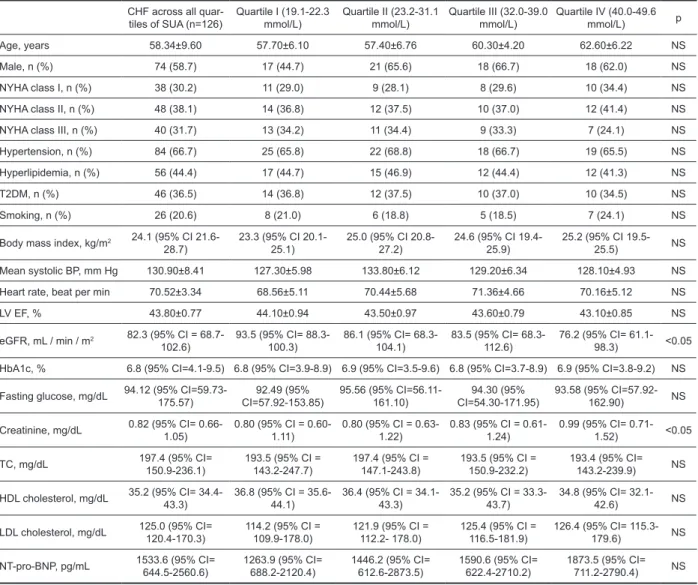
Table 1. General characteristics of study patients
CHF across all quar
-tiles of SUA (n=126) Quartile I (19.1-22.3 mmol/L) Quartile II (23.2-31.1 mmol/L) Quartile III (32.0-39.0 mmol/L) Quartile IV (40.0-49.6 mmol/L) p
Age, years 58.34±9.60 57.70±6.10 57.40±6.76 60.30±4.20 62.60±6.22 NS
Male, n (%) 74 (58.7) 17 (44.7) 21 (65.6) 18 (66.7) 18 (62.0) NS
NYHA class I, n (%) 38 (30.2) 11 (29.0) 9 (28.1) 8 (29.6) 10 (34.4) NS
NYHA class II, n (%) 48 (38.1) 14 (36.8) 12 (37.5) 10 (37.0) 12 (41.4) NS NYHA class III, n (%) 40 (31.7) 13 (34.2) 11 (34.4) 9 (33.3) 7 (24.1) NS Hypertension, n (%) 84 (66.7) 25 (65.8) 22 (68.8) 18 (66.7) 19 (65.5) NS Hyperlipidemia, n (%) 56 (44.4) 17 (44.7) 15 (46.9) 12 (44.4) 12 (41.3) NS
T2DM, n (%) 46 (36.5) 14 (36.8) 12 (37.5) 10 (37.0) 10 (34.5) NS
Smoking, n (%) 26 (20.6) 8 (21.0) 6 (18.8) 5 (18.5) 7 (24.1) NS
Body mass index, kg/m2 24.1 (95% CI
21.6-28.7) 23.3 (95% CI 20.1-25.1) 25.0 (95% CI 20.8-27.2) 24.6 (95% CI 19.4-25.9) 25.2 (95% CI 19.5-25.5) NS Mean systolic BP, mm Hg 130.90±8.41 127.30±5.98 133.80±6.12 129.20±6.34 128.10±4.93 NS Heart rate, beat per min 70.52±3.34 68.56±5.11 70.44±5.68 71.36±4.66 70.16±5.12 NS
LV EF, % 43.80±0.77 44.10±0.94 43.50±0.97 43.60±0.79 43.10±0.85 NS
eGFR, mL / min / m2 82.3 (95% CI =
68.7-102.6) 93.5 (95% CI= 88.3-100.3) 86.1 (95% CI= 68.3-104.1) 83.5 (95% CI= 68.3-112.6) 76.2 (95% CI= 61.1-98.3) <0.05 HbA1c, % 6.8 (95% CI=4.1-9.5) 6.8 (95% CI=3.9-8.9) 6.9 (95% CI=3.5-9.6) 6.8 (95% CI=3.7-8.9) 6.9 (95% CI=3.8-9.2) NS
Fasting glucose, mg/dL 94.12 (95% CI=59.73-175.57) CI=57.92-153.85)92.49 (95% 95.56 (95% CI=56.11-161.10) CI=54.30-171.95)94.30 (95% 93.58 (95% CI=57.92-162.90) NS
Creatinine, mg/dL 0.82 (95% CI= 0.66- 1.05) 0.80 (95% CI = 0.60-1.11) 0.80 (95% CI = 0.63-1.22) 0.83 (95% CI = 0.61-1.24) 0.99 (95% CI= 0.71-1.52) <0.05
TC, mg/dL 197.4 (95% CI= 150.9-236.1) 193.5 (95% CI = 143.2-247.7) 197.4 (95% CI = 147.1-243.8) 193.5 (95% CI = 150.9-232.2) 193.4 (95% CI= 143.2-239.9) NS
HDL cholesterol, mg/dL 35.2 (95% CI= 34.4- 43.3) 36.8 (95% CI = 35.6-44.1) 36.4 (95% CI = 34.1-43.3) 35.2 (95% CI = 33.3-43.7) 34.8 (95% CI= 32.1-42.6) NS
LDL cholesterol, mg/dL 125.0 (95% CI= 120.4-170.3) 114.2 (95% CI = 109.9-178.0) 121.9 (95% CI = 112.2- 178.0) 125.4 (95% CI = 116.5-181.9) 126.4 (95% CI= 115.3-179.6) NS
NT-pro-BNP, pg/mL 1533.6 (95% CI= 644.5-2560.6) 1263.9 (95% CI= 688.2-2120.4) 1446.2 (95% CI= 612.6-2873.5) 1590.6 (95% CI= 622.4-2710.2) 1873.5 (95% CI= 711.2-2790.4) NS
Note: CI-conidence interval, T2DM-type 2 diabetes mellitus, eGFR-estimated glomerular iltration ratio, TC-total cho
-lesterol, HbA1c-glycated haemoglobin, LDL-low-density cho-lesterol, HDL-high-density cho-lesterol, BP-blood pressure, LV EF-left ventricular ejection fraction, U-unit, NS-not signiicant.
Association between SUA level and biomarkers
The univariable linear correlation between SUA and CD45+CD34+ MPCs, CD45-CD34+ MPCs, CD14+CD309+ MPCs, CD14+CD309+Tie2+ MPCs, NT-pro-BNP concentration, NYHA class, LVEF,
T2DM, eGFR was evaluated. A signiicant posi
-tive relationship was found between SUA level and NYHA class (r=0.612; P=0.001); T2DM (r=0.462; P=0.001), NT-pro-BNP (r=0.612; P=0.001), diureti
= 0.30, P<0.01), and inverse association was ob
-tained between SUA level with eGFR (r=-0.476; P=0.002), LVEF (r=-0.42; P=0.001), CD45+CD34+ MPCs (r=-0.388; P=0.001); CD45-CD34+ MPCs 0.41; P=0.001); CD14+CD309+ MPCs 0.397; P=0.001); CD14+CD309+Tie2+ MPCs (r=-0.442; P=0.001). We did not ind a signiicant as
-sociation with the other biomarkers examined. Multivariable linear regression analyses were per
-formed for CD34+ phenotypes of MPCs, adjusted for eGFR, BMI, LVEF, NYHA, diuretics, and T2DM. We found an independent impact of SUA on counts of CD14+CD309+ MPCs (r=-0.388; P=0.001) and CD14+CD309+Tie2+ MPCs (r=-0.414; P=0.001).
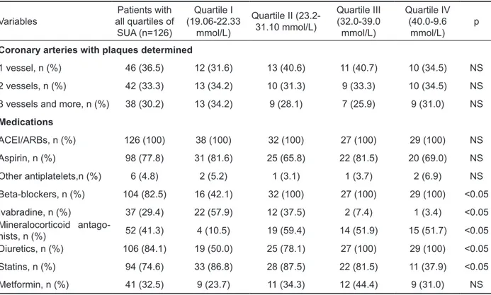
Table 2. Baseline angiographic and treatment characteristics of the patients with CHF depending quartiles of serum uric acid
Variables all quartiles of Patients with SUA (n=126)
Quartile I (19.06-22.33
mmol/L)
Quartile II (23.2-31.10 mmol/L)
Quartile III (32.0-39.0 mmol/L)
Quartile IV (40.0-9.6
mmol/L) p Coronary arteries with plaques determined
1 vessel, n (%) 46 (36.5) 12 (31.6) 13 (40.6) 11 (40.7) 10 (34.5) NS 2 vessels, n (%) 42 (33.3) 13 (34.2) 10 (31.3) 9 (33.3) 10 (34.5) NS 3 vessels and more, n (%) 38 (30.2) 13 (34.2) 9 (28.1) 7 (25.9) 9 (31.0) NS
Medications
ACEI/ARBs, n (%) 126 (100) 38 (100) 32 (100) 27 (100) 29 (100) NS Aspirin, n (%) 98 (77.8) 31 (81.6) 25 (65.8) 22 (81.5) 20 (69.0) NS Other antiplatelets,n (%) 6 (4.8) 2 (5.2) 1 (3.1) 1 (3.7) 2 (6.9) NS Beta-blockers, n (%) 104 (82.5) 16 (42.1) 32 (100) 27 (100) 29 (100) <0.05 Ivabradine, n (%) 37 (29.4) 22 (57.9) 12 (37.5) 2 (7.4) 1 (3.4) <0.05 Mineralocorticoid antago
-nists, n (%) 52 (41.3) 4 (10.5) 19 (59.4) 14 (51.9) 15 (51.7) <0.05 Diuretics, n (%) 106 (84.1) 19 (50.0) 25 (78.1) 27 (100) 29 (100) <0.05 Statins, n (%) 94 (74.6) 33 (86.8) 28 (87.5) 22 (81.5) 11 (37.9) <0.05 Metformin, n (%) 41 (32.5) 9 (23.7) 11 (34.3) 12 (44.4) 9 (31.0) NS CI: conidence interval, ACEI: angiotensin-converting enzyme inhibitor, ARBs: angiotensin-2 receptor blockers, NS: not signiicant
Table 3. Concentrations of MPCs in relation to serum uric acid (SUA) quartiles
Cell phenotypes,
% (median; IQR) CHF patients (n=126)
SUA quartiles (mmol/l)
p Quartile I
(19.06-22.33 mmol/L)
Quartile II (23.2-31.10
mmol/L)
Quartile III (32.0-39.0 mmol/L)
Quartile IV (40.00-49.60
mmol/L)
CD45+CD34+×10-4, % 1.282 (1.21-1.528) 1.77 (1.58-1.93) 1.72 (1.53-1.91) 1.45 (1.21-1.68) 1.05 (0.80-1.17) <0.001
CD45-CD34+×10-4, % 0.727 (0.54-0.91) 1.01 (0.91-1.15) 0.91 (0.81-1.01) 0.83 (0.72-0.93) 0.63 (0.33-0.86) <0.001
CD14+CD309+ ×10-4, % 29.18 (15.0-34.5) 43.9 (33.7-54.12) 37.2 (28.8-45.6) 28.0 (17.5-37.2) 14.0 (11.1-19.9) <0.001
CD14+CD309+Tie2+×10-4, % 0.67 (0.21-1.10) 0.86 (0.74-0.98) 0.82 (0.73- 0.92) 0.67 (0.58-0.76) 0.37 (0.29-0.56) <0.001
Cox proportional adjusted Odds Ratios analy
-ses for CD14+CD309+ and CD14+CD309+Tie2+ MPCs by SUA Quartiles (Q) has showed that high Q (Q3 and Q4) of SUA versus low Q (Q1 and Q2)
Table 4. Cox proportional adjusted Odds Ratios analyses for CD14+CD309+ and CD14+CD309+Tie2+ MPCs by serum uric acid quartiles
SUA Quartiles SUA, mmol/L OddsRatio 95% CI valuep
Mean value 95% CI
For CD14+CD309+ MPCs
Q1 20.11 19.06-22.33 1.0 -
-Q2 27.53 23.2-31.10 1.02 0.88-1.11 0.24
Q3 35.80 32.0-39.0 1.18 1.06-1.29 0.001
Q4 44.9 40.00-49.60 1.24 1.12-1.46 0.002
For CD14+CD309+Tie2+ MPCs
Q1 20.11 19.06-22.33 1.0 -
-Q2 27.53 23.2-31.10 1.08 1.00-1.20 0.054
Q3 35.80 32.0-39.0 1.22 1.11-1.34 0.001
Q4 44.9 40.00-49.60 1.38 1.20-1.55 0.001
Note: All the respective biomarker-models are adjusted for eGFR, BMI, LVEF, NYHA, diuretics, and T2DM
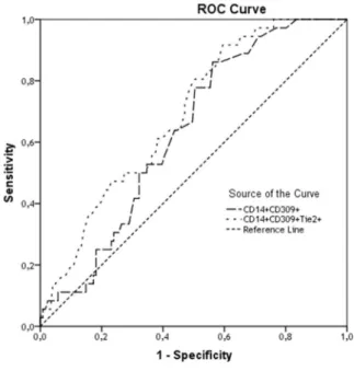
The predictive value of SUA level with respect to the MPCs with phenotypes CD14+CD309+ and CD14+CD309+Tie2+ in the patients with CHF was performed using ROC-analysis, the results of which are presented in Fig. 1. The indings suggest a high predictive power of SUA in the both models for de
-clining of CD14+CD309+ and CD14+CD309+Tie2+ MPCs in CHF patients. The estimated AUCs (area under curves) were 0.631 (sensitivity = 63.9%;
speciicity = 56.2%) and 0.687 (sensitivity = 72.2%; speciicity = 52.9%) respectively. In this case, the cut-off point for the SUA level that had the best prognostic potential on the risk of decreasing MPCs in both models was 31.5 mmol / L. Thus, these data suggest that for the CHF patient elevation of SUA might be considered as a predictor of lowed proan
-giogenic MPCs.
Figure 1. The predictive power of SUA in the both models for declining of CD14+CD309+ and CD14+CD309+Tie2+ MPCs in CHF patients. Results of the Receive Operation Characteristics analysis
Area under the curve
Test Result Variables Area Std. Errora Asymptotic Sig.b Asymptotic 95% Conidence Interval
Lower Bound Upper Bound
CD14+CD309+ 0.631 0.047 0.017 0.540 0.722
CD14+CD309+Tie2+ 0.687 0.046 0.001 0.598 0.776
The test result variables: CD14+CD309+, CD14+CD309+Tie2+ has at least one tie between the positive actual state
group and the negative actual state group.
a. Under the nonparametric assumption b. Null hypothesis: true area = 0.5
DISCUSSION
Previously reports have been predominantly eluci
-dated a relationship between cardiovascular out
-comes and documented hyperuricemia in patients with acute and chronic heart failure [3,21,22]. The effects of SUA on all-cause mortality at different SUA cut-offs in CHF patient population was evalu
-ated using meta-regression. There was a linear
association between SUA after 7 mg/dL and mor
-gressively declining proangiogenic MPCs, which have a tremendous tissue repair capacity. Probably, these indings might be taken into consideration to be explaining controversial role of SUA in CHF evolution and outcomes. Really, signiicant associa
-tion between high SUA level and BMI, diuretic use, some biomarkers, such as NT-pro-BNP, as wells as with hemodynamic performances (E/Ea and LVEF) even beyond declining eGFR was frequently noted in recent investigations [23,25]. Amin et al. [26] reported that mild elevated SUA levels in patients with systolic CHF is associated with impaired clini
-cal and hemodynamic proile and might be used as a noninvasive indicator of elevated left ventricular illing pressures. Misra et al. have been evaluated the independent impact of CHF status (compensa
-tion or decompensa-tion) on SUA levels among men with high cardiovascular risk proile [27]. Investiga
-tors found that mild elevated SUA associated with increased risk of CHF decompensation (OR = 1.67; 95% CI 1.21 to 2.32).
Although hyperuricemia predominantly affects men, in our study we have not received a conirma
-tion of differences in SUA between men and women with CHF. Noted, that there was not a documented hyperuricemia (SUA ≥6 mg/dL for women and ≥8 mg/dL for men) especially required treatment in patients enrolled in the study. However, the inter
-pretation of SUA levels for individual CHF patients may be confusing, but even small increased SUA levels at symptomatic CHF might be discussed as a marker of endothelial dysfunction and, probably, as an indicator of tissue repair disorders. Current evidence suggests that SUA could be a marker of oxidative damage in several settings distinguished CHF, such as overweight, obesity, diuretic use. We conirmed a slight linear association SUA with BMI and diuretic use, but direct effect of BMI and diuretic use on number of circulating MPCs was not found. It has predisposed that SUA may real
-ize their capacity for modulating tissue damage through other mechanisms irrespective SUA clear
-ance. Therefore, it is not clear whether this increase in SUA levels may be a counter-regulatory process or a pathophysiological detrimental factor. Because SUA is a product of xanthine oxidase (XO), apop
-tosis and tissue hypoxia that are suitable for CHF lead to increased purine catabolism, which, in turn, increases XO activity and subsequently SUA levels [28]. Indeed, has a signiicant association with poor outcomes in CHF patients without CKD but not in those with CKD [28-30], suggesting that hyperurice
-mia may predict poor outcomes when it is primarily
a marker of increased XO activity, but not when it is primarily due to impaired renal excretion of uric acid [29]. In controversy of data presented by Filippatos et al (2011) [28], no association between SUA and BMI was found in our study. Diuretics, widely used to treat CHF, increase SUA by stimulating the reab
-sorption of sodium and urate in the proximal tubule. Although we obtained an association between SUA and diuretics administration, the direct effect of di
-uretics in depletion of MPCs in patient study popu
-lation was not determined. Additionally, increased SUA might also associated with coronary artery disease and with its risk factors, such as obesity, hypertension, hypertriglyceridemia, dyslipidemia and T2DM, and worsen renal function. Multivariable linear regression analyses that was performed for CD34+ phenotypes of MPCs with adjustment for eGFR, LVEF, NYHA, diuretics, and T2DM, has been showed an independent impact of SUA on counts of CD14+CD309+ MPCs and CD14+CD309+Tie2+ MPCs. We suggest that tissue ischemia determines an increase in XO, which leads to an increase in SUA levels, and mediates suppression of recruit
-ment, mobbing, differentiation and functional status of MPCs through Akt / STAT /MAP-kinase mecha
-nisms, that is relection of chronic inlammatory, oxi
-dative stress and, probably, catabolic state suitable for CHF [31,32].
Study Restrictions
This study has some restrictions. The authors be
-lieve that a greater cohort is to be desirable to im
-prove the power of the study. There is a variation in the deinition of EPCs, the number of existing cardiovascular risk factors in various patients, and in the interaction between EPCs and other hema
-topoietic progenitor, inlammatory cells or platelets. The authors suppose that these restrictions might have no signiicant impact on the study data inter
-pretation.
Finally, signiicant confounder impacting SUA levels on population of proangiogenic MPCs with in
-volving several pathogenetic mechanisms are pre
-disposed. It is possible to address to new investiga
-tions whether rela-tionships between SUA and pro
-angiogenic MPCs are multidimensional, or if they can be associated with clinical outcomes.
In conclusion, circulating level of proangiogenic MPCs is declined progressively depended on quar
Abbreviations
BMI: Body Mass Index; CAD: Coronary Artery Dis
-ease; T2DM: Type two Diabetes Mellitus; eGFR: Estimated glomerular iltration rate; HbA1c: Glycat
-ed hemoglobin; CHF: Chronic Heart Failure; LVEF: Left Ventricular Ejection Fraction; SUA: serum uric acid; MPCs-mononuclear progenitor cells; LDL: low-density cholesterol, HDL: high-density choles
-terol.
Acknowledgement
We thank all patients for their participation in the investigation, staff of the Regional Zaporozhye Hospital (Ukraine) and the doctors, nurses, and ad
-ministrative staff in City hospital # 6 (Zaporozhye, Ukraine), general practices, and site-managed or
-ganizations that assisted with the study.
ETHICAL PRINCIPLES
The investigators followed strictly all the require
-ments to clinical trials in conformity with the World Medical Association (WMA) Declaration of Helsinki, 1964, Good Clinical Practice provided by Interna
-tional Conference on Harmonization (GCP-ICH), Council of Europe Convention for the Protection of Human Rights and Dignity of the Human Being in view of using achievements in biology and medi
-cine, Convention on Human Rights and Biomedi
-cine, including Additional Protocol to the Conven
-tion on Human Rights and Biomedicine, concerning Biomedical Research, and legislation of Ukraine.
Funding
This research received no speciic grant from any funding agency in the public, commercial, or not-for-proit sectors.
The author contributions
AB conceptualized and designed the study and car
-ried out the analysis and interpretation of data. AK performed visualization procedures, carried out the biochemical analysis including the determination of proangiogenic MPCs, and participated to the acqui
-sition and analysis of data. All authors read and ap
-proved the inal manuscript.
REFERENCES
1. Hartupee J, Mann DL. Positioning of inlammatory bio
-markers in the heart failure landscape. J Cardiovasc Transl Res 2013;6:485-492.
2. Gustafsson D, Unwin R. The pathophysiology of hyper
-uricaemia and its possible relationship to cardiovas
-cular disease, morbidity and mortality. BMC Nephrol 2013;14:164.
3. Huang H, Huang B, Li Y, et al. Uric acid and risk of heart failure: a systematic review and meta-analysis. Eur J Heart Fail. 2013 Aug 9. [Epub ahead of print] 4. Jeemon P, Prabhakaran D. Does uric acid qualify as an
independent risk factor for cardiovascular mortality? Clin Sci (Lond) 2013;124:255-257.
5. Kuo CF, See LC, Yu KH, et al. Signiicance of serum uric acid levels on the risk of all-cause and cardiovas
-cular mortality. Rheumatology (Oxford) 2013;52:127-134.
6. Rehman J, Li J, Orschell CM, March KL. Peripheral blood “endothelial progenitor cells” are derived from monocyte/macrophages and secrete angiogenic growth factors. Circulation 2003;107:1164-1169. 7. Schmidt-Lucke C, Roёssig L, Fichtlscherer S, et al.
Reduced number of circulating endothelial progeni
-tor cells predicts future cardiovascular events: proof of concept for the clinical importance of endogenous vascular repair. Circulation 2005;111:2981-2987. 8. Hill JM, Zalos G, Halcox JP, et al. Circulating endothe
-lial progenitor cells, vascular function, and cardiovas
-cular risk. N Engl J Med 2003;348: 593-600.
9. Valgimigli M, Rigolin GM, Fucili A, et al. CD34+ and endothelial progenitor cells in patients with vari
-ous degrees of congestive heart failure. Circulation 2004;110:1209-1212.
10. Kissel CK, Lehmann R, Assmus B, et al. Selective functional exhaustion of hematopoietic progenitor cells in the bone marrow of patients with postinfarction heart failure. J Am Coll Cardiol 2007;49:2341-2349 11. Fadini GP, Maruyama S, Ozaki T, et al. Circulating pro
-genitor cell count for cardiovascular risk stratiication: a pooled analysis. PLoS ONE 2010; 5, e11488. 12. Fritzenwanger M, Lorenz F, Jung C, et al. Differential
number of cd34+, cd133+ and cd34+/cd133+ cells in peripheral blood of patients with congestive heart fail
-ure. Eur J Med Res 2009;14:113-117.
13. Bakogiannis C, Tousoulis D, Androulakis E, et al. Cir
-culating endothelial progenitor cells as biomarkers for prediction of cardiovascular outcomes. Curr Med Chem 2012;19:2597-2604.
14. McMurray JJV, Adamopoulos S, Anker SD, et al. ESC Guidelines for the diagnosis and treatment of acute and chronic heart failure 2012. Eur Heart J 2012;33:1787-1847
15. Bluemke DA, Achenbach S, Budoff M, et al. Nonin
-vasive coronary artery imaging: magnetic resonance angiography and multidetector computed tomography angiography: a scientiic statement from the American Heart Association Committee on Cardiovascular Im
-aging and Intervention of the Council on Cardiovas
16. Schiller NB., Shah PM., Crawford M, et al. Recom
-mendations for quantitation of the left ventricle by two-dimensional echocardiography. American Society of Echocardiography Committee on Standards, Sub
-committee on Quantitation of Two-Dimensional Echo
-cardiograms. J Am Soc Echocardiogr 1989;2:358-367.
17. Pellerin D., Sharma R., Elliott P., Veyrat C. Tissue Doppler, strain, and strain rate echocardiography for the assessment of left and right systolic ventricular function. Heart 2003;89(90003):iii9-17
18. Levey AS, Stevens LA, Schmid CH, et al. for the CKD-EPI (Chronic Kidney Disease Epidemiology Collabo
-ration). A new equation to estimate glomerular iltra
-tion rate. Ann Intern Med 2009;150:604-612.
19. Friedewald WT, Levy RI, Fredrickson DS. Estimation of the concentration of low-density lipoprotein choles
-terol in plasma, without use of the preparative ultra
-centrifuge. Clin Chem 1972;18:499-502.
20. Tung JW, Parks DR, Moore WA, et al. New approach
-es to luor-escence compensation and visualization of FACS data. Clin Immunol 2004;110:277-283.
21. Málek F, Ošťádal P, Pařenica J, et al. Uric acid, allopu
-rinol therapy, and mortality in patients with acute heart failure-results of the Acute HEart FAilure Database registry. J Crit Care 2012;27:737.e11-24.
22. Manzano L, Babalis D, Roughton M, et al; SE
-NIORS Investigators. Predictors of clinical outcomes in elderly patients with heart failure. Eur J Heart Fail 2011;13:528-536.
23. Tamariz L, Harzand A, Palacio A, et al. Uric acid as a predictor of all-cause mortality in heart failure: a meta-analysis. Congest Heart Fail 2011;17:25-30.
24. Alcaíno H, Greig D, Castro P, et al. The role of uric acid in heart failure. Rev Med Chil 2011;139:505-515. 25. Hsieh MC, Su HM, Wang SY, et al. Signiicant cor
-relation between left ventricular systolic and diastolic dysfunction and decreased glomerular iltration rate. Ren Fail 2011;33:977-982.
26. Amin A, Vakilian F, Maleki M. Serum uric acid levels correlate with illing pressures in systolic heart failure. Congest Heart Fail 2011;17:80-84.
27. Misra D, Zhu Y, Zhang Y, Choi HK. The independent impact of congestive heart failure status and diuretic use on serum uric acid among men with a high cardio
-vascular risk proile: a prospective longitudinal study. Semin Arthritis Rheum 2011;41:471-476.
28. Filippatos GS, Ahmed MI, Gladden JD, et al. Hyper
-uricaemia, chronic kidney disease, and outcomes in heart failure: potential mechanistic insights from epi
-demiological data. Eur Heart J 2011;32:712-720. 29. Wu AH, Ghali JK, Neuberg GW et al. Uric acid
level and allopurinol use as risk markers of mortal
-ity and morbid-ity in systolic heart failure. Am Heart J 2010;160:928-933.
30. Kim H, Shin HW, Son J, et al. Uric Acid as prognostic marker in advanced nonischemic dilated cardiomyop
-athy: comparison with N-terminal pro B-type natriuret
-ic peptide level. Congest Heart Fail 2010;16:153-158. 31. Waring WS, Webb DJ, Maxwell SRJ. Effect of lo
-cal hyperuricemia on endothelial function in the hu
-man forearm vascular bed. Br J Clin Pharmacol 2000;49:511-515.