RBCCV 44205-1573 DOI 10.5935/1678-9741.20140091
Direct intramyocardial transthoracic
transplantation of bone marrow mononuclear
cells for non-ischemic dilated cardiomyopathy:
INTRACELL, a prospective randomized
controlled trial
Ensaio clínico prospectivo randomizado sobre terapia com células mononucleares da medula óssea para
cardiomiopatia dilatada idiopática: INTRACELL
Roberto T. Sant’Anna
1, MD; James Fracasso
1, MD, MSc; Felipe H. Valle
1, MD; Iran Castro
1, MD,
PhD; Nance B. Nardi
1, PhD; João Ricardo M. Sant’Anna
1, MD, PhD; Ivo Abrahão Nesralla
1, MD;
Renato A. K. Kalil
1, MD, PhD
1Cardiology Institute/University Foundation of Cardiology (IC/FUC), Car-diovascular Surgery Service, Porto Alegre, RS, Brazil.
This study was carried out at the Cardiology Institute/University Foundation of Cardiology (IC/FUC), Federal University of Health Sciences of Porto Alegre (UFCSPA), Porto Alegre, RS, Brazil.
Financial support from Brazilian government agencies CNPq, CAPES, Min-istry of Health and FAPERGS.
Correspondence address: Renato Abdala Karam Kalil
Instituto de Cardiologia /Fundação Universitária de Cardiologia (IC/FUC). Avenida Princesa Isabel, 395 – Santana – Porto Alegre, RS
Brazil – Zip Code: 90620-000 E-mail: [email protected]
Article received on February 20th, 2014 Article accepted on July 7th, 2014 Abstract
Objective: We tested the hypothesis that direct intramyo-cardial injection of bone marrow mononuclear cells in patients with non-ischemic dilated cardiomyopathy can improve left ventricular function and physical capacity.
Methods: Thirty non-ischemic dilated cardiomyopathy patients with left ventricular ejection fraction <35% were ran-domized at a 1:2 ratio into two groups, control and treated. The bone marrow mononuclear cells group received 1.06±108 bone marrow mononuclear cells through mini-thoracotomy. There was no intervention in the control group. Assessment was carried out through clinical evaluations as well as a 6-min walk test, nuclear magnectic resonance imaging and echocardiogram.
tracoronary route, showed small, but signiicant increase in
left ventricular ejection fraction (LVEF) after treatment[5,6].
Recently, intracoronary injection of CD34+ was associated with medium-term improvement of ventricular function, ex-ercise tolerance, and long-term survival in randomized trial of patients with DCM[7].
Intracoronary injection is associated with a small percent-age of transplanted cells effectively retained in the myocardi-um, which could limit results when that route is used for cell therapy[10]. In a pilot study, we found that direct application of
BMMC through a small thoracotomy was safe and
associat-ed with a signiicant, although transitory, increase in LVEF in
patients with non-ischemic dilated cardiomyopathy (NIDCM)
[11]. In the present study, we tested the hypothesis that direct
intramyocardial injection of BMMC in patients with NIDCM could improve LVEF assessed by cardiac magnetic resonance imaging (MRI) and echocardiogram, physical capacity evalu-ated by the six-minute walk test (6WT), and heart failure class functional (NYHA), when compared to conventional treatment. Abbreviations, acronyms & symbols
BMMC Bone marrow mononuclear cells DCM Dilated cardiomyopathy LVEF Left ventricular ejection fraction MLHF Minnesota Living with Heart Failure MRI Magnetic resonance cardiac imaging NIDCM Non-ischemic dilated cardiomyopathy NYHA New York Heart Association PO Post-operatively
INTRODUCTION
Dilated cardiomyopathy is a leading cause of heart failure and the primary indication for heart transplantation[1,2]. The
prevalence of this disease tends to increase according to pop-ulation ageing and survival improvement achieved through advances in pharmacological treatment and implantable de-vices[3]. However, current therapeutic approaches are
pallia-tive in the sense that they are unable to directly address the underlying problem of the loss of cardiac tissue[4].
Cell therapy with bone marrow mononuclear cells
(BMMC) has been associated with beneicial effects in pa -tients with acute myocardial infarction and ischemic heart failure. Data for non-ischemic dilated cardiomyopathy (NIDCM), however, is more limited[5-7]. Experimental
stud-ies suggest that stem cells improve heart function through both paracrine regulation of cytokines production and cell transdifferentiation, albeit in a small proportion of the trans-planted cells[8,9]. Two small trials, using BMMC through in-showed improvement in New York Heart Association functional class, from 3.40±0.50 to 2.41±0.79 (P=0.002); patients in the con-trol group showed no change (3.37±0.51 to 2.71±0.95; P=0.17).
Six-minute walk test improved in the bone marrow
mononucle-ar cells group (348.00±93.51m at baseline to 370.41±91.56m at 12 months, P=0.66) and there was a non-signiicant decline in the control group (361.25±90.78m to 330.00±123.42m after 12 months, P=0.66). Group comparisons were non-signiicant.
Conclusion: The trend of intragroup functional and
sub-jective improvement was not conirmed when compared to
the control group. Direct intramyocardial application of bone marrow mononuclear cells in non-ischemic dilated
cardiomy-opathy was not associated with signiicant changes in left ven -tricular function. Differences observed within the bone marrow mononuclear cells group could be due to placebo effect or low statistical power.
Descriptors: Cardiomyopathy, Dilated. Heart Failure. Cells. Myocardial Contraction. Transplantation.
Resumo
Objetivo: Testamos a hipótese de que a injeção intramiocárdi-ca direta de células mononucleares de medula óssea em pacientes
portadores de cardiomiopatia dilatada não-isquêmica pode me-lhorar a função ventricular e a capacidade física.
Métodos: Trinta pacientes com cardiomiopatia dilatada não isquêmica e fração de ejeção 35% foram randomizados na razão 1:2 em grupos controle e tratado. Grupo células mononucleares de medula óssea recebeu 1,06 ± 108 células mononucleares de medula óssea por mini-toracotomia. Grupo controle não recebeu inter-venção. Avaliação foi feita clinicamente e por teste de caminhada 6 minutos (T6m), ressonância magnética e ecocardiogramas.
Resultados: Grupo células mononucleares de medula óssea mostrou tendência de melhora da Fração de ejeção - ressonância magnética aos 3 meses, 27,80±6,86% para 30,13±9,06% (P=0,08), re-tornando ao basal aos 9 meses (28,78%, P=0,77). Grupo controle não apresentou variação (28,00±4,32%; 27,42±7,41% e 29,57±4,50%). Ecocardiogramas - fração de ejeção melhorou no grupo células mononucleares de medula óssea aos 3 meses: 25,09±3,98 para 30,94±9,16 (P=0,01) e aos 12 meses (30,07±7,25%, P=0,001), enquan-to o controle não variou: 26,1±4,4 vs. 26,5±4,7 e 30,2±7,39%, P=0,25 e 0,10, respectivamente). Células mononucleares de medula óssea melhorou classe funcional New York Heart Association: 3,40±0.50 para 2,41±0,79 (P=0,002); controles não mudaram (3,37±0,51 para 2,71±0,95; P=0,17). T6m melhorou no grupo células mononucleares de medula óssea (348,00±93,51 m inicial para 370,41±91,56 m aos 12 m, P=0,66) e declinou sem signiicância no controle (361,25±90,78 m
para 330,00±123,42 m aos 12 meses, P=0,66). Não houve diferenças
signiicativas entre os grupos.
Conclusão: A tendência intragrupo de melhora funcional e
subjetiva não se conirmou quando comparado com controle.
Portanto, a injeção direta intramiocárdica de células
mononu-cleares de medula óssea não se associou a mudança signiicativa
na função ventricular. As diferenças observadas no grupo tratado poderiam ser devidas a efeito placebo ou a baixo poder estatístico.
Descritores: Cardiomiopatia dilatada. Insuiciência Cardíaca.
METHODS
Study design
We conducted an open, parallel-group, explanatory randomized study in a single center to evaluate the
safe-ty and eficacy of BMMC in patients NIDCM. Thirsafe-ty pa
-tients that fulilled the criteria for inclusion were random -ly assigned to each of the groups by means of a computer software for simple randomization: BMMC or control, in a 2:1 proportion.
After randomization, functional status of both groups was assessed at baseline using (6WT), New York Heart As-sociation (NYHA) and Minnesota Living with Heart Failure (MLHF) questionnaires, which had been previously validat-ed for the Brazilian population[12]. We used cardiac magnetic
resonance imaging (MRI) to measure left ventricular diam-eters and ejection fraction. Functional status was reassessed every three months and MRI repeated three and nine months after inclusion. All patients were treated by the same physi-cian at a dedicated outpatient clinic, according to ACC/AHA Guidelines for the Diagnosis and Management of Heart Fail-ure in Adults[1]. Functional status was evaluated by a separate
team. In this article, we present the results of the irst year of
follow-up. We were unable to perform a blind study because of the invasive nature of the procedure proposed. The study was submitted to the National Comission in Research (CO-NEP) and approved. All patients signed an informed consent. Patients in the BMMC group, in addition to conventional treatment, were submitted to BMMC transplantation as de-scribed below up to one month after randomization. In this
group, we also conducted low citometry and imunohisto -chemistry to characterize bone marrow cells.
Our primary objective was to evaluate changes in left ventricular ejection fraction assessed with MRI. Secondary objectives consisted of evaluation of: 1) Safety; 2) New York Heart Association (NYHA) functional class and quality of life measured with Minnesota Living with Heart Failure Questionnaire; 3) Effects in mortality; 4) Left ventricular di-ameters using MRI.
Inclusion and exclusion criteria
The study was approved by the Ethics Review Board of Instituto de Cardiologia do Rio Grande do Sul/ Fundação Universitária de Cardiologia (IC/FUC), as well as by the Na-tional Ethics Committee in Research of Brazil, under proto-col number 10376 and registered in ClinicalTrials.gov under number: NCT00743639. Both the study and the registry were conducted in accordance with the Declaration of Helsinki. Written informed consent was obtained from each patient.
We screened for patients with heart failure, LVEF less then 35% and functional class III or IV, despite full medical
treatment. Speciic inclusion criteria were: (1) Age between
20 and 65 years; (2) Diagnosis of non-ischemic
cardiomy-opathy for at least 12 months before enrollment; (3) Coro-nary angiography with normal coroCoro-nary arteries, which
de-ined cardiomyopathy as non-ischemic for the purposes of
this trial; (4) negative serology for Chagas Disease; and (5)
Absence of other signiicant systemic disease limiting
mid-term prognosis. We excluded patients with: (1) Documented episodes of ventricular tachycardia; (2) Moderate to severe
mitral regurgitation or any other signiicant valve disease; (3)
History of myocardial infarction; and (4) Previous cardiac surgery.
Bone marrow mononuclear cells isolation and trans-plantation
Approximately four hours before the operation and with the patient under sedation, a volume of about 80 mL of bone marrow was aspirated from the anterior iliac crest and placed in an anticoagulated medium. Mononuclear cell fraction was separated by density centrifugation over Ficoll-Hypaque-1077 (Sigma Diagnostics, St Louis, MO) and washed in a heparin-ized saline solution containing 5% autologous serum. Cells were counted and suspended in 5 mL saline solution for in-tramyocardial injection. A small fraction was utilized for ste-rility and viability tests and for immunophenotyping. Viability greater than 90% was considered acceptable.
For detection of surface antigens the cells were trypsinized, centrifuged, and incubated for 30 minutes at 4°C with
phy-coerythrin (PE)- or luorescein isothiocyanate (FITC)-con -jugated antibodies against human CD3, CD4, CD14, CD34, CD38 and CD45 (Pharmingen BD, San Diego, CA). Excess antibody was removed by washing. The cells were analyzed using a FACScalibur cytometer equipped with 488 nm argon laser (Becton Dickinson, San Diego, CA) with the CellQuest software. At least 10,000 events were collected. WinMDI 2.8 software was used for building histograms.
Surgical approach was through a left mini-thoracotomy, consisting of an approximately 5 cm incision in the
antero-lateral portion of the ifth left intercostal space to expose the
pericardium. A T-shaped pericardial incision was made to access the free wall of the left ventricle. Coronary arteries
were identiied and the cell suspension was directly injected, using a 21F Butterly needle that was introduced about 5mm
intramyocardially and connected to an extension managed by the surgical assistant. Twenty 0.25 mL injections were given in the myocardium and in the anterior, lateral, posterior, and apical faces of the left ventricular free wall. After reviewing the hemostasis, pericardium was approximated, the thoracic cavity was drained and the chest wall closed.
Echocardiogram
treatment group and was not part of the study team. It should be noted, however, that treated patients had a visible scar in the left chest, which made it impossible to guarantee blindness to the echo examinator.
Magnetic Resonance Imaging
All MRI image analyses were performed by the same in-vestigator, which was blind to the assignment of the patient. We used a 1.5-T scanner (GE Excite HDx) with ECG gating and a 4-element phased-array surface coil. Scan planes were planned according to standard procedures. Endocardial and epicardial borders were traced in all end-diastolic and end-sys-tolic short-axis slices to determine LV end-diasend-sys-tolic and end-systolic volumes (LVEDV and LVESV). Global LVEF was calculated [(LVEDV-LVESV)/LVEDV]/100 LVEDV. Systolic ventricular wall thickening was calculated for differ-ent heart segmdiffer-ents and it was used for an already published sub-study[11].
Statistical analysis
Continuous variables are presented as mean±SD, if not stated otherwise. Categorical variables were compared with the x2 test or Fisher’s exact test. Statistical comparisons between
initial and follow-up data were performed in a nonparametric paired fashion using the Wilcoxon signed rank test. Nonpara-metric Mann–Whitney U and Kruskal–Wallis tests were used to compare continuous and categorical variable, as well as the results between different groups. All tests were performed as
2-sided tests at a signiicance level of 0.05.
In the irst three months of follow-up, 25% of the patients
from the BMMC group had either died or been withdrawn from the study, resulting in a decrease in the number of treat-ed cases available for late follow-up. Since those patients had a lower mean ejection fraction than the whole group (18.26 % vs. 21.75%), we excluded them from comparative analy-sis, in order to avoid overestimation of treatment effect. In other words, outcome analysis was performed in as-treated basis, not as intention-to-treat.
RESULTS
Baseline
Baseline characteristics are detailed in Table 1. There
were no signiicant differences in baseline data between
BMMC and control groups. All patients had severe heart fail-ure and were highly symptomatic by the time of inclusion, despite maximum medical therapy.
Cell analysis
A mean of 1.06±0.43 x 10⁸ mononuclear cells per patient
were available for injection using this method. Cell viability was greater than 99%, and fungal and bacterial cultures were negative.
Cells from the irst ive samples were analyzed in greater
detail. Immunophenotyping of the mononuclear cell fraction showed the following composition: CD34+ cells (1.5±0.7%), CD45+ (74.6±8.5%), CD14+ (8.4±4.7%), CD3+CD4+ (22.8±4.6%), CD3+CD8+ (8.2±6.1%), and CD34+CD38- cells (0.7±0.5%).
Procedural safety and clinical outcomes
Surgical procedure was effectively performed in 19 pa-tients, since one patient asked to be withdrawn soon after randomization. Table 2 summarizes surgical results. Four
subjects died in the irst month after the procedure. Two died of refractory cardiogenic shock in the irst 72 hours
post-operatively (PO). One of them presented with cardiac tamponade 7 hours after surgery and was submitted to re-operation. The patient's hemodynamic condition continued to deteriorate and he died due to cardiogenic shock 3 days PO. Another patient died on the 15th day PO due to
inces-sant ventricular tachycardia. Finally, one patient died on the 28th day PO: he had been discharged from hospital, but was
readmitted one week later due to heart failure, which was refractory to treatment. All of the patients above had LVEF below 21% before operation.
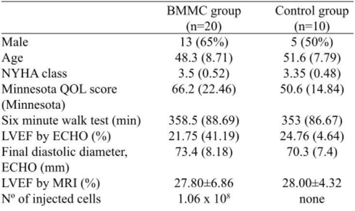
Table 1. Baseline patients characteristics.
Male Age NYHA class
Minnesota QOL score (Minnesota)
Six minute walk test (min) LVEF by ECHO (%) Final diastolic diameter, ECHO (mm)
LVEF by MRI (%) Nº of injected cells
BMMC group (n=20) 13 (65%) 48.3 (8.71)
3.5 (0.52) 66.2 (22.46)
358.5 (88.69) 21.75 (41.19) 73.4 (8.18)
27.80±6.86 1.06 x 10⁸
Control group (n=10) 5 (50%) 51.6 (7.79) 3.35 (0.48) 50.6 (14.84)
353 (86.67) 24.76 (4.64) 70.3 (7.4)
28.00±4.32 none
Data expressed as mean (% or SD);
LVEF by ECHO=left ventricular ejection fraction by echocardiogram; MRI=nuclear magnetic resonance imaging; QOL=quality of life by Minnesota Living with Heart Failure Questionnaire. There were
no statistically signiicant differences between baseline groups
characteristics
Table 2. Surgical results.
Surgery
Thoracotomy procedure (min) Post-operative, %
Length of ICU Stay (days) Length of hospital stay (days)
Mean ±DP 84.7±29
1.86±1.3 4.50±2.0
Max value 120
Three patients from the BMMC group died between 30 days and 12 months after surgery. In two patients, death was sudden, at 10 and 11 months PO. Another patient died 6 months PO due to heart failure.
In the control group, one patient died three months after randomization due to heart failure and another patient was lost for follow-up two months after randomization. There
was no statistically signiicant difference in safety outcomes
between BMMC and control groups: Fischer´s test showed
no difference in mortality between groups in the irst 30 days
(P=0.371) or up to 12 months (P=1.000).
Effects of BMMC on LV Function MRI evaluations
Patients of the BMMC group maintained LVEF at 3 months, from 27.80±6.86% to 30.13±9.06% (P=0.08), and at 9 months (28.78%, P=0.77). The control group also maintained systol-ic function during follow-up (28±4.32% vs. 27.4±7.4%, at 3 months, P=0.79, and vs. 29.57±4.50 at 9 months, P=0.46,
re-spectively). Differences between groups were non-signiicant.
Both groups maintained end-systolic and end-diastolic volumes during follow-up. (Table 3).
Echocardiographic evaluations
Patients of the BMMC group had a signiicant improve -ment in left ventricular ejection fraction 3 months after the
pro-cedure, from 25.09±3.98 to 30.94±9.16 (P=0.01). This
ben-eit was maintained after one year of follow-up (30.07±7.25,
P=0.001). The control group has showed no change in the same period (26.1±4.4 vs. 26.5±4.7 and 30.2±7.39, P=0.25 and 0.10, respectively). Differences between treated and
con-trol groups, however, were not statistically signiicant. Left
ventricular fractional shortening evaluation showed similar results. End-systolic and end-diastolic diameters had a small reduction in both groups. (Figure 1, Table 4).
Quality of life and 6-minute walk test
Quality of life, evaluated by MLHFQ (Minnesota Living
with Heart Failure Questionnaire), improved signiicantly in the
BMMC group three months after the procedure (69.00±20.71 vs. 43.13±27.92 points, P=0.005), a beneit maintained during 6 and 12 months of follow-up (48.92±24.26 points, P=0.018; 37.08±21.15 points, P=0.001; respectively). Patients of the
control group showed a non-signiicant improvement, with
smaller differences between baseline and 12 months follow-up (minus 31.92 points for BMMC group and minus 7.16 points for Control group). Differences between groups were not
sta-tistically signiicant. Functional class, evaluated using NYHA Classiication, showed similar results with a statistically sig
-niicant improvement in the BMMC group, no change in the
control group. However, once again, there were no statistically
signiicant differences between groups. (Figure 2, Table 5).
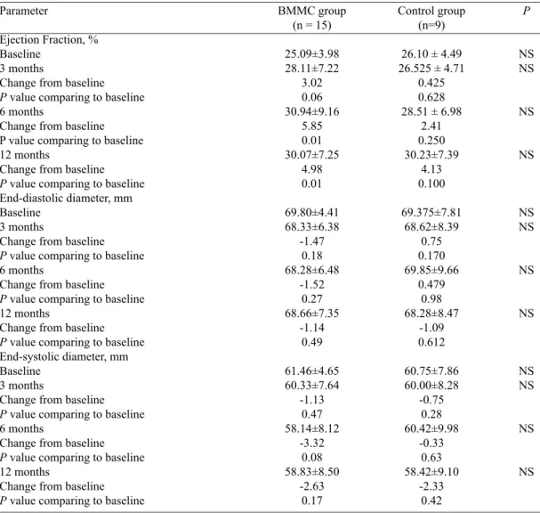
Table 3. Left ventricular volumes and function as assessed by Magnetic Resonance Imaging (MRI).
Parameter
Ejection Fraction, % Baseline
3 months
Change from baseline
P value comparing to baseline 9 months
Change from baseline
P value comparing to baseline End-diastolic volume, mL Baseline
3 months
Change from baseline
P value comparing to baseline 9 months
Change from baseline
P value comparing to baseline End-systolic volume, mL Baseline
3 months
Change from baseline
P value comparing to baseline 9 months
Change from baseline
P value comparing to baseline
BMMC group (n = 15)
27.80±6.86 30.13±9.06
2.33 0.08 28.78±9.97
0.98 0.77
272.73±63.41 264.86±72.24
-7.87 0.41 258.64±82.97
-14.09 0.54
200.40±61.59 195.93±78.49
-4.47 0.66 190.00±82.18
-10.40 0.73
Control group (n=9)
28.00±4.32 27.42±7.41
-0.58 0.79 29.57±4.50
+1.57 0.46
250.85±67.00 222.42±96.19
-28.43 0.32 258.85±75.55
+8.00 0.71
182.00±55.70 161.67±67.83
- 20.43 0.18 184.14±58.55
2.14 0.91
P
NS NS
NS
NS NS
NS
NS NS
Fig.1 - A) Left ventricular ejection fraction and B) end-systolic diameter by echocardiography at baseline and during follow-up
Six-minute-walk test results were maintained in the BMMC group (from 348.00±93.51min in baseline to 370.41±91.56 min after 12 months, P=0.66) and in control group (from 361.25±90.78 min in baseline to 330.00±123.42 min after 12 months, P=0.66). Again, differences between
groups were not statistically signiicant. (Table 5).
DISCUSSION
In this study, we tested the hypothesis that transplantation of BMMC through a small thoracotomy could improve left ventricular function in patients with refractory heart failure
due to NIDCM. Our main indings were: (1) Mortality asso -ciated with the procedure was directly related to preoperative disease severity. In particular, patients with LVEF below 21% had a poor surgical result. Early deaths were related to post-operative pump-failure and congestive heart failure, while deaths at medium term were sudden, probably caused by cardiac arrhythmias. These results differ from our previous pilot study, in which there had been no deaths[11]. (2)
Survi-vors treated with BMMC maintained left ventricular function relative to baseline according to MRI. (3) Overall, patients had symptomatic improvement during follow-up, which was
more pronounced in the BMMC group, but there was no sta-tistical difference between groups in the outcomes studied.
The seven deaths (35%) observed in patients who received BMMC were due to pump-failure in the early post-operative pe-riod (2 cases; 28.5%), congestive heart failure (2 cases, 28.5%) and documented ventricular arrhythmia or sudden death (3 cas-es, 42.8%). Using the Seattle Heart Failure Model, a tool with excellent accuracy for predicting survival among patients with heart failure[13], we could anticipate one-year mortality between
20% and 25%. Incidence of death attributable to arrhythmia (42,8%) was higher than expected for patients in NYHA func-tional class III (10.5%) or IV (18.6%) at one year[14].
This higher than expected risk of ventricular tachyar-rhythmia after surgery could be related to the intrinsic high risk of any procedure in patients with severe heart failure or
to a more speciic mechanism secondary to BMMC injection.
Stem cell pro-arrhythmic potential is a matter of intense de-bate[15,16]. Clinically, bone marrow progenitor cells were not
associated with increased risk of arrhythmias using differ-ent routes of delivery in differdiffer-ent clinical scenarios[17-19]. Yet,
experimental studies have shown that mesenchymal stem cells (MSCs) implantation altered cardiac conduction[20] and
Table 4. Left ventricular volumes and function as assessed by echocardiography (ECO).
Parameter
Ejection Fraction, % Baseline
3 months
Change from baseline
P value comparing to baseline 6 months
Change from baseline P value comparing to baseline 12 months
Change from baseline
P value comparing to baseline End-diastolic diameter, mm Baseline
3 months
Change from baseline
P value comparing to baseline 6 months
Change from baseline
P value comparing to baseline 12 months
Change from baseline
P value comparing to baseline End-systolic diameter, mm Baseline
3 months
Change from baseline
P value comparing to baseline 6 months
Change from baseline
P value comparing to baseline 12 months
Change from baseline
P value comparing to baseline
BMMC group (n = 15)
25.09±3.98 28.11±7.22 3.02 0.06 30.94±9.16 5.85 0.01 30.07±7.25 4.98 0.01 69.80±4.41 68.33±6.38 -1.47 0.18 68.28±6.48 -1.52 0.27 68.66±7.35 -1.14 0.49 61.46±4.65 60.33±7.64 -1.13 0.47 58.14±8.12 -3.32 0.08 58.83±8.50 -2.63 0.17 Control group (n=9)
26.10 ± 4.49 26.525 ± 4.71
0.425 0.628 28.51 ± 6.98
2.41 0.250 30.23±7.39 4.13 0.100 69.375±7.81 68.62±8.39 0.75 0.170 69.85±9.66 0.479 0.98 68.28±8.47 -1.09 0.612 60.75±7.86 60.00±8.28 -0.75 0.28 60.42±9.98 -0.33 0.63 58.42±9.10 -2.33 0.42 P NS NS NS NS NS NS NS NS NS NS NS NS
episodes[21,22]. Intramyocardial injections can create clusters
of cells leading to heterogeneity in conduction and have been associated with greater risk of ventricular premature com-plexes than intracoronary injection[23]. How this translates
into clinical practice, where patients are treated with
anti-ar-rhythmic drugs, needs to be further clariied. At this point,
however, we cannot dismiss the possibility of a pro-arrhyth-mic effect related the newly implanted cells.
BMMC treated patients who survived for more than
one month after the procedure showed signiicant im -provements in both symptoms and quality of life. Left ventricular ejection fraction was maintained one year af-ter the procedure (P=0.77). Results with echocardiogra-phy are more pronounced as they showed improvement in LVEF of patients treated with BMMC after 6 months (from 25.09±3.98 to 30.94±9.16, P=0.01). Nevertheless, since operators were not blind for patients' group, results from this method should be interpreted with caution.
Clinical experience with mononuclear cells in patients with DCM is limited. Fischer-Rasokat et al performed infu-sion of BMMC in 33 patients with DCM using an over-the-wire balloon catheter in the left descending coronary artery[8].
After 3 months, regional wall motion of the target area and global left ventricular ejection fraction had a small (about
10%) but statistically signiicant improvement. Increase of
regional contractile function was directly related to the func-tionality of infused cells as measured by their colony-forming capacity. Safety data was remarkably good, with no proce-dure related complications and no deaths, stroke or myocar-dial infarction up to one year follow-up. Compared to our study, patients were less symptomatic (67% in NYHA class II) and had better ventricular function (mean LEVF=0.2%).
Vrtovec et al.[7] conducted a study with 110 patients with
Fig. 2 - A) Quality-of-Life score (Minnesota), B) Six-minute walk test and C) NYHA Functional class at baseline and during follow-up
in the artery supplying segments with the greatest perfusion defect. At 5 years, stem cell therapy was associated with increased LVEF (from 24.3±6.5% to 30.0±5.1%; P=0.02), increased 6-minute walk distance (from 344±90 m to 477± 30 m; P<0.001), and decreased N-terminal B-type natriuret-ic peptide (from 2322±1234 pg/mL to 1011±893 pg/mL; P<0.01). Left ventricular ejection fraction improvement was
more signiicant in patients with higher myocardial homing
of injected cells. Total mortality was lower in patients treated with CD 34+ cells (14% vs. 35%; P=0.01) and the procedure was considered safe, with low morbimortality.
Several limitations of our study are recognized. First: the trial was projected with a small sample that was further re-duced due to early mortality. This drawback was unexpected, since we had no early major complications or mortality in a previous safety study[11]. High mortality could be attributed to
Table 5. Minnesota Living with Heart Failure Questionnaire, New York Heart Association Functional Class and 6-Minutes-Walk Test results.
Parameter
Minnesota LHFQ Baseline 3 months
Change from baseline
P value comparing to baseline 6 months
Change from baseline
P value comparing to baseline 12 months
Change from baseline
P value comparing to baseline 6-minutes-walk test
Baseline 3 months
Change from baseline
P value comparing to baseline 6 months
Change from baseline
P value comparing to baseline 12 months
Change from baseline
P value comparing to baseline NYHA
Baseline 3 months
Change from baseline
P value comparing to baseline 6 months
Change from baseline
P value comparing to baseline 12 months
Change from baseline
P value comparing to baseline
BMMC group (n = 15)
69.00±20.71 43.13±27.92 -25.87 0.005 48.92±24.26 -20.08 0.018 37.08±21.15 -31.92 0.001 348.00±93.51 338.66±116.48 -9.34 0.73 355.35±67.77 7.35 0.81 370.41±91.56 22.41 0.66 3.40±0.50 2.60±0.82 -0.8 0.005 2.50±0.75 -0.9 0.005 2.41±0.79 -0.99 0.002 Control group (n=9) 48.87±16.32 54.37±23.71 +5.50 0.57 40.28±23.59 -8.58 0.31 41.71±27.46 -7.16 0.48 361.25±90.78 336.25±114.38 - 25 0.44 365.00±126.45 3.75 0.98 330.00±123.42 -31.25 0.39 3.37±0.51 3.00±0.75 - 0.37 0.19 2.57±0.78 -0.8 0.04 2.71±0.95 - 0.66 0.17 P NS NS NS NS NS NS NS NS NS NS NS NS
could have employed special measures of support during and after treatment. Besides, since observed increments in contrac-tility reported on several studies with cell therapy are small (in the range of 5 to 10%), the clinical impact of a small increase in heart function in a very ill population may be negligible. Due to these reasons, we can conclude that, in further stud-ies, patients with LVEF<25% should be avoided. Secondly, we opted to analyze data by protocol instead of intention to treat, since patients who died had lower LVEF and maintaining their baseline data would have overestimated treatment effect. Thirdly, this was a non-blinded trial: it is possible that differ-ences observed in quality of life and functional class could be due to placebo effect. Results from the 6-minute walk test would have been useful to assess more objectively functional capacity, but they were highly variable among patients,
creat-ing high values of standard deviation that precluded an appro-priate analysis. Finally, high dispersion of data, from a wide range of parameters, such as LVEF and LV diameters, resulted in increased standard deviations in most outcome parameters, which in this small sample could have also precluded
achieve-ment of statistically signiicant differences between groups.
In conclusion, besides intragroup improvement in echo-cardiographic data, quality of life and NYHA class, when compared to control direct intramyocardial application of
Authors’ roles & responsibilities
RTSA Analysis and/or interpretation of data, statistical analysis,
inal approval of the manuscript, conception and design
of the study, realization of operations and/or experiments, drafting of the manuscript and critical review of the content
JF Analysis and/or interpretation of data, realization of operations and/or experiments
FHV Analysis and/or interpretation of data, statistical analysis,
inal approval of the manuscript conception and design of
the study, realization of operations and/or experiments, Drafting of the manuscript and critical review of the content IC Analysis and/or interpretation of data, realization of
operations and/or experiments
NBN Analysis and/or interpretation of data, statistical analysis,
inal approval of the manuscript, realization of operations
and/or experiments
JRMSA Analysis and/or interpretation of data, realization of operations and/or experiments
IAN Analysis and/or interpretation of data, inal approval of the
manuscript conception and design of the study, realization of operations and/or experiments
RAKK Analysis and/or interpretation of data, statistical analysis,
inal approval of the manuscript conception and design of
the study, realization of operations and/or experiments, drafting of the manuscript and critical review of the content
REFERENCES
1. Hunt SA, Abraham WT, Chin MH, Feldman AM, Francis GS, Ganiats TG, et al. 2009 focused update incorporated into the ACC/AHA 2005 Guidelines for the Diagnosis and Management of Heart Failure in Adults: a report of the American College of Cardiology Foundation/American Heart Association Task Force on Practice Guidelines: developed in collaboration with the International Society for Heart and Lung Transplantation. Circulation. 2009;119(14):e391-479.
2. Manolio TA, Baughman KL, Rodeheffer R, Pearson TA, Bristow JD, Michels VV, et al. Prevalence and etiology of idiopathic dilated cardiomyopathy (summary of a National Heart, Lung, and Blood Institute workshop. Am J Cardiol. 1992;69(17):1458-66.
3. Kubo T, Matsumura Y, Kitaoka H, Okawa M, Hirota T, Hamada T, et al. Improvement in prognosis of dilated cardiomyopathy in the elderly over the past 20 years. J Cardiol. 2008;52(2):111-7.
4. Lloyd-Jones D, Adams R, Carnethon M, De Simone G, Ferguson TB, Flegal K, et al. Heart disease and stroke statistics–2009 update: a report from the American Heart Association statistics committee and stroke statistics subcommittee. Circulation. 2009;119(3):e21-181.
5. Seth S, Bhargava B, Narang R, Ray R, Mohanty S, Gulati G, et al. The ABCD Autologous Bone Marrow Cells in Dilated Cardiomyopathy) trial a long-term follow-up study. J Am Coll Cardiol. 2010;55(15):1643-4.
6. Fischer-Rasokat U, Assmus B, Seeger FH, Honold J, Leistner D, Fichtlscherer S, et al. A pilot trial to assess potential effects of selective intracoronary bone marrow-derived progenitor cell infusion in patients with nonischemic dilated cardiomyopathy:
inal 1-year results of the transplantation of progenitor cells
and functional regeneration enhancement pilot trial in patients with nonischemic dilated cardiomyopathy. Circ Heart Fail. 2009;2(5):417-23.
7. Vrtovec B, Poglajen G, Lezaic L, Sever M, Domanovic D, Cernelc P, et al. Effects of intracoronary CD34+ stem cell transplantation in nonischemic dilated cardiomyopathy patients: 5-year follow-up. Circ Res. 2013;112(1):165-73.
8. Ishida M, Tomita S, Nakatani T, Fukuhara S, Hamamoto M, Nagaya N, et al. Bone marrow mononuclear cell transplantation
had beneicial effects on doxorubicin-induced cardiomyopathy.
J Heart Lung Transplant. 2004;23(4):436-45.
9. Nagaya N, Kangawa K, Itoh T, Iwase T, Murakami S, Miyahara Y, et al. Transplantation of mesenchymal stem cells improves cardiac function in a rat model of dilated cardiomyopathy. Circulation. 2005;112(8):1128-35.
10. Hofmann M, Wollert KC, Meyer GP, Menke A, Arseniev L, Hertenstein B, et al. Monitoring of bone marrow cell homing into the infarcted human myocardium. Circulation. 2005;111(17):2198-202.
11. Sant’anna RT, Kalil RA, Pretto Neto AS, Pivatto Júnior F, Fracasso J, Sant’anna JR, et al. Global contractility increment in nonischemic dilated cardiomyopathy after free wall-only intramyocardial injection of autologous bone marrow mononuclear cells: an insight over stem cells clinical mechanism of action. Cell Transplant. 2010;19(8):959-64.
12. Carvalho VO, Guimarães GV, Carrara D, Bacal F, Bocchi EA. Validation of the Portuguese version of the Minnesota Living with Heart Failure Questionnaire. Arq Bras Cardiol. 2009;93(1):39-44.
13. Levy WC, Mozaffarian D, Linker DT, Sutradhar SC, Anker SD, Cropp AB, et al. The Seattle Heart Failure Model: prediction of survival in heart failure. Circulation. 2006;113(11):1424-33.
14. Effect of metoprolol CR/XL in chronic heart failure: Metoprolol CR/XL Randomised Intervention Trial in Congestive Heart Failure (MERIT-HF). Lancet. 1999;353(9169):2001-7.
15. Ly HQ, Nattel S. Stem cells are not proarrhythmic: letting the genie out of the bottle. Circulation. 2009;119(13):1824-31.
17. Abdel-Latif A, Bolli R, Tleyjeh IM, Montori VM, Perin EC, Hornung CA, et al. Adult bone marrow-derived cells for cardiac repair: a systematic review and meta-analysis. Arch Intern Med. 2007;167(10):989-97.
18. van Ramshorst J, Bax JJ, Beeres SL, Dibbets-Schneider P, Roes SD, Stokkel MP, et al. Intramyocardial bone marrow cell injection for chronic myocardial ischemia: a randomized controlled trial. JAMA 2009;301(19):1997-2004.
19. Zhao Q, Sun Y, Xia L, Chen A, Wang Z. Randomized study of mononuclear bone marrow cell transplantation in patients with coronary surgery. Ann Thorac Surg. 2008;86(6):1833-40.
20. Chang MG, Tung L, Sekar RB, Chang CY, Cysyk J, Dong P, et al. Proarrhythmic potential of mesenchymal stem cell transplantation revealed in an in vitro coculture model. Circulation. 2006;113(15):1832-41.
21. Beeres SL, Atsma DE, van der Laarse A, Pijnappels DA, van Tuyn J, Fibbe WE, et al. Human adult bone marrow mesenchymal stem cells repair experimental conduction block in rat cardiomyocyte cultures. J Am Coll Cardiol. 2005;46(10):1943-52.
22. Pijnappels DA, Schalij MJ, van Tuyn J, Ypey DL, de Vries AA, van der Wall EE, et al. Progressive increase in conduction velocity across human mesenchymal stem cells is mediated by enhanced electrical coupling. Cardiovasc Res. 2006;72(2):282-91.